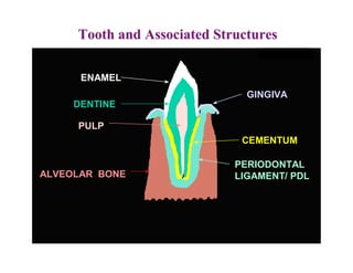
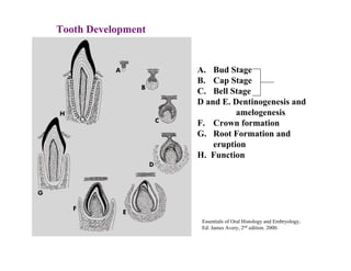
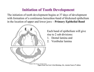
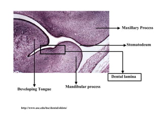
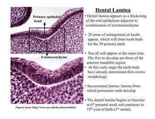
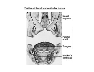
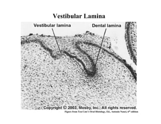
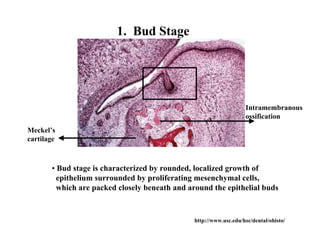
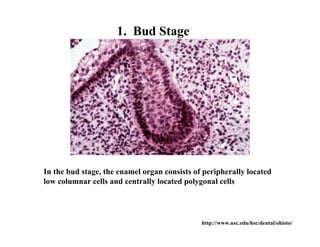
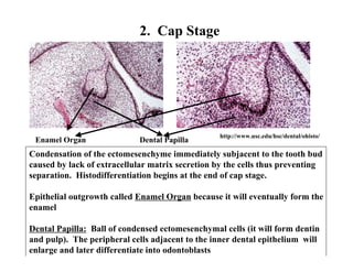
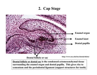
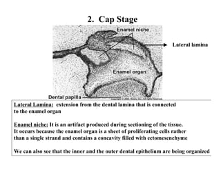
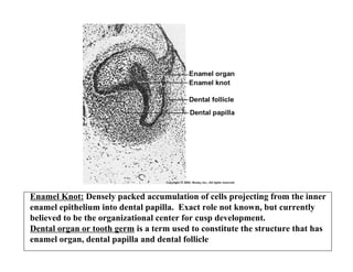
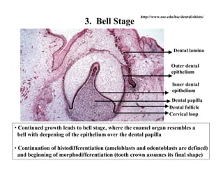
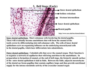
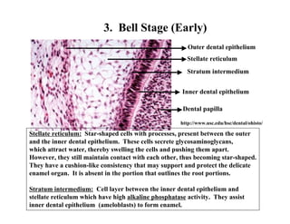
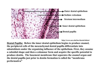
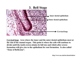
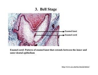
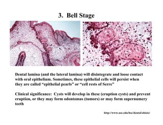
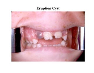
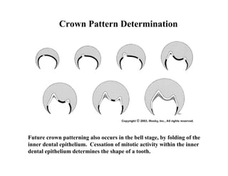
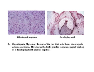
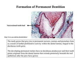
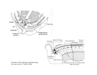
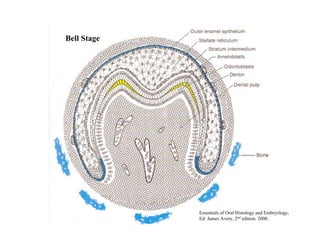
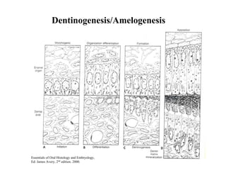
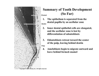
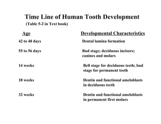
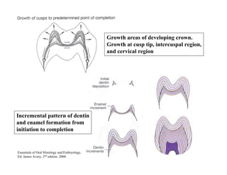
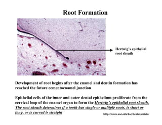
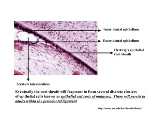
The document describes the development of teeth and supporting tissues. It discusses the initiation of tooth development from the primary epithelial band, the formation of the dental lamina and vestibular lamina. Tooth development occurs in 3 stages: the bud stage is characterized by localized epithelial growth, the cap stage shows epithelial outgrowth forming the enamel organ and dental papilla, and the bell stage resembles a bell shape with deepening epithelium over the dental papilla and histodifferentiation of ameloblasts and odontoblasts. The enamel organ and dental papilla develop further in the bell stage in preparation for enamel and dentin formation.
Cementum forms a functional unit which is designed to maintain tooth support, integrity, and protection.
Minor, non-pathological resorption defects on the root surface are generally reversible and heal by reparative cementum formation.
Irreversible damage may occur when the cementum is exposed to the environment of a pocket or oral cavity.
BE UPDATE TO IT,, AS IT IS 3 years back from 2017
Kindly mail me if you feel, needy of this presentation
you can find my mail id @ slide share,,, if not mail me @
sukesh3567@gmail.com.
Good luck
Upload By : Ahmed Ali Abbas
Babylon University College of Dentistry
download this file from Website on google theoptimalsmile.wix.com/dentistry
Oral histology
A Brief Description about the development of teeth. Understanding the process of tooth development is of particular importance for the dentist; as developmental disturbances may occur at any stage of this process resulting in different types of tooth anomalies.
Tooth development can be classified either based on morphology or histology
Morphological stages:
Bud stage
Cap stage
Bell stage:
* Early
* Advanced
Physiological stages:
Initiation
Proliferation
Histodifferentiation
Morphodifferentiation
Apposition
Knee anatomy and clinical tests 2024.pdfvimalpl1234
This includes all relevant anatomy and clinical tests compiled from standard textbooks, Campbell,netter etc..It is comprehensive and best suited for orthopaedicians and orthopaedic residents.
Title: Sense of Smell
Presenter: Dr. Faiza, Assistant Professor of Physiology
Qualifications:
MBBS (Best Graduate, AIMC Lahore)
FCPS Physiology
ICMT, CHPE, DHPE (STMU)
MPH (GC University, Faisalabad)
MBA (Virtual University of Pakistan)
Learning Objectives:
Describe the primary categories of smells and the concept of odor blindness.
Explain the structure and location of the olfactory membrane and mucosa, including the types and roles of cells involved in olfaction.
Describe the pathway and mechanisms of olfactory signal transmission from the olfactory receptors to the brain.
Illustrate the biochemical cascade triggered by odorant binding to olfactory receptors, including the role of G-proteins and second messengers in generating an action potential.
Identify different types of olfactory disorders such as anosmia, hyposmia, hyperosmia, and dysosmia, including their potential causes.
Key Topics:
Olfactory Genes:
3% of the human genome accounts for olfactory genes.
400 genes for odorant receptors.
Olfactory Membrane:
Located in the superior part of the nasal cavity.
Medially: Folds downward along the superior septum.
Laterally: Folds over the superior turbinate and upper surface of the middle turbinate.
Total surface area: 5-10 square centimeters.
Olfactory Mucosa:
Olfactory Cells: Bipolar nerve cells derived from the CNS (100 million), with 4-25 olfactory cilia per cell.
Sustentacular Cells: Produce mucus and maintain ionic and molecular environment.
Basal Cells: Replace worn-out olfactory cells with an average lifespan of 1-2 months.
Bowman’s Gland: Secretes mucus.
Stimulation of Olfactory Cells:
Odorant dissolves in mucus and attaches to receptors on olfactory cilia.
Involves a cascade effect through G-proteins and second messengers, leading to depolarization and action potential generation in the olfactory nerve.
Quality of a Good Odorant:
Small (3-20 Carbon atoms), volatile, water-soluble, and lipid-soluble.
Facilitated by odorant-binding proteins in mucus.
Membrane Potential and Action Potential:
Resting membrane potential: -55mV.
Action potential frequency in the olfactory nerve increases with odorant strength.
Adaptation Towards the Sense of Smell:
Rapid adaptation within the first second, with further slow adaptation.
Psychological adaptation greater than receptor adaptation, involving feedback inhibition from the central nervous system.
Primary Sensations of Smell:
Camphoraceous, Musky, Floral, Pepperminty, Ethereal, Pungent, Putrid.
Odor Detection Threshold:
Examples: Hydrogen sulfide (0.0005 ppm), Methyl-mercaptan (0.002 ppm).
Some toxic substances are odorless at lethal concentrations.
Characteristics of Smell:
Odor blindness for single substances due to lack of appropriate receptor protein.
Behavioral and emotional influences of smell.
Transmission of Olfactory Signals:
From olfactory cells to glomeruli in the olfactory bulb, involving lateral inhibition.
Primitive, less old, and new olfactory systems with different path
These simplified slides by Dr. Sidra Arshad present an overview of the non-respiratory functions of the respiratory tract.
Learning objectives:
1. Enlist the non-respiratory functions of the respiratory tract
2. Briefly explain how these functions are carried out
3. Discuss the significance of dead space
4. Differentiate between minute ventilation and alveolar ventilation
5. Describe the cough and sneeze reflexes
Study Resources:
1. Chapter 39, Guyton and Hall Textbook of Medical Physiology, 14th edition
2. Chapter 34, Ganong’s Review of Medical Physiology, 26th edition
3. Chapter 17, Human Physiology by Lauralee Sherwood, 9th edition
4. Non-respiratory functions of the lungs https://academic.oup.com/bjaed/article/13/3/98/278874
- Video recording of this lecture in English language: https://youtu.be/lK81BzxMqdo
- Video recording of this lecture in Arabic language: https://youtu.be/Ve4P0COk9OI
- Link to download the book free: https://nephrotube.blogspot.com/p/nephrotube-nephrology-books.html
- Link to NephroTube website: www.NephroTube.com
- Link to NephroTube social media accounts: https://nephrotube.blogspot.com/p/join-nephrotube-on-social-media.html
Basavarajeeyam is an important text for ayurvedic physician belonging to andhra pradehs. It is a popular compendium in various parts of our country as well as in andhra pradesh. The content of the text was presented in sanskrit and telugu language (Bilingual). One of the most famous book in ayurvedic pharmaceutics and therapeutics. This book contains 25 chapters called as prakaranas. Many rasaoushadis were explained, pioneer of dhatu druti, nadi pareeksha, mutra pareeksha etc. Belongs to the period of 15-16 century. New diseases like upadamsha, phiranga rogas are explained.
Explore natural remedies for syphilis treatment in Singapore. Discover alternative therapies, herbal remedies, and lifestyle changes that may complement conventional treatments. Learn about holistic approaches to managing syphilis symptoms and supporting overall health.
Lung Cancer: Artificial Intelligence, Synergetics, Complex System Analysis, S...Oleg Kshivets
RESULTS: Overall life span (LS) was 2252.1±1742.5 days and cumulative 5-year survival (5YS) reached 73.2%, 10 years – 64.8%, 20 years – 42.5%. 513 LCP lived more than 5 years (LS=3124.6±1525.6 days), 148 LCP – more than 10 years (LS=5054.4±1504.1 days).199 LCP died because of LC (LS=562.7±374.5 days). 5YS of LCP after bi/lobectomies was significantly superior in comparison with LCP after pneumonectomies (78.1% vs.63.7%, P=0.00001 by log-rank test). AT significantly improved 5YS (66.3% vs. 34.8%) (P=0.00000 by log-rank test) only for LCP with N1-2. Cox modeling displayed that 5YS of LCP significantly depended on: phase transition (PT) early-invasive LC in terms of synergetics, PT N0—N12, cell ratio factors (ratio between cancer cells- CC and blood cells subpopulations), G1-3, histology, glucose, AT, blood cell circuit, prothrombin index, heparin tolerance, recalcification time (P=0.000-0.038). Neural networks, genetic algorithm selection and bootstrap simulation revealed relationships between 5YS and PT early-invasive LC (rank=1), PT N0—N12 (rank=2), thrombocytes/CC (3), erythrocytes/CC (4), eosinophils/CC (5), healthy cells/CC (6), lymphocytes/CC (7), segmented neutrophils/CC (8), stick neutrophils/CC (9), monocytes/CC (10); leucocytes/CC (11). Correct prediction of 5YS was 100% by neural networks computing (area under ROC curve=1.0; error=0.0).
CONCLUSIONS: 5YS of LCP after radical procedures significantly depended on: 1) PT early-invasive cancer; 2) PT N0--N12; 3) cell ratio factors; 4) blood cell circuit; 5) biochemical factors; 6) hemostasis system; 7) AT; 8) LC characteristics; 9) LC cell dynamics; 10) surgery type: lobectomy/pneumonectomy; 11) anthropometric data. Optimal diagnosis and treatment strategies for LC are: 1) screening and early detection of LC; 2) availability of experienced thoracic surgeons because of complexity of radical procedures; 3) aggressive en block surgery and adequate lymph node dissection for completeness; 4) precise prediction; 5) adjuvant chemoimmunoradiotherapy for LCP with unfavorable prognosis.
Tom Selleck Health: A Comprehensive Look at the Iconic Actor’s Wellness Journeygreendigital
Tom Selleck, an enduring figure in Hollywood. has captivated audiences for decades with his rugged charm, iconic moustache. and memorable roles in television and film. From his breakout role as Thomas Magnum in Magnum P.I. to his current portrayal of Frank Reagan in Blue Bloods. Selleck's career has spanned over 50 years. But beyond his professional achievements. fans have often been curious about Tom Selleck Health. especially as he has aged in the public eye.
Follow us on: Pinterest
Introduction
Many have been interested in Tom Selleck health. not only because of his enduring presence on screen but also because of the challenges. and lifestyle choices he has faced and made over the years. This article delves into the various aspects of Tom Selleck health. exploring his fitness regimen, diet, mental health. and the challenges he has encountered as he ages. We'll look at how he maintains his well-being. the health issues he has faced, and his approach to ageing .
Early Life and Career
Childhood and Athletic Beginnings
Tom Selleck was born on January 29, 1945, in Detroit, Michigan, and grew up in Sherman Oaks, California. From an early age, he was involved in sports, particularly basketball. which played a significant role in his physical development. His athletic pursuits continued into college. where he attended the University of Southern California (USC) on a basketball scholarship. This early involvement in sports laid a strong foundation for his physical health and disciplined lifestyle.
Transition to Acting
Selleck's transition from an athlete to an actor came with its physical demands. His first significant role in "Magnum P.I." required him to perform various stunts and maintain a fit appearance. This role, which he played from 1980 to 1988. necessitated a rigorous fitness routine to meet the show's demands. setting the stage for his long-term commitment to health and wellness.
Fitness Regimen
Workout Routine
Tom Selleck health and fitness regimen has evolved. adapting to his changing roles and age. During his "Magnum, P.I." days. Selleck's workouts were intense and focused on building and maintaining muscle mass. His routine included weightlifting, cardiovascular exercises. and specific training for the stunts he performed on the show.
Selleck adjusted his fitness routine as he aged to suit his body's needs. Today, his workouts focus on maintaining flexibility, strength, and cardiovascular health. He incorporates low-impact exercises such as swimming, walking, and light weightlifting. This balanced approach helps him stay fit without putting undue strain on his joints and muscles.
Importance of Flexibility and Mobility
In recent years, Selleck has emphasized the importance of flexibility and mobility in his fitness regimen. Understanding the natural decline in muscle mass and joint flexibility with age. he includes stretching and yoga in his routine. These practices help prevent injuries, improve posture, and maintain mobilit
micro teaching on communication m.sc nursing.pdfAnurag Sharma
Microteaching is a unique model of practice teaching. It is a viable instrument for the. desired change in the teaching behavior or the behavior potential which, in specified types of real. classroom situations, tends to facilitate the achievement of specified types of objectives.
NVBDCP.pptx Nation vector borne disease control programSapna Thakur
NVBDCP was launched in 2003-2004 . Vector-Borne Disease: Disease that results from an infection transmitted to humans and other animals by blood-feeding arthropods, such as mosquitoes, ticks, and fleas. Examples of vector-borne diseases include Dengue fever, West Nile Virus, Lyme disease, and malaria.
The Gram stain is a fundamental technique in microbiology used to classify bacteria based on their cell wall structure. It provides a quick and simple method to distinguish between Gram-positive and Gram-negative bacteria, which have different susceptibilities to antibiotics
2. Tooth and Associated Structures
TOOTH & RELATED TISSUES: Developmental goal
WABeresford
ENAMEL
GINGIVA
DENTINE
PULP
CEMENTUM
PERIODONTAL
ALVEOLAR BONE LIGAMENT/ PDL
3. Tooth Development
A. Bud Stage
B. Cap Stage
C. Bell Stage
D and E. Dentinogenesis and
amelogenesis
F. Crown formation
G. Root Formation and
eruption
H. Function
Essentials of Oral Histology and Embryology,
Ed: James Avery, 2nd edition. 2000.
4. Initiation of Tooth Development
The initiation of tooth development begins at 37 days of development
with formation of a continuous horseshoe-band of thickened epithelium
in the location of upper and lower jaws – Primary Epithelial Band
Each band of epithelium will give
rise to 2 sub divisions:
1. Dental lamina and
2. Vestibular lamina
Figure from Ten Cate’s Oral Histology, Ed., Antonio Nanci, 6th edition
5. Maxillary Process
Stomatodeum
Dental lamina
Mandibular process
Developing Tongue
http://www.usc.edu/hsc/dental/ohisto/
6. Dental Lamina
Primary epithelial • Dental lamina appears as a thickening
band of the oral epithelium adjacent to
condensation of ectomesenchyme
• 20 areas of enlargement or knobs
appear, which will form tooth buds
for the 20 primary teeth
• Not all will appear at the same time.
Ectomesenchyme The first to develop are those of the
anterior mandible region
• At this early stage the tooth buds
have already determined their crown
morphology
• Successional lamina: lamina from
which permanent teeth develop
• The dental lamina begins to function
at 6th prenatal week and continues to
Figures from: http://www.usc.edu/hsc/dental/ohisto/
15th year of birth (3rd molar)
8. Vestibular Lamina
Figure from Ten Cate’s Oral Histology, Ed., Antonio Nanci, 6th edition
9. Tooth development is a continuous process, however can be
divided into 3 stages:
1. Bud Stage
2. Cap Stage
3. Bell Stage
10. 1. Bud Stage
Intramembranous
ossification
Meckel’s
cartilage
• Bud stage is characterized by rounded, localized growth of
epithelium surrounded by proliferating mesenchymal cells,
which are packed closely beneath and around the epithelial buds
http://www.usc.edu/hsc/dental/ohisto/
11. 1. Bud Stage
In the bud stage, the enamel organ consists of peripherally located
low columnar cells and centrally located polygonal cells
http://www.usc.edu/hsc/dental/ohisto/
13. 2. Cap Stage
http://www.usc.edu/hsc/dental/ohisto/
Enamel Organ Dental Papilla
Condensation of the ectomesenchyme immediately subjacent to the tooth bud
caused by lack of extracellular matrix secretion by the cells thus preventing
separation. Histodifferentiation begins at the end of cap stage.
Epithelial outgrowth called Enamel Organ because it will eventually form the
enamel
Dental Papilla: Ball of condensed ectomesenchymal cells (it will form dentin
and pulp). The peripheral cells adjacent to the inner dental epithelium will
enlarge and later differentiate into odontoblasts
14. 2. Cap Stage
Enamel organ
Enamel knot
Dental papilla
http://www.usc.edu/hsc/dental/ohisto/
Dental follicle or sac
Dental follicle or dental sac is the condensed ectomesenchymal tissue
surrounding the enamel organ and dental papilla. This gives rise to
cementum and the periodontal ligament (support structures for tooth)
15. 2. Cap Stage
Lateral lamina
Lateral Lamina: extension from the dental lamina that is connected
to the enamel organ
Enamel niche: It is an artifact produced during sectioning of the tissue.
It occurs because the enamel organ is a sheet of proliferating cells rather
than a single strand and contains a concavity filled with ectomesenchyme
We can also see that the inner and the outer dental epithelium are being organized
16. Enamel Knot: Densely packed accumulation of cells projecting from the inner
enamel epithelium into dental papilla. Exact role not known, but currently
believed to be the organizational center for cusp development.
Dental organ or tooth germ is a term used to constitute the structure that has
enamel organ, dental papilla and dental follicle
17. Enamel knots are clusters of nondividing epithelial cells visible
in sections of molar cap stage
Enamel knot precursor cells are first noted by expression of
p21 gene expression
Enamel knot and enamel cord are temporary structures that
disappear before enamel formation begins. It has been speculated
that the function of the enamel knot and cord may be to act as a
reservoir of dividing cells for the growing enamel organ
18. http://www.usc.edu/hsc/dental/ohisto/
3. Bell Stage
Dental lamina
Outer dental
epithelium
Inner dental
epithelium
Dental papilla
Dental follicle
Cervical loop
• Continued growth leads to bell stage, where the enamel organ resembles a
bell with deepening of the epithelium over the dental papilla
• Continuation of histodifferentiation (ameloblasts and odontoblasts are defined)
and beginning of morphodifferentiation (tooth crown assumes its final shape)
19. 3. Bell Stage (Early)
Outer dental epithelium
Stellate reticulum
Stratum intermedium
Inner dental epithelium
Dental papilla
http://www.usc.edu/hsc/dental/ohisto/
Inner dental epithelium: Short columnar cells bordering the dental papilla.
These will eventually become ameloblasts that will form the enamel of the
tooth crown by differentiating into tall columnar cells. The cells of inner dental
epithelium exert an organizing influence on the underlying mesenchymal cells
in the dental papilla, which later differentiate into odontoblasts.
Outer dental epithelium: Cuboidal cells that cover the enamel organ. Their function is
to organize a network of capillaries that will bring nutrition to the ameloblasts. In
preparation to formation of enamel, at the end of bell stage, the formerly smooth surface
of the outer dental epithelium is laid in folds. Between the folds, adjacent mesenchyme
of the dental sac forms papillae that contain capillary loops and thus provide nutritional
supply for the intense metabolic activity of the avascular enamel organ
20. 3. Bell Stage (Early)
Outer dental epithelium
Stellate reticulum
Stratum intermedium
Inner dental epithelium
Dental papilla
http://www.usc.edu/hsc/dental/ohisto/
Stellate reticulum: Star-shaped cells with processes, present between the outer
and the inner dental epithelium. These cells secrete glycosaminoglycans,
which attract water, thereby swelling the cells and pushing them apart.
However, they still maintain contact with each other, thus becoming star-shaped.
They have a cushion-like consistency that may support and protect the delicate
enamel organ. It is absent in the portion that outlines the root portions.
Stratum intermedium: Cell layer between the inner dental epithelium and
stellate reticulum which have high alkaline phosphatase activity. They assist
inner dental epithelium (ameloblasts) to form enamel.
21. Outer dental epithelium
Stellate reticulum
Stratum intermedium
Inner dental epithelium
Dental papilla
http://www.usc.edu/hsc/dental/ohisto/
Dental Papilla: Before the inner dental epithelium begins to produce enamel,
the peripheral cells of the mesenchymal dental papilla differentiate into
odontoblasts under the organizing influence of the epithelium. First, they assume
a cuboidal shape and then a columnar form and acquire the specific potential to
produce dentin. The basement membrane that separates the enamel organ and
the dental papilla just prior to dentin formation is called the “membrana
preformativa”
23. 3. Bell Stage
Inner dental epithelium
Outer dental epithelium
Cervical loop
Cervical loop: Area where the inner and the outer dental epithelium meet at
the rim of the enamel organ. This point is where the cells will continue to
divide until the tooth crown attains its full size and which after crown
formation will give rise to the epithelium for root formation. Is also called
“Zone of Reflexion”.
http://www.usc.edu/hsc/dental/ohisto/
24. 3. Bell Stage
Enamel knot
Enamel cord
Enamel cord: Pattern of enamel knot that extends between the inner and
outer dental epithelium
http://www.usc.edu/hsc/dental/ohisto/
25. 3. Bell Stage
Dental lamina (and the lateral lamina) will disintegrate and loose contact
with oral epithelium. Sometimes, these epithelial cells will persist when
they are called “epithelial pearls” or “cell rests of Serre”
Clinical significance: Cysts will develop in these (eruption cysts) and prevent
eruption, or they may form odontomas (tumors) or may form supernumery
teeth
http://www.usc.edu/hsc/dental/ohisto/
27. Crown Pattern Determination
Future crown patterning also occurs in the bell stage, by folding of the
inner dental epithelium. Cessation of mitotic activity within the inner
dental epithelium determines the shape of a tooth.
28. Vascular and Nerve Supply during Tooth Development
Vascular Supply: Clusters of blood vessels in dental follicle and papilla
Clustering of vessels in papilla coincide with position
of root formation
Enamel organ is avascular, however vessels seen in
close association in the follicle
Nerve Supply: Initially noted in the dental follicle during bud to cap stage
However after start of dentinogenesis, seen in dental papilla
Nerve fibers do not enter enamel organ
29. Clinical Correlation. Several odontogenic cysts and tumors can arise
from developing tooth structures. Two such conditions are:
1. Ameloblastoma – which are tumors of odontogenic epithelium that
may arise from cell rests of enamel organ or from the developing
enamel organ among other things
30. Ameloblastoma Enamel Organ
Histology resembles enamel organ epithelium with peripheral columnar
ameloblast-like cells surrounding loosely arranged stellate-reticulum-like cells
31. Odontogenic myxoma Developing tooth
2. Odontogenic Myxoma: Tumor of the jaw that arise from odontogenic
ectomesenchyme. Histologically, looks similar to mesenchymal portion
of a developing tooth (dental papilla).
32. Formation of Permanent Dentition
Successional tooth bud
http://www.usc.edu/hsc/dental/ohisto/
The tooth germs that give rise to permanent incisors, canines and premolars form
as a result of further proliferative activity within the dental lamina, lingual to the
deciduous tooth germ
The developing permanent molars have no deciduous predecessor and their tooth
germs originate from the dental lamina that extends posteriorly beneath the oral
epithelium after the jaws have grown
33. Essentials of Oral Histology and Embryology,
Ed: James Avery, 2nd edition. 2000.
34. A timetable to remember
Entire primary dentition initiated between 6 and 8 weeks of embryonic
development.
Successional permanent teeth initiated between 20th week in utero
and 10th month after birth
Permanent molars between 20th week in utero (first molar) and 5th year of
life (third molar)
35. The development of hard tissues will also be discussed by
Dr. Sandra Myers, however
You must remember the following:
• Hard tissue formation starts at the late stages of the bell
stage
• Differentiation of cells into odontoblasts and ameloblasts
• Dentin is formed before enamel
• Dentin initiates the formation of enamel
36. Bell Stage
Essentials of Oral Histology and Embryology,
Ed: James Avery, 2nd edition. 2000.
37. Hard Tissue Formation
Deposition of dental hard tissues is
called “apposition”
After the crown attains its final
shape during cap to early bell stage,
the inner dental epithelial cells stop
to proliferate, except the cells at the
cervical loop
First layer of dentin appears at the
cusp tips and progresses cervically,
and the columnar cells of the inner
dental epithelium become elongated
and show reverse polarization,
with the nuclei adjacent to stratum
intermediate (ameloblasts)
The boundary between the odontoblasts
and inner dental epithelium defines the
http://www.usc.edu/hsc/dental/ohisto/ future dentino-enamel junction
38. For dentinogenesis and amelogenesis to take place normally,
the differentiating odontoblasts and ameloblasts will receive
signals form each other – “reciprocal induction”
Stages of Apposition
1. Elongation of inner dental epithelium
2. Differentiation of odontoblasts
3. Formation of dentin
4. Formation of enamel
39. Ameloblasts
First layer of enamel
Dentin
Odontoblasts
At the same time or soon after the first layer of dentin (mantle dentin) is formed,
the inner dental epithelial cells differentiate into ameloblasts and secrete enamel
proteins. These proteins further will help in the terminal differentiation of
odontoblasts. The ameloblasts will then start laying down organic matrix of
enamel against the newly formed dentinal surface. The enamel matrix will
mineralize immediately and form the first layer of enamel. The formation of
enamel is called amelogenesis. http://www.usc.edu/hsc/dental/ohisto/
40. Apposition
At the same time when the inner
dental epithelium is differentiating,
the undifferentiated ectomesenchymal
cells increase rapidly in size and
ultimately differentiate into odontoblasts
The increase in size of the papillary
cells leads to elimination of the acellular
zone between dental papilla and inner
dental epithelium
Differentiation of odontoblasts from
ectomesenchymal cells are induced by
influence from the inner dental epithelium
Experiments have shown that if there is
no inner dental epithelium, there is no
Odontoblasts Dentin Ameloblasts dentin formed
Enamel http://www.usc.edu/hsc/dental/ohisto/
41. Essentials of Oral Histology and Embryology,
Ed: James Avery, 2nd edition. 2000.
42. Dentinogenesis
Dentin is formed by odontoblasts that differentiate from ectomesenchymal
cells of dental papilla with influence from the inner dental epithelium
Differentiation of odontoblasts is mediated by expression of signaling
molecules and growth factors in the inner dental epithelial cells
http://www.usc.edu/hsc/dental/ohisto/
43. Immediately after the inner dental epithelial cells undergo reverse polarization,
ectomesenchymal cells immediately subjacent to the acellular layer, will rapidly
enlarge and elongate to become odontoblasts and appear like protein-producing
cell. The acellular layer is eliminated and the odontoblasts will occupy this zone
Odontoblasts are highly polarized with the nuclei away from inner dental epith.
Following differentiation of odontoblasts, first layer of dentin is produced,
characterized by appearance of large-diameter type III collagen fibrils (0.1
to 0.2 µm in dia) called von Korff’s fibers, followed by type I collagen fibers
– MANTLE DENTINE
At the same time as initial dentin deposition, the odontoblasts will develop stubby
processes at the side close to the inner dental epithelium which extend into
forming extracellular matrix
As the odontoblasts move pulpward, the odontoblast process (Tomes´ fiber) will
elongate and become active in dentine matrix formation
It is initially called predentin and following mineralization is called dentin
45. The odontoblasts as they differentiate will start elaborating organic matrix
of dentin, which will mineralize. As the organic matrix of dentin is deposited,
the odontoblasts move towards the center of the dental papilla, leaving behind
cytoplasmic extensions which will soon be surrounded by dentin. Therefore,
a tubular structure of dentin is formed.
ameloblasts
odontoblasts dentin
http://www.usc.edu/hsc/dental/ohisto/
46. Odontoblasts with cytoplasmic processes forming dentinal tubules
2 steps of dentinogenesis: 1. Formation of collagen matrix
2. Deposition of calcium and phosphate
(hydroxyapatite) crystals in the matrix
http://www.usc.edu/hsc/dental/ohisto/
47. Amelogenesis is also a two-step process:
1. First step produces a partially mineralized matrix (~ 30%)
2. Second step involves influx of additional mineral coincident
with removal of organic material and water to attain greater
than 96% mineral content
48. Amelogenesis
Essentials of Oral Histology and Embryology,
Ed: James Avery, 2nd edition. 2000.
50. Amelogenesis
Amelogenesis begins after a few µm of dentin deposition at the
dentinoenamel junction
Ameloblasts goes through following functional stages:
1. Morphogenetic. During this stage the shape of the crown is determined.
2. Histodifferentiation. The cells of the inner dental epithelium is
differentiating into ameloblasts. The above two stages are the presecretory
stages, where the cells differentiate, acquire phenotype, change polarity,
develop an extensive protein synthesis machinery, and prepare to secrete an
organic matrix of enamel.
3. Secretory stage: Ameloblasts elaborate and organize the entire enamel
thickness. Short conical processes called Tomes´ processes develop at the
apical end of the ameloblasts. The main protein that accumulates is
amelogenin.
51. Essentials of Oral Histology and Embryology,
Ed: James Avery, 2nd edition. 2000.
52. Amelogenesis
4. Maturation stage: Ameloblasts modulate and transport specific ions
required for the concurrent accretion of mineral. At this stage,
ameloblast becomes more active in absorption of the organic matrix
and water, which allows mineralization to proceed. After the
ameloblasts have completed their contributions to the mineralization
phase, they secrete an organic cuticle on the surface of the enamel,
which is called developmental or primary cuticle
5. Protection: The ameloblast are shorter and contact the stratum
intermedium and outer dental epithelium and fuse to form the
reduced dental (enamel) epithelium. The reduced enamel epithelium
remains until the tooth erupts. As the tooth erupts and passes
through the oral epithelium, the incisal part of the reduced dental
epithelium is destroyed but the epithelium present cervically
interacts with oral epithelium to become the junctional epithelium
55. Summary of Tooth Development
(So Far)
1. The epithelium is separated from the
dental papilla by an acellular zone
2. Inner dental epithelial cells are elongated,
and the acellular zone is lost by
differentiation of odontoblasts
3. Odontoblasts retreat toward the center
of the pulp, leaving behind dentin
4. Ameloblasts begin to migrate outward and
leave behind formed enamel
56. Time Line of Human Tooth Development
(Table 5-2 in Text book)
Age Developmental Characteristics
42 to 48 days Dental lamina formation
55 to 56 days Bud stage; deciduous incisors;
canines and molars
14 weeks Bell stage for deciduous teeth; bud
stage for permanent teeth
18 weeks Dentin and functional ameloblasts
in deciduous teeth
32 weeks Dentin and functional ameloblasts
in permanent first molars
57. Growth areas of developing crown.
Growth at cusp tip, intercuspal region,
and cervical region
Incremental pattern of dentin
and enamel formation from
initiation to completion
Essentials of Oral Histology and Embryology,
Ed: James Avery, 2nd edition. 2000.
58. Root Formation
Hertwig’s epithelial
root sheath
Development of root begins after the enamel and dentin formation has
reached the future cementoenamel junction
Epithelial cells of the inner and outer dental epithelium proliferate from the
cervical loop of the enamel organ to form the Hertwig’s epithelial root sheath.
The root sheath determines if a tooth has single or multiple roots, is short or
long, or is curved ir straight http://www.usc.edu/hsc/dental/ohisto/
59. Inner dental epithelium
Outer dental epithelium
Hertwig’s epithelial
root sheath
Stratum intermedium
Eventually the root sheath will fragment to form several discrete clusters
of epithelial cells known as epithelial cell rests of malassez. These will persist in
adults within the periodontal ligament
http://www.usc.edu/hsc/dental/ohisto/
60. Epithelial Cell Rests of Malassez
The epithelial rests appear as small clusters of epithelial cells which
are located in the periodontal ligament adjacent to the surface of
cementum. They are cellular residues of the embryonic structure
known as Hertwig's epithelial root sheath.
http://www.usc.edu/hsc/dental/ohisto/
61. Epithelial diaphragm: the proliferating
end of the root sheath bends at a near
45-degree angle. The epithelial
diaphragm will encircle the apical
opening of the dental pulp during root
development
Primary apical formen
http://www.usc.edu/hsc/dental/ohisto/
62. Secondary apical foramen form as a result of two or three tongues of
epithelium growing inward toward each other resulting in multirooted teeth
Essentials of Oral Histology and Embryology,
Ed: James Avery, 2nd edition. 2000
63. Direction of root growth versus eruptive movement of tooth
Essentials of Oral Histology and Embryology,
Ed: James Avery, 2nd edition. 2000.
64. Tooth eruption and Development of
supporting structures
Soon after root formation begins, tooth begins to erupt until it reaches
its final position
While roots are forming, the supporting structures of tooth also
develop – periodontal ligament and cementum
As the root sheath fragments, the dental follicle cells will penetrate between
the epithelial cells and lie close to the newly formed root dentin
These cells will differentiate into cementoblasts, which will make cementum
Fibers of the periodontal ligament, which will also form from the cells of the
dental follicle will get anchored in the organic matrix of the cementum which
will later get mineralized
65. Oral Ectoderm and Tooth Patterning
Is the epithelium or the mesenchyme responsible for tooth morphology?
Recombination experiments show that
at early (E10.5) time point the epithelium
directs patterning, however at later time
(E11.0) mesenchyme directs patterning –
Reciprocal Signaling
66. What are the molecular mediators of this patterning information?
Future oral ectoderm has acquires a concept of “pre-pattern” through the nested
expression of fibroblast growth factors (Fgf), sonic hedgehog (Shh) and Bmps.
These signals are then interpreted and refined by the underlying mesenchyme
into spatially restricted domains of homeobox gene expression, which in turn
regulate other signaling molecules (Bmp, Wnt and Fgf) that induce the epithelial
folding and invagination that signal the initiation of tooth development
Proximal-distal patterning (what decides a tooth is incisor or molar)?
Rostral – caudal patterning
Tooth Number
Shape of the resulting tooth
67. Dlx and Barx – Molar region
Msx – Incisor region
68. FGF8 activates LIM homeobox genes (Lhx 6 and 7),
which is unaffected by BMP4. So Lhx6 and 7 are
expressed throughout the oral half of the mesenchyme.
Lhx 6/7 expressed (oral portion of mesenchyme).
Marks the region where tooth buds form
Goosecoid (Gsc) expression.
Marks the aboral portion of the mesenchyme
Therefore the jaw is divided into a tooth-forming
LHX-positive domain and a non-tooth-forming
GSC-positive domain. In mice, in which Gsc
has been knocked out, the teeth form normally
but the supporting skeletal structures in the
aboral region are absent – thus defining the
rostral-caudal patterning
Overall called Odontogenic Homeobox Code
69. What are the signals responsible for initiation of tooth bud?
In the epithelium: SHH
expression marks the sites of
tooth development
In the mesenchyme, PAX9
and Activin expression
marks the sites of future
epithelial invagination
Pax9 KO - tooth development arrested at the bud stage
Runx2 KO – arrest at bud stage however upper molars make it to an aberrant
cap stage. Difference explained by the expression of Runx3 in the maxilla
and not mandible.
70. What is responsible for shape of tooth?
Enamel knots express a wealth of signaling molecules such as SHH, members of
FGFs, BMPs and WNT families along with p21(cell cycle) and
MSX2 (transcription factor)
High levels of apoptosis also occurs at the enamel knot thereby leading to loss of
structure and signaling center at late-cap to early bell stage
Secondary and tertiary enamel knots develop in molar tooth germs but not
in incisor tooth germs – thereby more cusps
These extra enamel knots express Fgf4