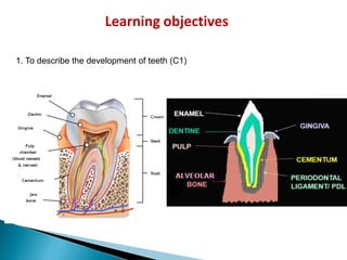
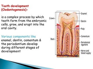
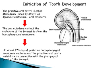
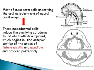
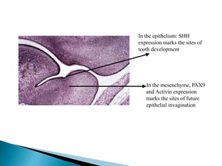
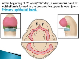
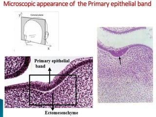
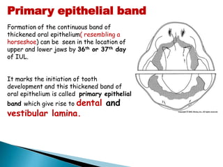
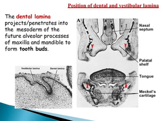
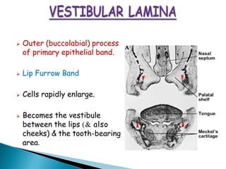
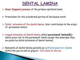
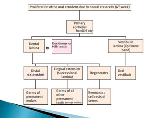
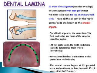
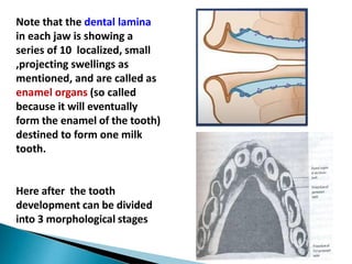
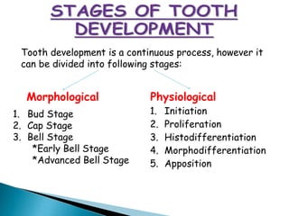
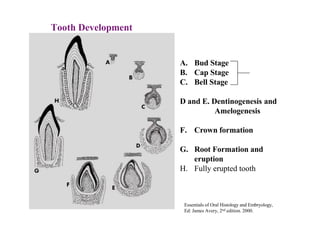
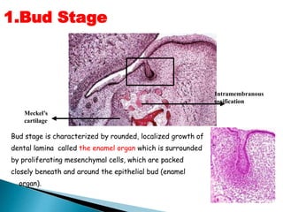
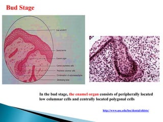
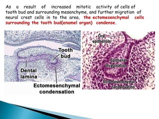
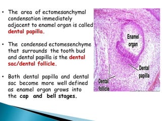
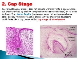
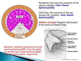
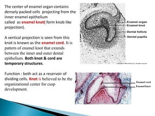
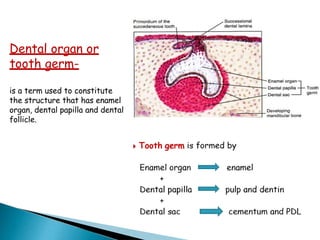
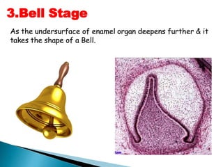
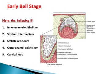
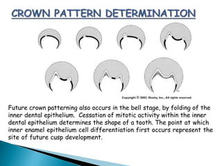
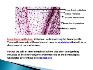
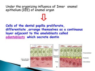
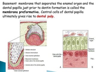
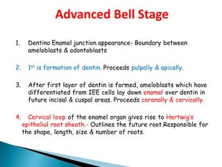
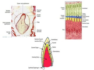
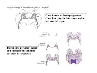
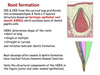
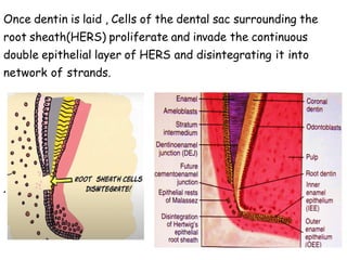
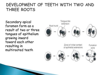
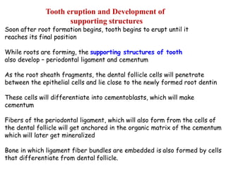
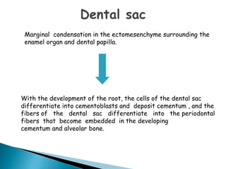
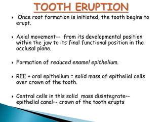
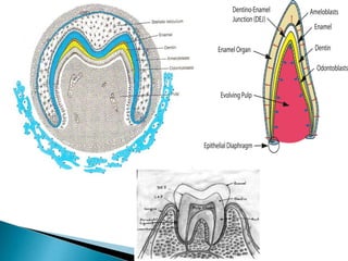
1. Development of teeth
(Odontogenesis)
All the information, including the images and pics
collected from different sources is strictly for teaching
purposes only.
G R N 1
3. Tooth development
(Oodontogenesis)-
is a complex process by which
teeth form from the embryonic
cells, grow, and erupt into the
oral cavity.
Various components like
enamel, dentin, cementum &
the periodontium develop
during different stages of
development
4. The primitive oral cavity is called
stomodeum – lined by stratified
squamous epithelium - oral ectoderm.
The oral ectoderm contact the
endoderm of the foregut to form the
buccopharyngeal membrane.
At about 27th day of gestation buccopharyngeal
membrane ruptures and the primitive oral cavity
establishes a connection with the pharyngeal
part of the foregut.
5. Most of mesoderm cells underlying
the oral ectoderm are of neural
crest origin.
These mesodermal cells
induce the overlying ectoderm
to initiate tooth development,
which begins in the anterior
portion of the areas of
future maxilla and mandible
and prooced posteriorly
6. What are the signals responsible for initiation of tooth bud
formation?
In the epithelium: SHH
expression marks the sites of
tooth development
In the mesenchyme, PAX9
and Activin expression
marks the sites of future
epithelial invagination
7. At the beginning of 6th week(~36th day), a continuous band of
epithelium is formed in the presumptive upper & lower jaws-
Primary epithelial band.
9. Formation of the continuous band of
thickened oral epithelium( resembling a
horseshoe) can be seen in the location of
upper and lower jaws by 36th or 37th day
of IUL.
It marks the initiation of tooth
development and this thickened band of
oral epithelium is called primary epithelial
band which give rise to dental and
vestibular lamina.
Primary epithelial band
10. Position of dental and vestibular lamina
The dental lamina
projects/penetrates into
the mesoderm of the
future alveolar processes
of maxilla and mandible to
form tooth buds.
11. ➢ Outer (buccolabial) process
of primary epithelial band.
➢ Lip Furrow Band
➢ Cells rapidly enlarge.
➢ Becomes the vestibule
between the lips (& also
cheeks) & the tooth-bearing
area.
12. ➢ Inner (lingular) process of the primary epithelial band.
➢ Primordium for the ectodermal portion of deciduous teeth.
➢ Distal extension of the dental lamina, later contributes to the origin
of permanent molars
➢ Lingual extension of dental lamina called successional lamina(SL)
which gives rise to the permanent teeth except the molars(as they
are given by distal extension of dental lamina).
➢ Remnants of dental lamina persists as epithelial pearls or islands
within the jaw as well as gingiva-- Cell rests of Serres.
13.
14. 20 areas of enlargement(rounded swellings)
or knobs appear(10 in each jaw) which
will form tooth buds for the 20 primary/milk
teeth. These epithelial part of the tooth
germs/buds are known as the enamel
organs.
•Not all will appear at the same time. The
first to develop are those of the anterior
mandible region
• At this early stage, the tooth buds have
already determined their crown
morphology
•Successional lamina: lamina from which
permanent teeth develop
•The dental lamina begins at 6th prenatal
week and continues to function until 15-18
years of birth (3rd molar)
15. Note that the dental lamina
in each jaw is showing a
series of 10 localized, small
,projecting swellings as
mentioned, and are called as
enamel organs (so called
because it will eventually
form the enamel of the tooth)
destined to form one milk
tooth.
Here after the tooth
development can be divided
into 3 morphological stages
16. Morphological Physiological
1. Initiation
2. Proliferation
3. Histodifferentiation
4. Morphodifferentiation
5. Apposition
1. Bud Stage
2. Cap Stage
3. Bell Stage
*Early Bell Stage
*Advanced Bell Stage
Tooth development is a continuous process, however it
can be divided into following stages:
17. Tooth Development
A. Bud Stage
B. Cap Stage
C. Bell Stage
D and E. Dentinogenesis and
Amelogenesis
F. Crown formation
G. Root Formation and
eruption
H. Fully erupted tooth
Essentials of Oral Histology and Embryology,
Ed: James Avery, 2nd edition. 2000.
18. 1.Bud Stage
Intramembranous
ossification
Meckel’s
cartilage
Bud stage is characterized by rounded, localized growth of
dental lamina called the enamel organ which is surrounded
by proliferating mesenchymal cells, which are packed
closely beneath and around the epithelial bud (enamel
organ).
19. Bud Stage
In the bud stage, the enamel organ consists of peripherally located
low columnar cells and centrally located polygonal cells
http://www.usc.edu/hsc/dental/ohisto/
20. As a result of increased mitotic activity of cells of
tooth bud and surrounding mesenchyme, and further migration of
neural crest cells in to the area, the ectomesenchymal cells
surrounding the tooth bud(enamel organ) condense.
21. • The area of ectomesanchymal
condensation immediately
adjacent to enamel organ is called
dental papilla.
• The condensed ectomesenchyme
that surrounds the tooth bud
and dental papilla is the dental
sac/dental follicle.
• Both dental papilla and dental
sac become more well defined
as enamel organ grows into
the cap and bell stages.
22. Tooth bud(Enamel organ) does not expand uniformly into a large sphere,
but characterised by shallow invagination (assumes cup shape) on its deep
surface . The dental Papilla (condensed mass of ectomesenchymal
cells) occupy this cup of enamel organ. At this stage the developing
tooth looks like a cap ,hence called cap stage of development
2. Cap Stage
24. Peripheral cells lining the convexity of the
cap are cuboidal –Outer Enamel
Epithelium(OEE).
Cells lining the concavity of the cap
become tall, columnar– Inner Enamel
Epithelium(IEE)
Stellate reticulum: Polygonal cells located
in the centre of Enamel Organ.
Basement membrane separates the outer
enamel epithelium(OEE) from the dental
sac and the inner enamel epithelium(IEE)
from the dental papilla respectively.
25. The center of enamel organ contains
densely packed cells projecting from the
inner enamel epithelium
called as enamel knot( form knob like
projection).
A vertical projection is seen from this
knot is known as the enamel cord. It is
pattern of enamel knot that extends
between the inner and outer dental
epithelium. Both knot & cord are
temporary structures.
Function : both act as a reservoir of
dividing cells. Knot is believed to be the
organizational center for cusp
development.
26. Enamel niche: It is an artifact produced during sectioning of the tissue. It
occurs because the enamel organ/tooth bud is a sheet of proliferating cells
rather than a single strand and contains a concavity filled with
ectomesenchyme
We can also see that the inner and the outer dental epithelium are being organized
27. Dental organ or
tooth germ-
is a term used to constitute
the structure that has enamel
organ, dental papilla and dental
follicle.
28. As the undersurface of enamel organ deepens further & it
takes the shape of a Bell.
3.Bell Stage
29. Note the following !!!
1. Inner enamel epithelium
2. Stratum intermedium
3. Stellate reticulum
4. Outer enamel epithelium
5. Cervical loop
Early Bell Stage
30. The enamel organs of deciduous teeth in the bell stage show
successional lamina and their permanent successor teeth in
the bud stage.
31. • Consists of tall columnar cells(~ 40 µm
long, ~ 5 µm in diameter)
• Cells of IEE attached by desmosomes to
cells of stratum intermedium but
separated from cells of dental papilla by
basement membrane.(BM).
• IEE cells differentiate into ameloblasts
prior to enamel formation.
• Cells of IEE stimulate underlying
mesenchymal cells in the dental papilla,
which later differentiate into
odontoblasts.
1.Inner Enamel Epithelium
32. Future crown patterning also occurs in the bell stage, by folding of the
inner dental epithelium. Cessation of mitotic activity within the inner
dental epithelium determines the shape of a tooth. The point at which
inner enamel epithelium cell differentiation first occurs represent the
site of future cusp development.
33. Inner dental epithelium: Columnar cells bordering the dental papilla.
These will eventually differentiate and become ameloblasts that will form
the enamel of the tooth crown.
Further the cells of inner dental epithelium also exert an organizing
influence on the underlying mesenchymal cells of the dental papilla,
which later differentiate into odontoblasts
34. Inner enamel epithelium consists of
a single layer of columnar cells
Differentiate into ameloblasts
35. Under the organizing influence of Inner enamel
epithelium (IEE) of enamel organ
Cells of the dental papilla proliferate,
differentiate ,arrange themselves as a continuous
layer adjacent to the ameloblasts called
odontoblasts which secrete dentin
36. Basement membrane that separates the enamel organ and the
dental papilla just prior to dentin formation is called the
membrana preformativa. Central cells of dental papilla
ultimately gives rise to dental pulp.
37. 2. Stratum intermedium
Few layers of squamous cells
between the IEE and the stellate
reticulum form the stratum
intermedium.
Cells are closely attached by
desmosomes & gap junctions.
Metabolically very active cells-
work synergistically with cells of
IEE & form Enamel.
38. 3. Stellate Reticulum (SR)
• Polygonal(star shaped) cells located
between the outer and inner enamel
epithelia with long processes that
anastomose with adjacent cells.
• Cells attached to OEE and SI by
desmosomes.
• Expand further by an increase in
intercellular fluid and cells assume a
branched reticular form.
• The spaces in this reticular network are
filled with a mucoid fluid which gives
the stellate reticulum a cushion like
consistency that may support and
protect the delicate enamel-forming
39. Just before enamel formation SR collapses, reducing the
distance between the centrally situated ameloblasts and the
nutrient capillaries near the OEE
40. These cells are low cuboidal in form.
At the end of the bell stage, the
formerly smooth surface of the
outer enamel epithelium(OEE) is laid in
folds.
Between these folds, the adjacent
mesenchyme of the dental sac
forms papillae that contain capillary
loops to provide a rich nutritional
supply for the intense metabolic
activity of the avascular enamel
organ.
4.Outer Enamel Epithelium
41. 5. Cervical loop:
The region where the inner and outer enamel epithelia
meet at the rim of the enamel organ is known as the zone
of reflexion or cervical loop.
42.
43. 1. Dentino Enamel junction appearance- Boundary between
ameloblasts & odontoblasts
2. 1st is formation of dentin. Proceeds pulpally & apically.
3. After first layer of dentin is formed, ameloblasts which have
differentiated from IEE cells lay down enamel over dentin in
future incisal & cuspal areas. Proceeds coronally & cervically.
4. Cervical loop of the enamel organ gives rise to Hertwig’s
epithelial root sheath.- Outlines the future root.Responsible for
the shape, length, size & number of roots.
Advanced Bell Stage
44.
45. Incremental pattern of dentin
and enamel formation from
initiation to completion
Growth areas of developing crown.
Growth at cusp tip, intercuspal region,
and cervical region
46. IEE & OEE from the cervical loop proliferate
into ectomesenchyme & form a 2 layered
structure known as Hertwig’s epithelial root
sheath (HERS) which encloses more of dental
papilla cells.
HERS determines shape of the roots
➢short or long
➢Single or multiple
➢
Straight or curved,
and initiates radicular dentin formation.
Root develops after enamel & dentin formation
have reached future Cemento-Enamel Junction.
Note the structural components of the HERS in
the figure (outer and inner enamel epithelium)
Root formation
47. Epithelial diaphragm: the proliferating
end of the root sheath(HERS) bends at a
near 45-degree angle. The epithelial
diaphragm will encircle the apical
opening of the dental pulp during root
development
48. HERS induces differentiation of peripheral radicular dental
papilla cells into odontoblasts which begin to deposit
dentin.(deposition follows along the lengthening of root
sheath)
49. Once dentin is laid , Cells of the dental sac surrounding the
root sheath(HERS) proliferate and invade the continuous
double epithelial layer of HERS and disintegrating it into
network of strands.
.
50. Disintegration of the HERS allows contact of the dental
follicle cells with the outer surface of the dentin and they
differentiate into cementoblasts and deposit cementum covering the
root dentin( entire surface of the dentin along the root surface).
51. • Remnants of HERS persist as an epithelial network of strands
or clumps near the external surface of the root.
• They are found in the PDL of erupted teeth-rests of Malassez
52. The first layer of dentin has been laid down
the epithelial root sheath loses its structural
continuity & its close relation to the surface of the
root.
Its remnants persists as an epithelial network of
strands or tubules near the external surface of the
root.
These epithelial remnants are found in the periodontal
ligament of erupted teeth
called rests of malassez.
53. Direction of root growth versus eruptive movement of tooth
Essentials of Oral Histology and Embryology,
Ed: James Avery, 2nd edition. 2000.
54. Secondary apical
foramen form as a
result of two or three
tongues of epithelium
growing inward
toward each other
resulting in
multirooted teeth
55. Soon after root formation begins, tooth begins to erupt until it
reaches its final position
While roots are forming, the supporting structures of tooth
also develop – periodontal ligament and cementum
As the root sheath fragments, the dental follicle cells will penetrate
between the epithelial cells and lie close to the newly formed root dentin
These cells will differentiate into cementoblasts, which will make
cementum
Fibers of the periodontal ligament, which will also form from the cells of
the dental follicle will get anchored in the organic matrix of the cementum
which will later get mineralized
Bone in which ligament fiber bundles are embedded is also formed by cells
that differentiate from dental follicle.
Tooth eruption and Development of
supporting structures
56. With the development of the root, the cells of the dental sac
differentiate into cementoblasts and deposit cementum , and the
fibers of the dental sac differentiate into the periodontal
fibers that become embedded in the developing
cementum and alveolar bone.
Marginal condensation in the ectomesenchyme surrounding the
enamel organ and dental papilla.
57. Once root formation is initiated, the tooth begins to
erupt.
Axial movement-- from its developmental position
within the jaw to its final functional position in the
occlusal plane.
Formation of reduced enamel epithelium.
REE + oral epithelium = solid mass of epithelial cells
over crown of the tooth.
Central cells in this solid mass disintegrate–-
epithelial canal–- crown of the tooth erupts
58.
59. ➢ Blood vessels grow in the dental follicle and enter the
dental papilla in cap stage and the dental papilla
eventually forms in the pulp of a tooth.
➢ Enamel organ is avascular, however vessels seen in close
association in the follicle
➢ Nerve fibres approach developing tooth, during its bud
stage of development and ramify ,form a rich plexus
around the tooth germ . They penetrate dental papilla,
sensory for future pulp and PDL. Nerve fibers do not
enter enamel organ !
Vascular supply and Nerve supply