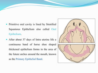
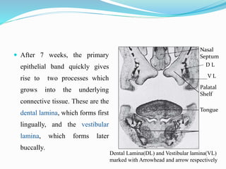
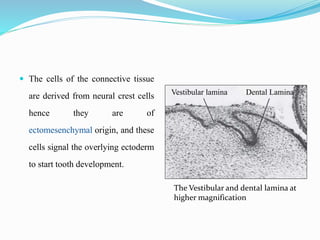
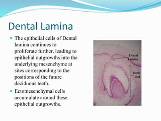
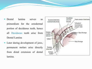
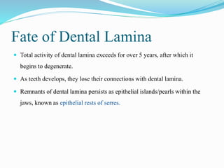
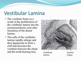
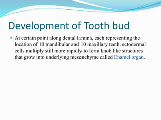
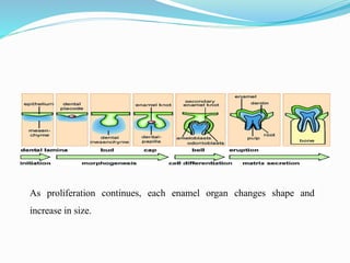
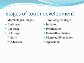
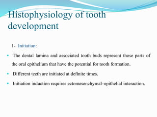
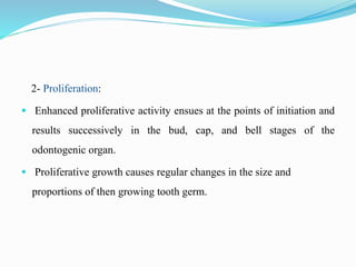
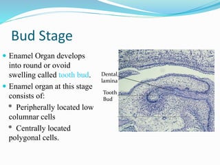
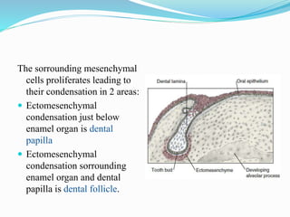
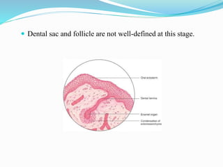
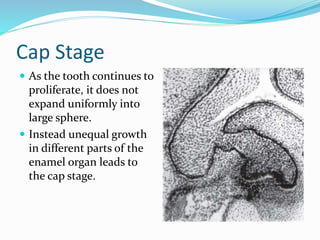
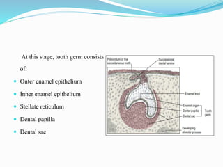
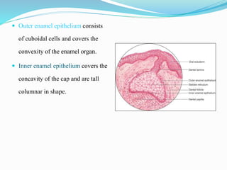
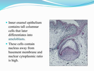
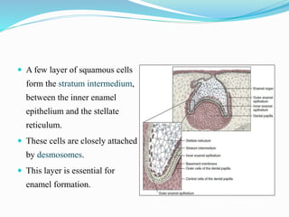
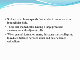
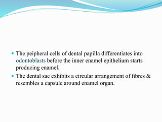
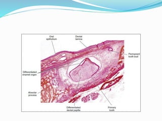
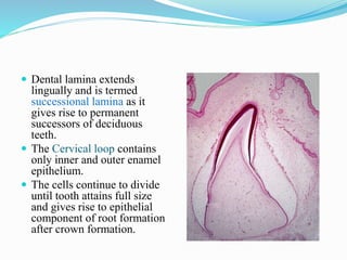
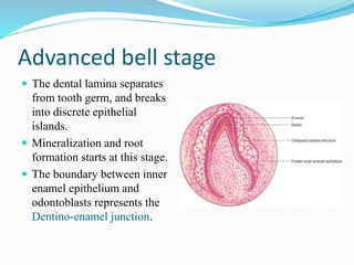
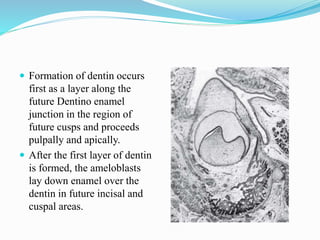
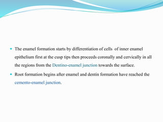
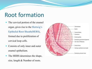
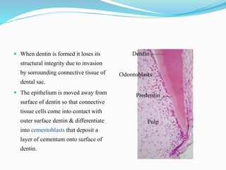
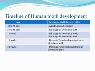
Jishnu Kinkor Goswami
Contents
 Introduction
 Dental Lamina
 Vestibular lamina
 Development of tooth bud
 Stages of tooth development
 Histophysiological Stages
 Bud stage
 Cap stage
 Bell stage
 Root formation
 References
Introduction
 The Process of tooth development is known as Odontogenesis
 Understanding the process of tooth development is of particular
importance for the dentist; as developmental disturbances may
occur at any stage of this process resulting in different types of
tooth anomalies.
 Primitive oral cavity is lined by Stratified
Squamous Epithelium also called Oral
Epithelium.
 After about 37 days of Intra uterine life a
continuous band of horse shoe shaped
thickened epithelium forms in the area of
the future arches around the mouth, known
as the Primary Epithelial Band.
 After 7 weeks, the primary
epithelial band quickly gives
rise to two processes which
grows into the underlying
connective tissue. These are the
dental lamina, which forms first
lingually, and the vestibular
lamina, which forms later
buccally.
Nasal
Septum
Palatal
Shelf
Tongue
Dental Lamina(DL) and Vestibular lamina(VL)
marked with Arrowhead and arrow respectively
D L
V L
 The cells of the connective tissue
are derived from neural crest cells
hence they are of
ectomesenchymal origin, and these
cells signal the overlying ectoderm
to start tooth development.
The Vestibular and dental lamina at
higher magnification
Vestibular lamina Dental Lamina
Dental Lamina
 The epithelial cells of Dental
lamina continues to
proliferate further, leading to
epithelial outgrowths into the
underlying mesenchyme at
sites corresponding to the
positions of the future
deciduous teeth.
 Ectomesenchymal cells
accumulate around these
epithelial outgrowths.
 Dental lamina serves as
primordium for the ectodermal
portion of deciduous teeth, hence
all Deciduous teeth arise from
Dental Lamina
 Later during development of jaws,
permanent molars arise directly
from distal extension of dental
lamina.
 The other permanent teeth develop from a lingual extension of the free end
of dental lamina( also called successional lamina) opposite to enamel organ
of each deciduous teeth.
Fate of Dental Lamina
 Total activity of dental lamina exceeds for over 5 years, after which it
begins to degenerate.
 As teeth develops, they lose their connections with dental lamina.
 Remnants of dental lamina persists as epithelial islands/pearls within the
jaws, known as epithelial rests of serres.
Vestibular Lamina
 The vestibule forms as a
result of the proliferation of
the vestibular lamina into the
ectomesenchyme soon after
formation of the dental
lamina.
 The cells of the vestibular
lamina rapidly enlarge and
then degenerate to form a
cleft that becomes the
vestibule between the cheek
and the tooth-bearing area. Vestibular
lamina
Enamel
Organ
Development of Tooth bud
 At certain point along dental lamina, each representing the
location of 10 mandibular and 10 maxillary teeth, ectodermal
cells multiply still more rapidly to form knob like structures
that grow into underlying mesenchyme called Enamel organ.
As proliferation continues, each enamel organ changes shape and
increase in size.
 As proliferation advances, each enamel organ takes a
shape that resembles a cap.
 On the inside of the cap, the ectomesenchymal cells
increase in number. The tissue appears more dense than
the sorrounding mesenchyme, and represents Dental
papilla.
 Sorrounding the combined Enamel organ and Dental
papilla, the third part of tooth bud develops, called as
Dental sac/follicle.
 The ectomesenchymal cells and fibres of dental sac
covers the enamel organ and dental papilla.
 Hence the tooth bud consists of Ectodermal
component, the Enamel organ; the ectomesenchymal
component, Dental Papilla and Dental sac/ follicle.
 The enamel is formed from the Enamel organ; the
dentin and pulp from Dental papilla; and the
supporting tissues namely the cementum, periodontal
ligaments and alveolar bones from Dental sac.
 Depression occupied by dental papilla deepens as
development continues, until enamel organ starts
resembling a bell.
 The dental lamina becomes longer and thinner till it
finally loses connection with the epithelium of primitive
oral cavity.
Stages of tooth development
Morphological stages:
 Bud stage
 Cap stage
 Bell stage:
* Early
* Advanced
Physiological stages:
 Initiation
 Proliferation
 Histodifferentiation
 Morphodifferentiation
 Apposition
Histophysiology of tooth
development
1- Initiation:
 The dental lamina and associated tooth buds represent those parts of
the oral epithelium that have the potential for tooth formation.
 Different teeth are initiated at definite times.
 Initiation induction requires ectomesenchymal–epithelial interaction.
2- Proliferation:
 Enhanced proliferative activity ensues at the points of initiation and
results successively in the bud, cap, and bell stages of the
odontogenic organ.
 Proliferative growth causes regular changes in the size and
proportions of then growing tooth germ.
3- Histodifferentiation: The formative cells of the tooth germs developing
during the proliferative stage undergo definite morphologic as well as
functional changes and acquire their functional assignment (the appositional
growth potential).
4- Morphodifferentiation: The morphologic pattern, or basic form and relative
size of the future tooth, is established by morphodifferentiation, that is, by
differential growth.
 Morphodifferentiation therefore is impossible without proliferation.
 The advanced bell stage marks not only active histodifferentiation but also
an important stage of morphodifferentiation in the crown, outlining the
future dentinoenamel junction.
5- Apposition: is the deposition of the matrix of the hard dental structures.
Bud Stage
 Enamel Organ develops
into round or ovoid
swelling called tooth bud.
 Enamel organ at this stage
consists of:
* Peripherally located low
columnar cells
* Centrally located
polygonal cells.
Dental
lamina
Tooth
Bud
The sorrounding mesenchymal
cells proliferates leading to
their condensation in 2 areas:
 Ectomesenchymal
condensation just below
enamel organ is dental
papilla
 Ectomesenchymal
condensation sorrounding
enamel organ and dental
papilla is dental follicle.
 Dental sac and follicle are not well-defined at this stage.
Cap Stage
 As the tooth continues to
proliferate, it does not
expand uniformly into
large sphere.
 Instead unequal growth
in different parts of the
enamel organ leads to
the cap stage.
At this stage, tooth germ consists
of:
 Outer enamel epithelium
 Inner enamel epithelium
 Stellate reticulum
 Dental papilla
 Dental sac
 Outer enamel epithelium consists
of cuboidal cells and covers the
convexity of the enamel organ.
 Inner enamel epithelium covers the
concavity of the cap and are tall
columnar in shape.
 Outer enamel epithelium is separated from dental sac and inner enamel
epithelium from dental papilla by a delicate basement membrane.
 Stellate reticulum contains polygonal cells located between inner and outer
enamel epithelium, they separate from one another and becomes star shaped
due to accumulation of intracellular fluid, but remains in contact through
cytoplasmic process.
 They protect and support the delicate enamel forming cells by cushioning
effect.
 The cells in the centre of
enamel organ are densely
packed and are called enamel
knot.
 Enamel knot may function as a
signaling centre as many
growth factors are expressed by
the cells, hence determining the
shape of the tooth.
 The Ectomesenchymal
condensation i.e. dental papilla
and dental sac becomes more
prominent during this stage.
Bell Stage
 Due to continued uneven growth of the enamel organ and
the deepened invagination of epithelium, it acquires a bell
shape.
 The crown shape is determined at this stage
 Subdivided into:
* Early bell stage
* Late bell stage
Early bell stage
 Inner enamel epithelium
 Outer enamel epithelium
 Stratum intermedium
 Stellate reticulum
 Cervical loop
 Dental papilla
 Dental sac
 Inner enamel epithelium
contains tall columnar
cells that later
differentiates into
ameloblasts.
 These cells contain
nucleus away from
basement membrane and
nuclear cytoplasmic ratio
is high.
 A few layer of squamous cells
form the stratum intermedium,
between the inner enamel
epithelium and the stellate
reticulum.
 These cells are closely attached
by desmosomes.
 This layer is essential for
enamel formation.
 Stellate reticulum expands further due to an increase in
intracellular fluid.
 These star shaped cells, having a large processes
anastomose with adjacent cells.
 When enamel formation starts, this zone starts collapsing
to reduce distance between inner and outer enamel
epithelium.
 The cells of outer enamel epithelium flattens to low
cuboidal form.
 Since the outer enamel epithelium is rich in capillary
network, it provides nutrition to the cells of enamel organ.
 The peipheral cells of dental papilla differentiates into
odontoblasts before the inner enamel epithelium starts
producing enamel.
 The dental sac exhibits a circular arrangement of fibres &
resembles a capsule around enamel organ.
 Dental lamina extends
lingually and is termed
successional lamina as it
gives rise to permanent
successors of deciduous
teeth.
 The Cervical loop contains
only inner and outer enamel
epithelium.
 The cells continue to divide
until tooth attains full size
and gives rise to epithelial
component of root formation
after crown formation.
Advanced bell stage
 The dental lamina separates
from tooth germ, and breaks
into discrete epithelial
islands.
 Mineralization and root
formation starts at this stage.
 The boundary between inner
enamel epithelium and
odontoblasts represents the
Dentino-enamel junction.
 Formation of dentin occurs
first as a layer along the
future Dentino enamel
junction in the region of
future cusps and proceeds
pulpally and apically.
 After the first layer of dentin
is formed, the ameloblasts
lay down enamel over the
dentin in future incisal and
cuspal areas.
 The enamel formation starts by differentiation of cells of inner enamel
epithelium first at the cusp tips then proceeds coronally and cervically in all
the regions from the Dentino-enamel junction towards the surface.
 Root formation begins after enamel and dentin formation have reached the
cemento-enamel junction.
Root formation
 The cervical portion of the enamel
organ, gives rise to the Hertwig’s
Epithelial Root Sheath(HERS),
formed due to proliferation of
cervical loop cells.
 Consists of only inner and outer
enamel epithelium.
 The HERS determines the shape,
size, length & Number of roots.
 When dentin is formed it loses its
structural integrity due to invasion
by sorrounding connective tissue of
dental sac.
 The epithelium is moved away from
surface of dentin so that connective
tissue cells come into contact with
outer surface dentin & differentiate
into cementoblasts that deposit a
layer of cementum onto surface of
dentin.
Dentin
Odontoblasts
Predentin
Pulp
 Remnants of Hertwig’s Epithelial Root Sheath are found in Periodontal
ligament and are known as cell rests of Malassez.
 Prior to the beginning of root formation, epithelial root sheath forms
epithelial diaphragm by bending at future Cemento-Enamel junction into a
horizontal plane, narrowing the wide cervical margin.
 Proliferation of cells of epithelial diaphragm is
accompanied by ectomesenchymal cell proliferation
adjacent to diaphragm.
 In the last stages of root development, the proliferation of
epithelium in diaphragm lags behind that of pulpal
connective tissue
 Thus wide apical foramen is reduced to width of
diaphragmatic opening itself, later by apposition of dentin
and cementum at the apex of root.
 In case of multirooted teeth, there is differential
growth of epithelial diaphragm in the form of tongue
like extensions which grow towards each other and
fuse causing division of trunk into two or three roots.
 Before division of the root trunk occurs, free ends of
the horizontal epithelial flaps grow towards each other
and fuse.
Timeline of Human tooth development
Age Developmental Characteristics
42 to 48 days Dental Lamina Formation
55 to 56 days Bud stage for Deciduous teeth
14 weeks Bell stage for Deciduous teeth;
Bud stage for Permanent teeth
18 weeks Dentin & Functional Ameloblasts in
deciduous teeth
32 weeks Dentin & Functional ameloblasts in
permanent teeth
References
o Antonio Nanci. Development of the Tooth and its Supporting Tissues.
Ten Cate’s Oral Histology Development Structure and Function. 8th Edition;
2012, 70-94.
o GS Kumar. Development and Growth of Teeth. Orban’s Oral
histology and Embryology. Elsevier, 13th Edition; 2013, Page no. 24-47.
1 Development of teeth.pptx

1 Development of teeth.pptx

  • 2.
  • 3.
    Contents  Introduction  DentalLamina  Vestibular lamina  Development of tooth bud  Stages of tooth development  Histophysiological Stages  Bud stage  Cap stage  Bell stage  Root formation  References
  • 4.
    Introduction  The Processof tooth development is known as Odontogenesis  Understanding the process of tooth development is of particular importance for the dentist; as developmental disturbances may occur at any stage of this process resulting in different types of tooth anomalies.
  • 5.
     Primitive oralcavity is lined by Stratified Squamous Epithelium also called Oral Epithelium.  After about 37 days of Intra uterine life a continuous band of horse shoe shaped thickened epithelium forms in the area of the future arches around the mouth, known as the Primary Epithelial Band.
  • 6.
     After 7weeks, the primary epithelial band quickly gives rise to two processes which grows into the underlying connective tissue. These are the dental lamina, which forms first lingually, and the vestibular lamina, which forms later buccally. Nasal Septum Palatal Shelf Tongue Dental Lamina(DL) and Vestibular lamina(VL) marked with Arrowhead and arrow respectively D L V L
  • 7.
     The cellsof the connective tissue are derived from neural crest cells hence they are of ectomesenchymal origin, and these cells signal the overlying ectoderm to start tooth development. The Vestibular and dental lamina at higher magnification Vestibular lamina Dental Lamina
  • 8.
    Dental Lamina  Theepithelial cells of Dental lamina continues to proliferate further, leading to epithelial outgrowths into the underlying mesenchyme at sites corresponding to the positions of the future deciduous teeth.  Ectomesenchymal cells accumulate around these epithelial outgrowths.
  • 9.
     Dental laminaserves as primordium for the ectodermal portion of deciduous teeth, hence all Deciduous teeth arise from Dental Lamina  Later during development of jaws, permanent molars arise directly from distal extension of dental lamina.
  • 10.
     The otherpermanent teeth develop from a lingual extension of the free end of dental lamina( also called successional lamina) opposite to enamel organ of each deciduous teeth.
  • 11.
    Fate of DentalLamina  Total activity of dental lamina exceeds for over 5 years, after which it begins to degenerate.  As teeth develops, they lose their connections with dental lamina.  Remnants of dental lamina persists as epithelial islands/pearls within the jaws, known as epithelial rests of serres.
  • 12.
    Vestibular Lamina  Thevestibule forms as a result of the proliferation of the vestibular lamina into the ectomesenchyme soon after formation of the dental lamina.  The cells of the vestibular lamina rapidly enlarge and then degenerate to form a cleft that becomes the vestibule between the cheek and the tooth-bearing area. Vestibular lamina Enamel Organ
  • 13.
    Development of Toothbud  At certain point along dental lamina, each representing the location of 10 mandibular and 10 maxillary teeth, ectodermal cells multiply still more rapidly to form knob like structures that grow into underlying mesenchyme called Enamel organ.
  • 14.
    As proliferation continues,each enamel organ changes shape and increase in size.
  • 15.
     As proliferationadvances, each enamel organ takes a shape that resembles a cap.  On the inside of the cap, the ectomesenchymal cells increase in number. The tissue appears more dense than the sorrounding mesenchyme, and represents Dental papilla.
  • 16.
     Sorrounding thecombined Enamel organ and Dental papilla, the third part of tooth bud develops, called as Dental sac/follicle.  The ectomesenchymal cells and fibres of dental sac covers the enamel organ and dental papilla.
  • 17.
     Hence thetooth bud consists of Ectodermal component, the Enamel organ; the ectomesenchymal component, Dental Papilla and Dental sac/ follicle.  The enamel is formed from the Enamel organ; the dentin and pulp from Dental papilla; and the supporting tissues namely the cementum, periodontal ligaments and alveolar bones from Dental sac.
  • 18.
     Depression occupiedby dental papilla deepens as development continues, until enamel organ starts resembling a bell.  The dental lamina becomes longer and thinner till it finally loses connection with the epithelium of primitive oral cavity.
  • 19.
    Stages of toothdevelopment Morphological stages:  Bud stage  Cap stage  Bell stage: * Early * Advanced Physiological stages:  Initiation  Proliferation  Histodifferentiation  Morphodifferentiation  Apposition
  • 20.
    Histophysiology of tooth development 1-Initiation:  The dental lamina and associated tooth buds represent those parts of the oral epithelium that have the potential for tooth formation.  Different teeth are initiated at definite times.  Initiation induction requires ectomesenchymal–epithelial interaction.
  • 21.
    2- Proliferation:  Enhancedproliferative activity ensues at the points of initiation and results successively in the bud, cap, and bell stages of the odontogenic organ.  Proliferative growth causes regular changes in the size and proportions of then growing tooth germ.
  • 22.
    3- Histodifferentiation: Theformative cells of the tooth germs developing during the proliferative stage undergo definite morphologic as well as functional changes and acquire their functional assignment (the appositional growth potential). 4- Morphodifferentiation: The morphologic pattern, or basic form and relative size of the future tooth, is established by morphodifferentiation, that is, by differential growth.
  • 23.
     Morphodifferentiation thereforeis impossible without proliferation.  The advanced bell stage marks not only active histodifferentiation but also an important stage of morphodifferentiation in the crown, outlining the future dentinoenamel junction. 5- Apposition: is the deposition of the matrix of the hard dental structures.
  • 24.
    Bud Stage  EnamelOrgan develops into round or ovoid swelling called tooth bud.  Enamel organ at this stage consists of: * Peripherally located low columnar cells * Centrally located polygonal cells. Dental lamina Tooth Bud
  • 25.
    The sorrounding mesenchymal cellsproliferates leading to their condensation in 2 areas:  Ectomesenchymal condensation just below enamel organ is dental papilla  Ectomesenchymal condensation sorrounding enamel organ and dental papilla is dental follicle.
  • 26.
     Dental sacand follicle are not well-defined at this stage.
  • 27.
    Cap Stage  Asthe tooth continues to proliferate, it does not expand uniformly into large sphere.  Instead unequal growth in different parts of the enamel organ leads to the cap stage.
  • 28.
    At this stage,tooth germ consists of:  Outer enamel epithelium  Inner enamel epithelium  Stellate reticulum  Dental papilla  Dental sac
  • 29.
     Outer enamelepithelium consists of cuboidal cells and covers the convexity of the enamel organ.  Inner enamel epithelium covers the concavity of the cap and are tall columnar in shape.
  • 30.
     Outer enamelepithelium is separated from dental sac and inner enamel epithelium from dental papilla by a delicate basement membrane.  Stellate reticulum contains polygonal cells located between inner and outer enamel epithelium, they separate from one another and becomes star shaped due to accumulation of intracellular fluid, but remains in contact through cytoplasmic process.  They protect and support the delicate enamel forming cells by cushioning effect.
  • 31.
     The cellsin the centre of enamel organ are densely packed and are called enamel knot.  Enamel knot may function as a signaling centre as many growth factors are expressed by the cells, hence determining the shape of the tooth.  The Ectomesenchymal condensation i.e. dental papilla and dental sac becomes more prominent during this stage.
  • 32.
    Bell Stage  Dueto continued uneven growth of the enamel organ and the deepened invagination of epithelium, it acquires a bell shape.  The crown shape is determined at this stage  Subdivided into: * Early bell stage * Late bell stage
  • 33.
    Early bell stage Inner enamel epithelium  Outer enamel epithelium  Stratum intermedium  Stellate reticulum  Cervical loop  Dental papilla  Dental sac
  • 34.
     Inner enamelepithelium contains tall columnar cells that later differentiates into ameloblasts.  These cells contain nucleus away from basement membrane and nuclear cytoplasmic ratio is high.
  • 35.
     A fewlayer of squamous cells form the stratum intermedium, between the inner enamel epithelium and the stellate reticulum.  These cells are closely attached by desmosomes.  This layer is essential for enamel formation.
  • 36.
     Stellate reticulumexpands further due to an increase in intracellular fluid.  These star shaped cells, having a large processes anastomose with adjacent cells.  When enamel formation starts, this zone starts collapsing to reduce distance between inner and outer enamel epithelium.
  • 37.
     The cellsof outer enamel epithelium flattens to low cuboidal form.  Since the outer enamel epithelium is rich in capillary network, it provides nutrition to the cells of enamel organ.
  • 38.
     The peipheralcells of dental papilla differentiates into odontoblasts before the inner enamel epithelium starts producing enamel.  The dental sac exhibits a circular arrangement of fibres & resembles a capsule around enamel organ.
  • 40.
     Dental laminaextends lingually and is termed successional lamina as it gives rise to permanent successors of deciduous teeth.  The Cervical loop contains only inner and outer enamel epithelium.  The cells continue to divide until tooth attains full size and gives rise to epithelial component of root formation after crown formation.
  • 41.
    Advanced bell stage The dental lamina separates from tooth germ, and breaks into discrete epithelial islands.  Mineralization and root formation starts at this stage.  The boundary between inner enamel epithelium and odontoblasts represents the Dentino-enamel junction.
  • 42.
     Formation ofdentin occurs first as a layer along the future Dentino enamel junction in the region of future cusps and proceeds pulpally and apically.  After the first layer of dentin is formed, the ameloblasts lay down enamel over the dentin in future incisal and cuspal areas.
  • 43.
     The enamelformation starts by differentiation of cells of inner enamel epithelium first at the cusp tips then proceeds coronally and cervically in all the regions from the Dentino-enamel junction towards the surface.  Root formation begins after enamel and dentin formation have reached the cemento-enamel junction.
  • 44.
    Root formation  Thecervical portion of the enamel organ, gives rise to the Hertwig’s Epithelial Root Sheath(HERS), formed due to proliferation of cervical loop cells.  Consists of only inner and outer enamel epithelium.  The HERS determines the shape, size, length & Number of roots.
  • 45.
     When dentinis formed it loses its structural integrity due to invasion by sorrounding connective tissue of dental sac.  The epithelium is moved away from surface of dentin so that connective tissue cells come into contact with outer surface dentin & differentiate into cementoblasts that deposit a layer of cementum onto surface of dentin. Dentin Odontoblasts Predentin Pulp
  • 46.
     Remnants ofHertwig’s Epithelial Root Sheath are found in Periodontal ligament and are known as cell rests of Malassez.  Prior to the beginning of root formation, epithelial root sheath forms epithelial diaphragm by bending at future Cemento-Enamel junction into a horizontal plane, narrowing the wide cervical margin.
  • 48.
     Proliferation ofcells of epithelial diaphragm is accompanied by ectomesenchymal cell proliferation adjacent to diaphragm.  In the last stages of root development, the proliferation of epithelium in diaphragm lags behind that of pulpal connective tissue  Thus wide apical foramen is reduced to width of diaphragmatic opening itself, later by apposition of dentin and cementum at the apex of root.
  • 49.
     In caseof multirooted teeth, there is differential growth of epithelial diaphragm in the form of tongue like extensions which grow towards each other and fuse causing division of trunk into two or three roots.  Before division of the root trunk occurs, free ends of the horizontal epithelial flaps grow towards each other and fuse.
  • 50.
    Timeline of Humantooth development Age Developmental Characteristics 42 to 48 days Dental Lamina Formation 55 to 56 days Bud stage for Deciduous teeth 14 weeks Bell stage for Deciduous teeth; Bud stage for Permanent teeth 18 weeks Dentin & Functional Ameloblasts in deciduous teeth 32 weeks Dentin & Functional ameloblasts in permanent teeth
  • 51.
    References o Antonio Nanci.Development of the Tooth and its Supporting Tissues. Ten Cate’s Oral Histology Development Structure and Function. 8th Edition; 2012, 70-94. o GS Kumar. Development and Growth of Teeth. Orban’s Oral histology and Embryology. Elsevier, 13th Edition; 2013, Page no. 24-47.