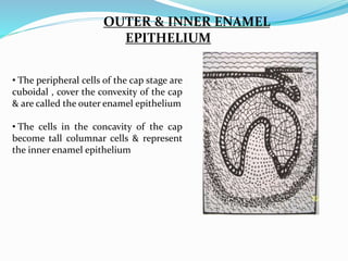
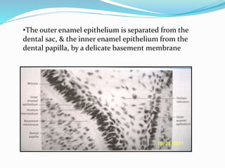
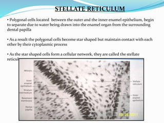
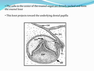
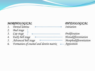
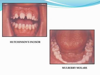
DEVELOPMENT OF
TOOTH
Dr. Muznah Sultan
Lecturer
Department of Oral
Biology, DIDC-DUHS
CONTENTS
1. Introduction
2. Dental Lamina
3. Vestibular Lamina
4. Tooth development
5. Developmental stages
• Bud stage
• Cap stage
• Bell stage
• Advanced bell stage
6. Hertwig’s epithelial root sheath and root formation
7. Conclusion
8. References
INTRODUCTION
•Tooth formation occurs in the 6th week of
intrauterine life with the formation of primary
epithelial band.
•At about 7th week the primary epithelial band
divides into a lingual process called dental lamina &
a buccal process called vestibular lamina.
DENTAL LAMINA
• 2- 3 weeks after the rupture of buccopharyngeal membrane, certain areas
of basal cells of oral ectoderm proliferate rapidly, leading to the formation
of primary epithelial band
• The band invades the underlying ectomesenchyme along each of the
horse-shoe shaped future dental arches.
• At about 7th week the primary epithelial band divides into an inner (lingual)
process called Dental Lamina & an outer ( buccal) process called Vestibular
Lamina
• The dental lamina serves as the primordium for the ectodermal portion
of the deciduous teeth
• Later during the development of jaws, permanent molars arise directly
from the distal extension of the dental lamina
FATE OF DENTAL LAMINA
• It is evident that total activity of dental lamina exceeds over a period of atleast 5 yrs
• As the teeth continue to develop, they loose their connection with the dental
lamina
•They later break up by mesenchymal invasion, which is at first incomplete and does
not perforate the total thickness of the lamina
• Fragmentation of the dental lamina progresses toward the developing
enamel organ
• Any particular portion of the dental lamina functions for a much briefer
period since only a relatively short time elapses after initiation of tooth
development before the dental lamina begins to degenerate
VESTIBULAR LAMINA
• Labial and buccal to the dental lamina in each dental arch, another epithelial
thickening develops independently
• It is Vestibular Lamina also termed as lip furrow band
• Subsequently hollows and form the oral vestibule between the alveolar portion
of the jaws and the lips and cheeks.
TOOTH DEVELOPMENT
• At certain points along the dental lamina each representing the location of one
of the 10 mandibular & 10 maxillary teeth, ectodermal cells multiply rapidly &
little knobs that grow into the underlying mesenchyme
• Each of these little down growths from the dental lamina represents the
beginning of the enamel organ of the tooth bud of a deciduous tooth
• First to appear are those of anterior mandibular region
• As the cell proliferation occurs each enamel organ takes a shape that resembles a
cap
DENTAL PAPILLA
On the inside of the cap, the ectomesenchymal cells increase in number.
The tissue appears more dense than the surrounding mesenchyme and
represents the beginning of the dental papilla
B = Dental Papilla
DENTAL SAC/ DENTAL
FOLLICLE
Surrounding the combined enamel organ or dental papilla, the third part of the
tooth bud forms. It is known as dental sac/follicle and it consists of
ectomesenchymal cells and fibres that surrounds the dental papilla and the enamel
organ.
C= Dental sac
• Thus the tooth germ consists of ectodermal
component- the enamel organ, the
ectomesenchymal components- the dental
papilla & the dental follicle
• The enamel is formed from the enamel organ,
the dentin and the pulp from the dental papilla
and the supporting tissues namely the
cementum, periodontal ligament & the alveolar
bone from the dental follicle
• During & after these developments the shape
of the enamel organ continues to change
• The depression occupied by the dental papilla
deepens until the enamel organ assumes a shape
resembling a bell
• The dental lamina becomes longer, thinner &
finally loses its connection with the epithelium
of the primitive oral cavity
DEVELOPMENTAL STAGES
MORPHOLOGICAL
1. Dental lamina
2. Bud stage
3. Cap stage
4. Early bell stage
5. Advanced bell stage
6. Formation of enamel and dentin matrix
PHYSIOLOGICAL
Initiation
Proliferation
Histodifferentiation
Morphodifferentiation
Apposition
BUD STAGE / PROLIFERATION
• This is the initial stage of tooth formation
where enamel organ resembles a small bud
• During the bud stage, the enamel organ
consists of peripherally located low
columnar cells & centrally located
polygonal cells
• The surrounding mesenchymal cells
proliferate, which results in their
condensation in two areas
• The area of condensation immediately
below the enamel organ is the dental
papilla
• The ectomesenchymal condensation that
surrounds the tooth bud & the dental
papilla is the tooth sac
• The dental papilla as well as the dental sac are not well defined during the bud
stage, they become more defined during the subsequent cap & bell stages
• The cells of the dental papilla form the dentin and pulp while the dental sac forms
cementum & periodontal ligament
CAP STAGE / PROLIFERATION
• As the tooth bud continues to proliferate, it does not expand uniformly into
a large sphere
• Instead unequal growth in different parts of the tooth bud leads to the cap
stage which is characterized by a shallow invagination on the deep surface of
the bud
OUTER & INNER ENAMEL
EPITHELIUM
• The peripheral cells of the cap stage are
cuboidal , cover the convexity of the cap
& are called the outer enamel epithelium
• The cells in the concavity of the cap
become tall columnar cells & represent
the inner enamel epithelium
•The outer enamel epithelium is separated from the
dental sac, & the inner enamel epithelium from the
dental papilla, by a delicate basement membrane
STELLATE RETICULUM
• Polygonal cells located between the outer and the inner enamel epithelium, begin
to separate due to water being drawn into the enamel organ from the surrounding
dental papilla
• As a result the polygonal cells become star shaped but maintain contact with each
other by their cytoplasmic process
• As the star shaped cells form a cellular network, they are called the stellate
reticulum
• The cells in the center of the enamel organ are densely packed and form
the enamel knot
• This knot projects toward the underlying dental papilla
• At the same time a vertical extension of the enamel knot,
called the enamel cord occurs
• The function of enamel knot &
cord may act as a reservoir of the
dividing cells for the growing
enamel organ
• The enamel knot act as a
signaling centers as many
important growth factors are
expressed by the cells of the
enamel knot & thus play an
important role in determining the
shape of the tooth
• The ectomesenchymal
condensation i.e the dental papilla
& the dental sac are pronounced
during this stage of dental
development
BELL STAGE / HISTODIFFERENTIATION
• Due to continued uneven growth of the
enamel organ it acquires a bell shape
• In bell stage crown shape is determined
• It was thought that the shape of the crown
is due to pressure exerted by the growing
dental papilla cells on the inner enamel
epithelium
• This pressure however was shown to be
opposed equally by the pressure exerted by
fluid present in the stellate reticulum
• The folding of enamel organ to cause
different crown shapes is shown to be due
to different rates of mitosis & difference in
cell differentiation time
INNER ENAMEL EPITHELIUM
• The inner enamel epithelium consists of a single layer of cells that differentiate
prior to amelogenesis into tall columnar cells called ameloblasts
• These elongated cells are attached to one another by junctional complexes
laterally & to cells in the stratum intermedium by desmosomes
• The cells of the inner enamel epithelium exert a strong influence on the
underlying mesenchymal cells of the dental papilla, which later differentiate into
odontoblasts
STRATUM INTERMEDIUM
• A few layers of squamous cells form the stratum intermedium , between the
inner enamel epithelium & the stellate reticulum
• These cells are closely attached by desmosomes & gap junctions
• This layer seems to be essential to enamel formation
STELLATE RETICULUM
• The stellate reticulum expands further due to continued accumulation of intra-
cellular fluid
• These star shaped cells, having a large processes anastomose with those of adjacent
cells
• As the enamel formation starts., the Stellate reticulum collapses to a narrow zone
thereby reducing the distance between the outer & inner enamel epithelium
OUTER ENAMEL EPITHELIUM
• The cells of the outer enamel epithelium flatten to form low cuboidal cells
• The outer enamel epithelium is thrown into folds which are rich in capillary
network, this provides a source of nutrition for the enamel organ
• Before the inner enamel epithelium begins to produce enamel. Peripheral cells
of the dental papilla differentiate into odontoblasts
• These cuboidal cells later assumes a columnar form & produce dentin
DENTAL LAMINA
• Dental lamina is seem to extend lingually and is termed successional dental
lamina as it gives rise to enamel organs of permanent successors of deciduous
teeth
• The enamel organs of deciduous teeth in the bell stage show successional lamina
& their permanent successor teeth in the bud stage
DENTAL SAC
• The dental sac exhibits a circular
arrangement of fibres & resembles
a capsule around the enamel
organ
• The fibres of the dental sac form
the periodontal ligament fibres
that span between the root & the
bone
• The junction between the inner
enamel epithelium & odontoblasts
outlines the future dentino-
enamel junction
ADVANCED BELL STAGE/MORPHODIFFERENTIATION
 Characterized by the
commencement of mineralization &
root formation
 The boundary between the inner
enamel epithelium & odontoblasts
outline the future dentinoenamel
junction
 Formation of dentin occurs first as a
layer along the future dentinoenamel
junction in the region of future cusps &
proceeds pulpally & apically
 After the first layer of dentin is
formed, the ameloblasts lay down
enamel over the dentin in the future
incisal & cuspal areas
 The enamel formation then proceeds
coronally & cervically in all the regions
from the dentinoenamel junction
toward the surface
 The cervical portion of enamel organ
gives rise to Hertwig Epithelial Root
Sheath (HERS)
 This HERS outlines the future root &
thus responsible for the size, shape ,
length & number of roots
CLINICAL
CONSIDERATIONS
HUTCHINSON’S INCISOR
MULBERRY MOLARS
FUSION
• The phenomenon of tooth
fusion arises through union of two
normally separated tooth germs, and
depending upon the stage of
development of the teeth at the time
of union, it may be either complete or
incomplete.
• However, fusion can also be the
union of a normal tooth bud to a
supernumerary tooth germ. In these
cases, the number of teeth is fewer if
the anomalous tooth is counted as one
tooth.
GEMINATION
Gemination arises when two
teeth develop from one tooth
bud and, as a result, the
patient has an extra tooth
FORMATION OF ENAMEL & DENTIN MATIX
( APPOSITION)
• Apposition is the deposition of the matrix of the hard enamel
structures
• Appositional growth of the enamel & dentin is a layer like
deposition of an extracellular matrix. This type of growth is
therefore additive
• Appositional growth is characterised by regular & rhythmic
deposition of the extracellular matrix, which is of itself
incapable of further growth
ROOT FORMATION
• The development of roots begin after
enamel & dentin formation has reached
the future cementoenamel junction
• The enamel organ plays an important
role in root development by forming
HERS, which models the shape of the
root
• HERS consists of outer & inner enamel
epithelium only
• As the first layer of the dentin has been
laid down, the epithelial root sheath
loses its structural continuity and is
close relation to the surface of the root
•Its remnants persists as an
epithelial network of strands or
clumps near the external surface
of the root
• These epithelial remnants are
found in the periodontal
ligament of erupted teeth and are
called as rests of mallasez
• Prior to the beginning of root formation,
the root sheath forms the epithelial
diaphragm
• The outer & the inner enamel epithelium
bend at the future cementoenamel
junction into a horizontal plane,
narrowing the wide cervical opening
• The proliferation of the cells of the
epithelial diaphragm is accompanied by
the proliferation of the cells of the
connective tissues of the pulp, adjacent to
the diaphragm
• The free end of diaphragm does not
grow into the connective tissue but the
epithelium proliferates coronal to the
epithelial diaphragm
• Connective tissue of the dental sac
surrounding the root sheath
proliferates & invades the continuous
double epithelial layer dividing it into
network of epithelial strands
• The rapid sequence of proliferation &
destruction of Hertwig’s root sheath
explains the fact that it cannot be seen
as a continuous layer on the surface of
developing root
• In the last stages of the root
development, the proliferation of the
epithelium in the diaphragm lags
behind that of the pulpal connective
tissue
• The wide apical foramen is reduced
first to the width of the diaphragmatic
opening itself & later is further
narrowed by opposition of dentin &
CLINICAL CONSIDERATIONS
DILACERATION
• Dilaceration refers to an angulation or a
sharp bend or curve anywhere along the
root portion of a tooth
• Condition probably occurs subsequent
to trauma or any other defect of
development which alters the angulation
of the tooth germ during root formation
• Can easily be detected by radiographs
• Care should be taken during extraction
since these teeth are more prone to
fracture
CONCRESCENCE
Concrescence is a condition of
teeth where
the cementum overlying the roots
of at least two teeth join together.
The cause can sometimes be
attributed to trauma or crowding
of teeth.
Radiographic diagnosis is
mandatory before attempting
tooth extraction
Development of tooth - DCPS

Development of tooth - DCPS

  • 1.
    DEVELOPMENT OF TOOTH Dr. MuznahSultan Lecturer Department of Oral Biology, DIDC-DUHS
  • 2.
    CONTENTS 1. Introduction 2. DentalLamina 3. Vestibular Lamina 4. Tooth development 5. Developmental stages • Bud stage • Cap stage • Bell stage • Advanced bell stage 6. Hertwig’s epithelial root sheath and root formation 7. Conclusion 8. References
  • 3.
  • 4.
    •Tooth formation occursin the 6th week of intrauterine life with the formation of primary epithelial band. •At about 7th week the primary epithelial band divides into a lingual process called dental lamina & a buccal process called vestibular lamina.
  • 5.
  • 6.
    • 2- 3weeks after the rupture of buccopharyngeal membrane, certain areas of basal cells of oral ectoderm proliferate rapidly, leading to the formation of primary epithelial band • The band invades the underlying ectomesenchyme along each of the horse-shoe shaped future dental arches.
  • 7.
    • At about7th week the primary epithelial band divides into an inner (lingual) process called Dental Lamina & an outer ( buccal) process called Vestibular Lamina • The dental lamina serves as the primordium for the ectodermal portion of the deciduous teeth • Later during the development of jaws, permanent molars arise directly from the distal extension of the dental lamina
  • 9.
    FATE OF DENTALLAMINA • It is evident that total activity of dental lamina exceeds over a period of atleast 5 yrs • As the teeth continue to develop, they loose their connection with the dental lamina •They later break up by mesenchymal invasion, which is at first incomplete and does not perforate the total thickness of the lamina
  • 10.
    • Fragmentation ofthe dental lamina progresses toward the developing enamel organ • Any particular portion of the dental lamina functions for a much briefer period since only a relatively short time elapses after initiation of tooth development before the dental lamina begins to degenerate
  • 11.
    VESTIBULAR LAMINA • Labialand buccal to the dental lamina in each dental arch, another epithelial thickening develops independently • It is Vestibular Lamina also termed as lip furrow band • Subsequently hollows and form the oral vestibule between the alveolar portion of the jaws and the lips and cheeks.
  • 12.
  • 13.
    • At certainpoints along the dental lamina each representing the location of one of the 10 mandibular & 10 maxillary teeth, ectodermal cells multiply rapidly & little knobs that grow into the underlying mesenchyme • Each of these little down growths from the dental lamina represents the beginning of the enamel organ of the tooth bud of a deciduous tooth • First to appear are those of anterior mandibular region • As the cell proliferation occurs each enamel organ takes a shape that resembles a cap
  • 14.
    DENTAL PAPILLA On theinside of the cap, the ectomesenchymal cells increase in number. The tissue appears more dense than the surrounding mesenchyme and represents the beginning of the dental papilla B = Dental Papilla
  • 15.
    DENTAL SAC/ DENTAL FOLLICLE Surroundingthe combined enamel organ or dental papilla, the third part of the tooth bud forms. It is known as dental sac/follicle and it consists of ectomesenchymal cells and fibres that surrounds the dental papilla and the enamel organ. C= Dental sac
  • 16.
    • Thus thetooth germ consists of ectodermal component- the enamel organ, the ectomesenchymal components- the dental papilla & the dental follicle • The enamel is formed from the enamel organ, the dentin and the pulp from the dental papilla and the supporting tissues namely the cementum, periodontal ligament & the alveolar bone from the dental follicle • During & after these developments the shape of the enamel organ continues to change • The depression occupied by the dental papilla deepens until the enamel organ assumes a shape resembling a bell • The dental lamina becomes longer, thinner & finally loses its connection with the epithelium of the primitive oral cavity
  • 17.
  • 18.
    MORPHOLOGICAL 1. Dental lamina 2.Bud stage 3. Cap stage 4. Early bell stage 5. Advanced bell stage 6. Formation of enamel and dentin matrix PHYSIOLOGICAL Initiation Proliferation Histodifferentiation Morphodifferentiation Apposition
  • 19.
    BUD STAGE /PROLIFERATION • This is the initial stage of tooth formation where enamel organ resembles a small bud • During the bud stage, the enamel organ consists of peripherally located low columnar cells & centrally located polygonal cells • The surrounding mesenchymal cells proliferate, which results in their condensation in two areas • The area of condensation immediately below the enamel organ is the dental papilla • The ectomesenchymal condensation that surrounds the tooth bud & the dental papilla is the tooth sac
  • 20.
    • The dentalpapilla as well as the dental sac are not well defined during the bud stage, they become more defined during the subsequent cap & bell stages • The cells of the dental papilla form the dentin and pulp while the dental sac forms cementum & periodontal ligament
  • 21.
    CAP STAGE /PROLIFERATION • As the tooth bud continues to proliferate, it does not expand uniformly into a large sphere • Instead unequal growth in different parts of the tooth bud leads to the cap stage which is characterized by a shallow invagination on the deep surface of the bud
  • 22.
    OUTER & INNERENAMEL EPITHELIUM • The peripheral cells of the cap stage are cuboidal , cover the convexity of the cap & are called the outer enamel epithelium • The cells in the concavity of the cap become tall columnar cells & represent the inner enamel epithelium
  • 23.
    •The outer enamelepithelium is separated from the dental sac, & the inner enamel epithelium from the dental papilla, by a delicate basement membrane
  • 24.
    STELLATE RETICULUM • Polygonalcells located between the outer and the inner enamel epithelium, begin to separate due to water being drawn into the enamel organ from the surrounding dental papilla • As a result the polygonal cells become star shaped but maintain contact with each other by their cytoplasmic process • As the star shaped cells form a cellular network, they are called the stellate reticulum
  • 25.
    • The cellsin the center of the enamel organ are densely packed and form the enamel knot • This knot projects toward the underlying dental papilla
  • 26.
    • At thesame time a vertical extension of the enamel knot, called the enamel cord occurs
  • 27.
    • The functionof enamel knot & cord may act as a reservoir of the dividing cells for the growing enamel organ • The enamel knot act as a signaling centers as many important growth factors are expressed by the cells of the enamel knot & thus play an important role in determining the shape of the tooth • The ectomesenchymal condensation i.e the dental papilla & the dental sac are pronounced during this stage of dental development
  • 28.
    BELL STAGE /HISTODIFFERENTIATION • Due to continued uneven growth of the enamel organ it acquires a bell shape • In bell stage crown shape is determined • It was thought that the shape of the crown is due to pressure exerted by the growing dental papilla cells on the inner enamel epithelium • This pressure however was shown to be opposed equally by the pressure exerted by fluid present in the stellate reticulum • The folding of enamel organ to cause different crown shapes is shown to be due to different rates of mitosis & difference in cell differentiation time
  • 29.
    INNER ENAMEL EPITHELIUM •The inner enamel epithelium consists of a single layer of cells that differentiate prior to amelogenesis into tall columnar cells called ameloblasts • These elongated cells are attached to one another by junctional complexes laterally & to cells in the stratum intermedium by desmosomes • The cells of the inner enamel epithelium exert a strong influence on the underlying mesenchymal cells of the dental papilla, which later differentiate into odontoblasts
  • 30.
    STRATUM INTERMEDIUM • Afew layers of squamous cells form the stratum intermedium , between the inner enamel epithelium & the stellate reticulum • These cells are closely attached by desmosomes & gap junctions • This layer seems to be essential to enamel formation
  • 31.
    STELLATE RETICULUM • Thestellate reticulum expands further due to continued accumulation of intra- cellular fluid • These star shaped cells, having a large processes anastomose with those of adjacent cells • As the enamel formation starts., the Stellate reticulum collapses to a narrow zone thereby reducing the distance between the outer & inner enamel epithelium
  • 32.
    OUTER ENAMEL EPITHELIUM •The cells of the outer enamel epithelium flatten to form low cuboidal cells • The outer enamel epithelium is thrown into folds which are rich in capillary network, this provides a source of nutrition for the enamel organ • Before the inner enamel epithelium begins to produce enamel. Peripheral cells of the dental papilla differentiate into odontoblasts • These cuboidal cells later assumes a columnar form & produce dentin
  • 33.
    DENTAL LAMINA • Dentallamina is seem to extend lingually and is termed successional dental lamina as it gives rise to enamel organs of permanent successors of deciduous teeth • The enamel organs of deciduous teeth in the bell stage show successional lamina & their permanent successor teeth in the bud stage
  • 34.
    DENTAL SAC • Thedental sac exhibits a circular arrangement of fibres & resembles a capsule around the enamel organ • The fibres of the dental sac form the periodontal ligament fibres that span between the root & the bone • The junction between the inner enamel epithelium & odontoblasts outlines the future dentino- enamel junction
  • 35.
    ADVANCED BELL STAGE/MORPHODIFFERENTIATION Characterized by the commencement of mineralization & root formation  The boundary between the inner enamel epithelium & odontoblasts outline the future dentinoenamel junction  Formation of dentin occurs first as a layer along the future dentinoenamel junction in the region of future cusps & proceeds pulpally & apically  After the first layer of dentin is formed, the ameloblasts lay down enamel over the dentin in the future incisal & cuspal areas
  • 36.
     The enamelformation then proceeds coronally & cervically in all the regions from the dentinoenamel junction toward the surface  The cervical portion of enamel organ gives rise to Hertwig Epithelial Root Sheath (HERS)  This HERS outlines the future root & thus responsible for the size, shape , length & number of roots
  • 37.
  • 38.
  • 39.
    FUSION • The phenomenonof tooth fusion arises through union of two normally separated tooth germs, and depending upon the stage of development of the teeth at the time of union, it may be either complete or incomplete. • However, fusion can also be the union of a normal tooth bud to a supernumerary tooth germ. In these cases, the number of teeth is fewer if the anomalous tooth is counted as one tooth.
  • 40.
    GEMINATION Gemination arises whentwo teeth develop from one tooth bud and, as a result, the patient has an extra tooth
  • 41.
    FORMATION OF ENAMEL& DENTIN MATIX ( APPOSITION) • Apposition is the deposition of the matrix of the hard enamel structures • Appositional growth of the enamel & dentin is a layer like deposition of an extracellular matrix. This type of growth is therefore additive • Appositional growth is characterised by regular & rhythmic deposition of the extracellular matrix, which is of itself incapable of further growth
  • 42.
  • 43.
    • The developmentof roots begin after enamel & dentin formation has reached the future cementoenamel junction • The enamel organ plays an important role in root development by forming HERS, which models the shape of the root • HERS consists of outer & inner enamel epithelium only • As the first layer of the dentin has been laid down, the epithelial root sheath loses its structural continuity and is close relation to the surface of the root
  • 44.
    •Its remnants persistsas an epithelial network of strands or clumps near the external surface of the root • These epithelial remnants are found in the periodontal ligament of erupted teeth and are called as rests of mallasez
  • 45.
    • Prior tothe beginning of root formation, the root sheath forms the epithelial diaphragm • The outer & the inner enamel epithelium bend at the future cementoenamel junction into a horizontal plane, narrowing the wide cervical opening • The proliferation of the cells of the epithelial diaphragm is accompanied by the proliferation of the cells of the connective tissues of the pulp, adjacent to the diaphragm • The free end of diaphragm does not grow into the connective tissue but the epithelium proliferates coronal to the epithelial diaphragm
  • 46.
    • Connective tissueof the dental sac surrounding the root sheath proliferates & invades the continuous double epithelial layer dividing it into network of epithelial strands • The rapid sequence of proliferation & destruction of Hertwig’s root sheath explains the fact that it cannot be seen as a continuous layer on the surface of developing root • In the last stages of the root development, the proliferation of the epithelium in the diaphragm lags behind that of the pulpal connective tissue • The wide apical foramen is reduced first to the width of the diaphragmatic opening itself & later is further narrowed by opposition of dentin &
  • 47.
  • 48.
    DILACERATION • Dilaceration refersto an angulation or a sharp bend or curve anywhere along the root portion of a tooth • Condition probably occurs subsequent to trauma or any other defect of development which alters the angulation of the tooth germ during root formation • Can easily be detected by radiographs • Care should be taken during extraction since these teeth are more prone to fracture
  • 49.
    CONCRESCENCE Concrescence is acondition of teeth where the cementum overlying the roots of at least two teeth join together. The cause can sometimes be attributed to trauma or crowding of teeth. Radiographic diagnosis is mandatory before attempting tooth extraction