Downloaded 127 times


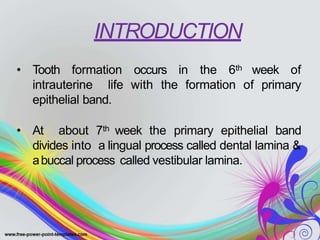
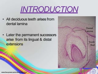

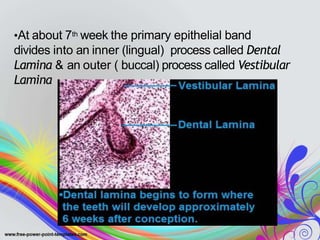


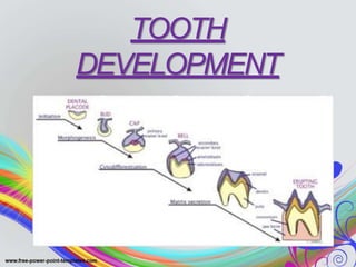
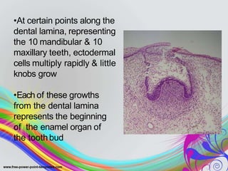


1. Tooth development begins around the 6th week of gestation with the formation of the primary epithelial band, which divides into the dental lamina and vestibular lamina. 2. Teeth develop through a series of stages from bud to bell shaped to advanced bell stage when mineralization begins and root formation commences. 3. The dental lamina gives rise to the tooth buds and plays a role in shaping tooth development through later stages. The enamel organ and dental papilla are structures that form within the developing tooth bud.


