Tissue
•Download as PPTX, PDF•
0 likes•20 views

Tissue is a group of cells that have similar structure and that function together as a unit. A nonliving material, called the intercellular matrix, fills the spaces between the cells.

More Related Content
What's hot
What's hot (20)
Similar to Tissue
Similar to Tissue (20)
More from Jagruti Marathe
More from Jagruti Marathe (20)
Recently uploaded
Recently uploaded (20)
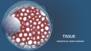
Tissue
- 1. TISSUE PRESENTED BY: JAGRUTI MARATHE
- 2. TISSUES FRENCH WORD “TO WEAVE” Definition : Tissue is a group of cells that have similar structure and that function together as a unit. A nonliving material, called the intercellular matrix, fills the spaces between the cells.
- 3. Size,Shape,Function,and their ,Constitute cell . 3 Classification Tissue Epithelia Connective Muscle Nervous
- 4. Epithelial tissue: Covering on all internal and external surface of your body, lines body cavities all hollow organ and is the major tissue in glands 4
- 5. Epithelialtissue: 5 Epithelial tissue has a variety of the functions depending on where its located in body, including ,protection, secretion and absorption. Epithelial tissue is made up of epithelial cells. The cells can be different in the shape and size and be arranged in a single and multiple layers depending on where they are in body and function they have.
- 9. 9 Protection Excretion Absorption Filtration Secretion Diffusion Sensory reception Functions:
- 10. Connective tissue : The tearm “connective tissue” (In german, Bindegewebe) was introduced in 1830 by Johannes Peter Muller. 10
- 11. Connective tissue : Tissue that supports, protects, and gives structure to other tissue and organ in the body. 11
- 12. Connectivetissue: 12 The common cell type in connective tissue include: fibroblast, mast cell , plasma cells , macrophages, adipocytes and leukocytes. Fibro blast are the most common cell type of connective tissue . They produce both fibres and amorphous ground substance. Specialize connective tissues include a number of different tissues with specialized cells and unique ground substance. Some of these are solid and strong, while others are fluid and flexible. Example include adipose, cartilage, bone, blood and lymph.
- 14. Tissue Purpose Components Location Collagen fibres Bind bones and other tissues to each other Alpha polypeptide chains Tendon ,ligament , skin, Cornea, cartilage , bone, blood vessels, gut and intervertebral Elastic fibres Allow organs like arteries and lungs to recoil Elastic microfibril and elastin Extracellular matrix Reticular fibres Form a scaffolding for other cells Type 3 collage Liver, bone marrow, and lymphatic organs
- 15. 15 Functions: • Connects tissues to one another—Tendons and ligaments Binding structure • Bones Provide support and movement • Bones , cells of the immune system Protection • Blood Transportation • Fat Storage • Fat Insulation
- 16. MUSCLETISSUE 16
- 17. • Muscle tissue is composed of cell that have the special ability to shorten or contraction in order to produce movement of the body parts. • The tissue is highly cellular and is well supplied with blood vessels. • The cells are long and slender so they are sometimes called muscle fibres, and these are usually arranged in bundles or layers that are surrounded by connective tissue. • Actin and myosin are contractile protein in muscle tissue.
- 20. • Skeletal muscles attach to and move bones by contracting and relaxing in response to voluntary messages from the nervous system. • Skeletal muscle tissue is composed of long cells called muscle fibers that have a striated appearance. • Muscle fibers are organized into bundles supplied by blood vessels and innervated by motor neurons.
- 21. • Smooth muscle is found in the walls of hollow organs throughout the body. • Smooth muscle contractions are involuntary movements triggered by impulses that travel through the autonomic nervous system to the smooth muscle tissue. • The arrangement of cells within smooth muscle tissue allows for contraction and relaxation with great elasticity.
- 22. • The smooth muscle in the walls of organs like the urinary bladder and the uterus allow those organs to expand and relax as needed. • The smooth muscle of the alimentary canal (the digestive tract) facilitates the peristaltic waves that move swallowed food and nutrients. • In the eye smooth muscle changes the shape of the lens to bring objects into focus. • Artery walls include smooth muscle that relaxes and contracts to move blood through the body
- 23. • The heart wall is composed of three layers. The middle layer, the myocardium, is responsible for the heart’s pumping action. • Cardiac muscle, found only in the myocardium, contracts in response to signals from the cardiac conduction system to make the heart beat. • Cardiac muscle is made from cells called cardiocytes. Like skeletal muscle cells cardiocytes have a striated appearance, but their overall structure is shorter and thicker. • Cardiocytes are branched, allowing them to connect with several other cardiocytes, forming a network that facilitates coordinated contraction.
- 24. 24 Functions: • Skeletal muscles pull on the bones causing movements at the joints. Movement • Muscles of the body wall support the internal organs Provide support • Skeletal muscles, particularly of the body wall, cushion the body's internal organs (abdominal cavity) from force applied to the exterior of the body. Protection • Heat is a waste product of muscle metabolism, which helps maintain an internal body temperature of 98.6 F. Heat generation • Cardiac muscles aid pumping action of the heart by aiding blood circulation. Blood circulation
- 25. NEVERSTISSUE 25 Nervous tissue is the main component of the nervous system, which includes the brain, spinal cord, and nerves. This Photo by Unknown Author is licensed under CC BY-SA
- 26. NEVERSTISSUE 26 This Photo by Unknown Author is licensed under CC BY-SA Neurons Neuroglia
- 27. •Nervous tissue is one of four major classes of tissues and makes up the central nervous system and the peripheral nervous system. •Integration and communication are the two major functions of nervous tissue. •Nervous tissue contains two categories of cells — neurons and neuroglia. •Neurons are highly specialized nerve cells that generate and conduct nerve impulses. •Neuroglia are supporting cells that provide physical sport, remove debris, and provide electrical insulation.
- 28. Nervous Tissue The nervous system is responsible for the control of the body and the communication among its parts. Nervous tissue contains two categories of cells—neurons and neuroglia. Neurons Neurons are highly specialized nerve cells that generate and conduct nerve impulses. A typical neuron consists of dendrites, the cell body, and an axon.
- 29. Dendrites Dendrites are responsible for responding to stimuli; they receive incoming signals towards the cell body. The axons are responsible for transmitting impulses over long distances from cell body. The cell body is like a factory for the neuron. It produces all the proteins and contains specialized organelles such as nucleus, granules and Nissl bodies. Dendrite The axon is surrounded by a whitish, fatty layer called the myelin sheath. Outside the myelin sheath there is a cellular layer called the neurilemma.
- 30. Schwann Cells In the peripheral nervous system, Schwann cells are neuroglia cells that support neuronal function by increasing the speed of impulse propagation. The Schwann cells are underlain by the medullary sheath. The medullary sheath is interrupted at intervals by the nodes of Ranvier.
- 31. Types of Nervous Tissue The nervous system consists of nervous tissue, which is composed of two principal types of cells called neuron and neuroglia. •Nervous tissue is composed of neurons and supporting cells called neuroglia, or ” glial cells.” •There are six types of neuroglia. Four are found in the central nervous system, while two are found in the peripheral nervous system. •The four types of neuroglia found in the central nervous system are astrocytes, microglial cells, ependymal cells, and oligodendrocytes.
- 32. •The two types of neuroglia found in the peripheral nervous system are satellite cells and Schwann cells. •Neurons are the other the other type of cell that comprise nervous tissue. •Neurons have cell bodies, dendrites, and axons.
- 33. Astrocytes Astrocytes are shaped like a star and are the most abundant glial cell in the CNS. They have many radiating processes which help in clinging to the neurons and capillaries. They support and brace the neurons and anchor them to the nutrient supply lines. They also help in the guiding the migration of young neurons. Astrocytes control the chemical environment around the neurons. Microglial Cells Microglial cells are small and ovoid un shape with thorny processes. They are found in the CNS. When invading microorganism or dead neurons are present, the microglial cells can transform into a phagocytic macrophage and help in cleaning the neuronal debris.
- 34. 34 Ependymal Cells Ependymal cells are ciliated and line the central cavities of the brain and spinal cord where they form a fairly permeable barrier between the cerebrospinal fluid that fills these cavities and the tissue cells of the CNS. Oligodendrocytes Oligodendrocytes line up along the nerves and produce an insulating cover called myelin sheath. They are found in the CNS. Satellite Cells Satellite cells surround neuron cell bodies in the peripheral nervous system (PNS). They are analogous to the astrocytes in the CNS. Schwann Cells Schwann cells surround all nerve fibers in the peripheral nervous system and form myelin sheaths around the nerve fibers. They are found in the PNS. Their function is similar to oligodendrocytes. This Photo by Unknown Author is licensed under CC BY-SA
- 35. 35 Neurons Neurons consist of cell body and one or more slender processes. The neuronal cell body consists of a nucleus and rough endoplasmic reticulum or Nissl Bodies. The cell body is the major biosynthetic center of a neuron and contains the usual organelles for the synthesis of proteins and other chemicals. Arm like processes extend from the cell body to all neurons. The two types of neuron processes are called dendrites and axons. Dendrites are motor neurons that are short and have a large surface area for receiving signals from other neurons. Dendrites convey incoming messages towards the cell body and are therefore called the receptive input region. This Photo by Unknown Author is licensed under CC BY-SA
- 36. 36 The axon arises from the cone shaped portion of the cell body called the axon hillock. Functionally, the axon is the conducting region of the neuron and is responsible for generating and transmitting impulses typically away from the cell body. A single axon routes the nerve impulse from the cell body to another neuron or an effector organ. The axon can have many terminal branches, so each time the nerve fires, it can stimulate more than one cell.