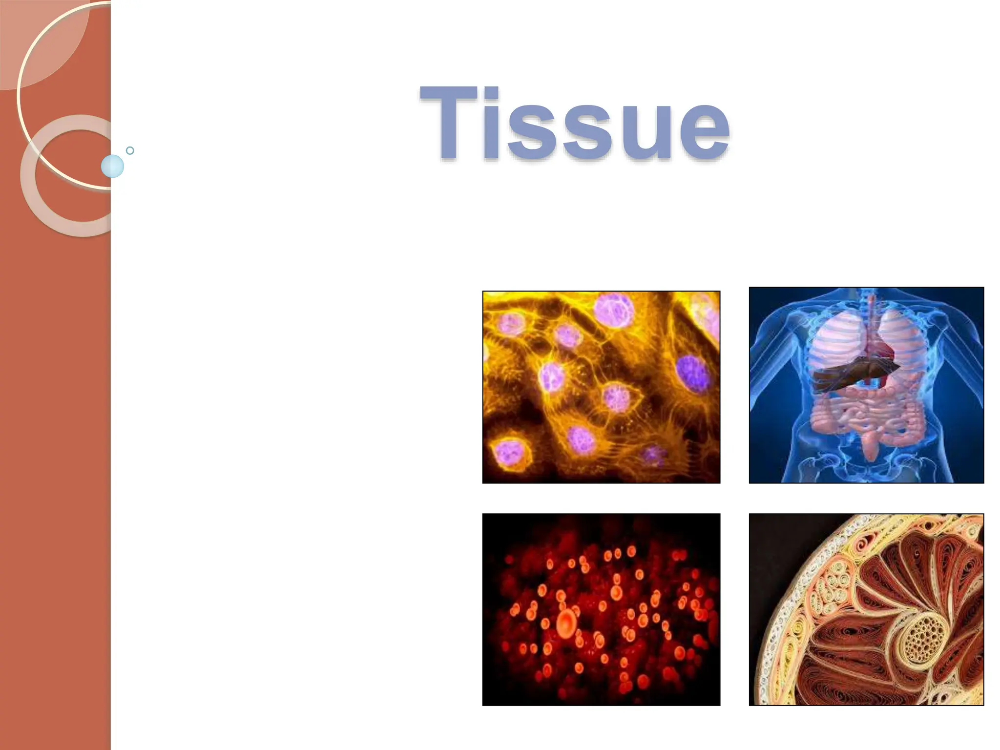
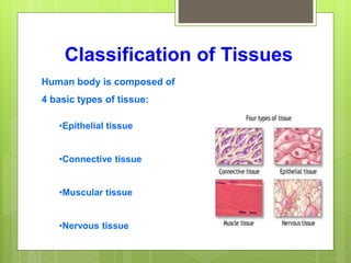
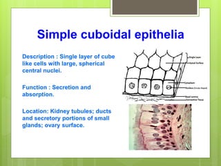
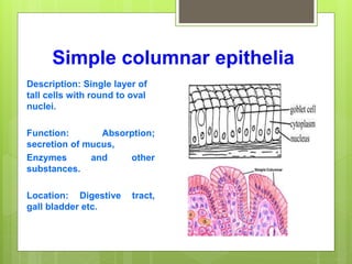
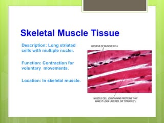
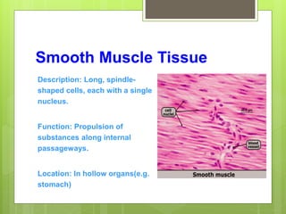
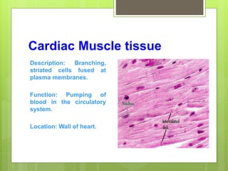
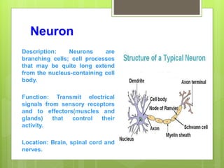
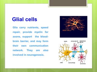
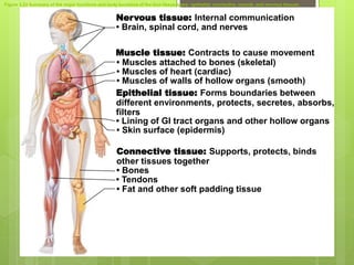
Tissue
Definition of Tissues
Biological tissue is a collection of interconnected
cells that perform a similar function within an
organism.
Classification of Tissues
Human body is composed of
4 basic types of tissue:
•Epithelial tissue
•Connective tissue
•Muscular tissue
•Nervous tissue
Epithelial Tissue
Epithelial cells cover or line all body surfaces, cavities and
tubes. So, These are called covering epithelia.
Epithelial cells form the functional units of secretory glands.
So, These are called glandular epithelia.
General Characteristic
 Closely attached to each other forming a protective barrier.
 Always has one free surface open to outside the body or inside
(cavity) an internal organ.
 Always has one fixed section attached to underlying connective
tissue.
 Has no blood vessels but can soak up nutrients from blood vessels
in connective tissue underneath.
 Can have lots of nerves in it.
 Very good at regenerating. i.e. sunburn, skinned knee.
Functions
 To protect the tissues that lie beneath it from radiation,
desiccation, toxins, invasion by pathogens, and physical trauma.
 The regulation and exchange of chemicals between the underlying
tissues and a body cavity.
 The secretion of hormones into the blood vascular system, and/or
the secretion of sweat, mucus, enzymes, and other products that are
delivered by ducts glandular epithelium.
 To provide sensation.
Classification of Epithelia
According to thickness
 “simple” - one cell layer
 “stratified” – more than one layer of cells
According to shape
 “squamous” – wider than tall
 “cuboidal” – as tall as wide
 “columnar” - taller than wide
Simple squamous epithelium
Description: Single layer of flattened
cells with disc-shaped central nuclei
and sparse cytoplasm; the simplest of
the epithelia.
Function: Passive transport of gases
and fluids.
Location:Alveoli of lungs, lining body
cavities, lining blood vessels
Simple cuboidal epithelia
Description : Single layer of cube
like cells with large, spherical
central nuclei.
Function : Secretion and
absorption.
Location: Kidney tubules; ducts
and secretory portions of small
glands; ovary surface.
Simple columnar epithelia
Description: Single layer of
tall cells with round to oval
nuclei.
Function: Absorption;
secretion of mucus,
Enzymes and other
substances.
Location: Digestive tract,
gall bladder etc.
Connective Tissues
The tissues that connect the different parts of the
body together are called connective tissues.
General characteristic
 The intercellular material is maximum where as the
cellular component is minimum.
 Unlike the other tissues, (e.g. epithelium, muscle and
nerve) which are formed mainly by cells, the major
constituent of connective tissue is ECM (Extra-cellular
matrix).
 Possess cells, fibers and ground substances.
Basic Functions
 Support and binding of other tissues
 Holding body fluids
 Defending the body against infection
 macrophages, plasma cells, mast cells, WBCs
 Storing nutrients as fat
Classification of connective
tissues
Connective tissue proper :
Description: Gel like matrix with
all three fiber types ; cells:
fibroblasts, macrophages, mast
cells, and white blood cells.
Function: Its macrophages
phagocytize bacteria ; plays
important role in inflammation ;
holds and conveys tissue fluid.
Location: Distributed under
epithelia of body; surrounds
capillaries.
Cartilage: Hyaline
Description: Amorphous but firm
matrix; chondroblasts produce
the matrix and when mature lie in
lacunae.
Function: Supports and
reinforces; resists compressive
stress.
Location: Forms most of the
embryonic skeleton; ends of long
bones; cartilages of nose.
Cartilage: Elastic
Description: Similar to hyaline
cartilage, but more elastic
fibers in matrix.
Function: Maintains the shape
and allows flexibility.
Location: Supports the external
ear(pinna).
Cartilage : Fibrocartilage
Description : Collagen fibers
are predominant ; matrix is
as hyaline but less firm.
Function: High tensile
strength, absorb
compressive shock.
Location: Intervertebral disc;
discs of knee joint.
Bone
Description: Hard, calcified
matrix containing many
collagen fibers. Very well
vascularized.
Function: Bone supports and
protects; provides levers for
the muscles to act on; stores
calcium and other minerals
and fat; bone marrow is the
site of blood cell formation.
Location: Skeleton.
Blood
Description: Liquid connective
tissue, red and white blood
cells in fluid matrix.
Function: Transport
respiratory gases, nutrients,
wastes and other substances.
Location: In the blood vessels.
Muscle Tissue
 Muscle is one of our 4 tissue types and muscle tissue
combined with nerves, blood vessels, and various
connective tissues.
3 Types of Muscle Tissue
General characteristics
1. Excitability
 The ability to receive and respond to a stimulus
 In skeletal muscle, the stimulus is a neurotransmitter
(chemical signal) release by a neuron (nerve cell).
 In smooth muscle, the stimulus could be a
neurotransmitter, a hormone, stretch, pH, Pco2, or
Po2.
 In cardiac muscle, the stimulus could be a
neurotransmitter, a hormone, or stretch.
 The response is the generation of an electrical impulse
that travels along the plasma membrane of the muscle
cell.
2. Contractility
 The ability to shorten forcibly when adequately
stimulated.
 This is the defining property of muscle tissue.
2. Extensibility
 The ability to be stretched (Extended)
4. Elasticity
 The ability to recoil and resume original length
after being stretched.
Functions
 Movement
 Locomotion
 Maintains posture
 Produces heat
 Facial expressions
 Pumps blood
Skeletal Muscle Tissue
Description: Long striated
cells with multiple nuclei.
Function: Contraction for
voluntary movements.
Location: In skeletal muscle.
Smooth Muscle Tissue
Description: Long, spindle-
shaped cells, each with a single
nucleus.
Function: Propulsion of
substances along internal
passageways.
Location: In hollow organs(e.g.
stomach)
Cardiac Muscle tissue
Description: Branching,
striated cells fused at
plasma membranes.
Function: Pumping of
blood in the circulatory
system.
Location: Wall of heart.
Nerve Tissue
By far the most complex tissue in the human body is
nerve tissue.
Formed by a network of more than 100 million nerve
cells, assisted by many more glial cells.
Each neuron has, on an average , at least a thousand
interconnection with other neurons forming a very
complex nervous system.
Functions
 Regulates & controls body functions
 Generates & transmits nerve impulses
 Supports, insulates and protects impulse
generating neurons.
Composition of Nerve Tissue
The nerve tissue is composed of two elements:
1. The nerve cell or neuron
2. The neuroglia
Neuron
Description: Neurons are
branching cells; cell processes
that may be quite long extend
from the nucleus-containing cell
body.
Function: Transmit electrical
signals from sensory receptors
and to effectors(muscles and
glands) that control their
activity.
Location: Brain, spinal cord and
nerves.
Glial cells
Glia carry nutrients, speed
repair, provide myelin for
axons, support the blood-
brain barrier, and may form
their own communication
network. They are also
involved in neurogenesis.
Figure 3.22 Summary of the major functions and body locations of the four tissue types: epithelial, connective, muscle, and nervous tissues.
Nervous tissue: Internal communication
• Brain, spinal cord, and nerves
Muscle tissue: Contracts to cause movement
Epithelial tissue: Forms boundaries between
different environments, protects, secretes, absorbs,
filters
Connective tissue: Supports, protects, binds
other tissues together
• Muscles attached to bones (skeletal)
• Muscles of heart (cardiac)
• Muscles of walls of hollow organs (smooth)
• Lining of GI tract organs and other hollow organs
• Skin surface (epidermis)
• Bones
• Tendons
• Fat and other soft padding tissue

L8.Tissues.ppt

  • 1.
  • 2.
    Definition of Tissues Biologicaltissue is a collection of interconnected cells that perform a similar function within an organism.
  • 3.
    Classification of Tissues Humanbody is composed of 4 basic types of tissue: •Epithelial tissue •Connective tissue •Muscular tissue •Nervous tissue
  • 4.
    Epithelial Tissue Epithelial cellscover or line all body surfaces, cavities and tubes. So, These are called covering epithelia. Epithelial cells form the functional units of secretory glands. So, These are called glandular epithelia.
  • 5.
    General Characteristic  Closelyattached to each other forming a protective barrier.  Always has one free surface open to outside the body or inside (cavity) an internal organ.  Always has one fixed section attached to underlying connective tissue.  Has no blood vessels but can soak up nutrients from blood vessels in connective tissue underneath.  Can have lots of nerves in it.  Very good at regenerating. i.e. sunburn, skinned knee.
  • 6.
    Functions  To protectthe tissues that lie beneath it from radiation, desiccation, toxins, invasion by pathogens, and physical trauma.  The regulation and exchange of chemicals between the underlying tissues and a body cavity.  The secretion of hormones into the blood vascular system, and/or the secretion of sweat, mucus, enzymes, and other products that are delivered by ducts glandular epithelium.  To provide sensation.
  • 7.
    Classification of Epithelia Accordingto thickness  “simple” - one cell layer  “stratified” – more than one layer of cells According to shape  “squamous” – wider than tall  “cuboidal” – as tall as wide  “columnar” - taller than wide
  • 8.
    Simple squamous epithelium Description:Single layer of flattened cells with disc-shaped central nuclei and sparse cytoplasm; the simplest of the epithelia. Function: Passive transport of gases and fluids. Location:Alveoli of lungs, lining body cavities, lining blood vessels
  • 9.
    Simple cuboidal epithelia Description: Single layer of cube like cells with large, spherical central nuclei. Function : Secretion and absorption. Location: Kidney tubules; ducts and secretory portions of small glands; ovary surface.
  • 10.
    Simple columnar epithelia Description:Single layer of tall cells with round to oval nuclei. Function: Absorption; secretion of mucus, Enzymes and other substances. Location: Digestive tract, gall bladder etc.
  • 11.
    Connective Tissues The tissuesthat connect the different parts of the body together are called connective tissues.
  • 12.
    General characteristic  Theintercellular material is maximum where as the cellular component is minimum.  Unlike the other tissues, (e.g. epithelium, muscle and nerve) which are formed mainly by cells, the major constituent of connective tissue is ECM (Extra-cellular matrix).  Possess cells, fibers and ground substances.
  • 13.
    Basic Functions  Supportand binding of other tissues  Holding body fluids  Defending the body against infection  macrophages, plasma cells, mast cells, WBCs  Storing nutrients as fat
  • 14.
  • 15.
    Connective tissue proper: Description: Gel like matrix with all three fiber types ; cells: fibroblasts, macrophages, mast cells, and white blood cells. Function: Its macrophages phagocytize bacteria ; plays important role in inflammation ; holds and conveys tissue fluid. Location: Distributed under epithelia of body; surrounds capillaries.
  • 16.
    Cartilage: Hyaline Description: Amorphousbut firm matrix; chondroblasts produce the matrix and when mature lie in lacunae. Function: Supports and reinforces; resists compressive stress. Location: Forms most of the embryonic skeleton; ends of long bones; cartilages of nose.
  • 17.
    Cartilage: Elastic Description: Similarto hyaline cartilage, but more elastic fibers in matrix. Function: Maintains the shape and allows flexibility. Location: Supports the external ear(pinna).
  • 18.
    Cartilage : Fibrocartilage Description: Collagen fibers are predominant ; matrix is as hyaline but less firm. Function: High tensile strength, absorb compressive shock. Location: Intervertebral disc; discs of knee joint.
  • 19.
    Bone Description: Hard, calcified matrixcontaining many collagen fibers. Very well vascularized. Function: Bone supports and protects; provides levers for the muscles to act on; stores calcium and other minerals and fat; bone marrow is the site of blood cell formation. Location: Skeleton.
  • 20.
    Blood Description: Liquid connective tissue,red and white blood cells in fluid matrix. Function: Transport respiratory gases, nutrients, wastes and other substances. Location: In the blood vessels.
  • 21.
    Muscle Tissue  Muscleis one of our 4 tissue types and muscle tissue combined with nerves, blood vessels, and various connective tissues.
  • 22.
    3 Types ofMuscle Tissue
  • 23.
    General characteristics 1. Excitability The ability to receive and respond to a stimulus  In skeletal muscle, the stimulus is a neurotransmitter (chemical signal) release by a neuron (nerve cell).  In smooth muscle, the stimulus could be a neurotransmitter, a hormone, stretch, pH, Pco2, or Po2.  In cardiac muscle, the stimulus could be a neurotransmitter, a hormone, or stretch.  The response is the generation of an electrical impulse that travels along the plasma membrane of the muscle cell.
  • 24.
    2. Contractility  Theability to shorten forcibly when adequately stimulated.  This is the defining property of muscle tissue. 2. Extensibility  The ability to be stretched (Extended) 4. Elasticity  The ability to recoil and resume original length after being stretched.
  • 25.
    Functions  Movement  Locomotion Maintains posture  Produces heat  Facial expressions  Pumps blood
  • 26.
    Skeletal Muscle Tissue Description:Long striated cells with multiple nuclei. Function: Contraction for voluntary movements. Location: In skeletal muscle.
  • 27.
    Smooth Muscle Tissue Description:Long, spindle- shaped cells, each with a single nucleus. Function: Propulsion of substances along internal passageways. Location: In hollow organs(e.g. stomach)
  • 28.
    Cardiac Muscle tissue Description:Branching, striated cells fused at plasma membranes. Function: Pumping of blood in the circulatory system. Location: Wall of heart.
  • 29.
    Nerve Tissue By farthe most complex tissue in the human body is nerve tissue. Formed by a network of more than 100 million nerve cells, assisted by many more glial cells. Each neuron has, on an average , at least a thousand interconnection with other neurons forming a very complex nervous system.
  • 30.
    Functions  Regulates &controls body functions  Generates & transmits nerve impulses  Supports, insulates and protects impulse generating neurons.
  • 31.
    Composition of NerveTissue The nerve tissue is composed of two elements: 1. The nerve cell or neuron 2. The neuroglia
  • 32.
    Neuron Description: Neurons are branchingcells; cell processes that may be quite long extend from the nucleus-containing cell body. Function: Transmit electrical signals from sensory receptors and to effectors(muscles and glands) that control their activity. Location: Brain, spinal cord and nerves.
  • 33.
    Glial cells Glia carrynutrients, speed repair, provide myelin for axons, support the blood- brain barrier, and may form their own communication network. They are also involved in neurogenesis.
  • 34.
    Figure 3.22 Summaryof the major functions and body locations of the four tissue types: epithelial, connective, muscle, and nervous tissues. Nervous tissue: Internal communication • Brain, spinal cord, and nerves Muscle tissue: Contracts to cause movement Epithelial tissue: Forms boundaries between different environments, protects, secretes, absorbs, filters Connective tissue: Supports, protects, binds other tissues together • Muscles attached to bones (skeletal) • Muscles of heart (cardiac) • Muscles of walls of hollow organs (smooth) • Lining of GI tract organs and other hollow organs • Skin surface (epidermis) • Bones • Tendons • Fat and other soft padding tissue