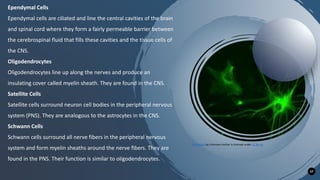
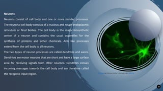
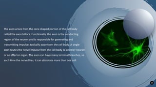
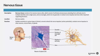
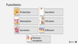
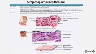
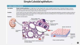
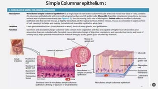
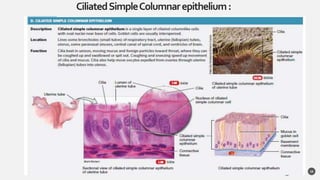
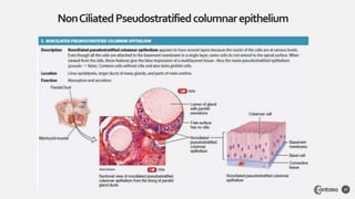
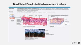
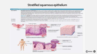
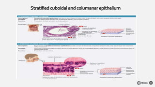
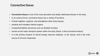
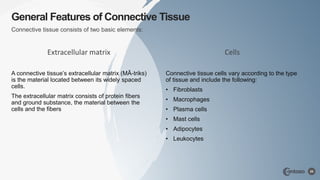
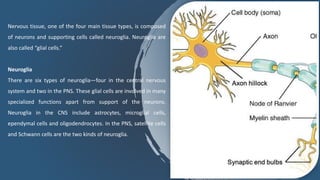
TISSUE
PRESENTED BY: JAGRUTI MARATHE
TISSUES
FRENCH WORD “TO WEAVE”
Definition :
Tissue is a group of cells that have similar
structure and that function together as a unit.
A nonliving material, called the intercellular
matrix, fills the spaces between the cells.
• Histology (his′-TOL-oˉ-jē; histo- = tissue;
logy = study of) is the science that deals
with the study of tissues.
• A pathologist (pa-THOL-oˉ - jist; patho- =
disease) is a physician who examines
cells and tissues to help other physicians
make accurate diagnoses.
Size,Shape,Function,and their
,Constitute cell .
3
Classification
Tissue
Epithelia Connective Muscle Nervous
Size,Shape,Function,and their
,Constitute cell .
4
Classification
1. Epithelial tissue covers body surfaces and lines hollow
organs, body cavities, and ducts; it also forms glands. This
tissue allows the body to interact with both its internal and
external environments.
2. Connective tissue protects and supports the body and its
organs. Various types of connective tissues bind organs
together, store energy reserves as fat, and help provide the
body with immunity to disease-causing organisms.
3. Muscular tissue is composed of cells specialized for
contraction and generation of force. In the process, muscular
tissue generates heat that warms the body.
4. Nervous tissue detects changes in a variety of conditions
inside and outside the body and responds by generating
electrical signals called nerve action potentials (nerve
impulses) that activate muscular contractions and glandular
secretions.
 Cell junctions are contact points
between the plasma membranes of
tissue cells.
The five most important types of cell
junctions:
• tight junctions,
• adherens junctions,
• desmosomes,
• hemidesmosomes,
• and gap junctions
CellJunctions
5
6
1. Tight junctions consist of weblike strands of transmembrane proteins that fuse together the outer
surfaces of adjacent plasma membranes to seal off passageways between adjacent cells . Cells of
epithelial tissue that lines the stomach, intestines, and urinary bladder have many tight junctions.
2. Adherens junctions (ad-HĒR-ens) contain plaque (PLAK), a dense layer of proteins on the inside of
the plasma membrane that attaches both to membrane proteins and to microfilaments of the
cytoskeleton.
3. Desmosomes Like adherens junctions, desmosomes (DEZ-moˉ-soˉms; desmo- = band) contain
plaque and have transmembrane glycoproteins (cadherins) that extend into the intercellular space
between adjacent cell membranes and attach cells to one another.
4. Hemidesmosomes (hemi- = half) resemble desmosomes, but they do not link adjacent cells. The
name arises from the fact that they look like half of a desmosome
5. At gap junctions, membrane proteins called connexins form tiny fluid-filled tunnels called connexons that
connect neighbouring cells .
Epithelial tissue:
Covering on all internal and
external surface of your body,
lines body cavities all hollow
organ and is the major tissue
in glands
7
Epithelialtissue:
8
• An epithelial tissue (ep-i-THĒ-lē-al) or epithelium (plural is epithelia) consists of cells arranged in
continuous sheets, in either single or multiple layers.
• Because the cells are closely packed and are held tightly together by many cell junctions, there is little
intercellular space between adjacent plasma membranes.
• Epithelial tissue is arranged in two general patterns in the body:
• (1) covering and lining various surfaces and
• (2) forming the secreting portions of glands.
• Epithelial tissue has a variety of the functions depending on where its located in body, including ,protection,
secretion and absorption.
• Epithelial tissue is made up of epithelial cells.
• The cells can be different in the shape and size and be arranged in a single and multiple layers depending
on where they are in body and function they have.
Arrangement of
layers
Simple
epithelium
Pseudostratified
epithelium
Stratified
epithelium
Types of covering and lining epithelial tissue are classified according to two characteristics:
• the arrangement of cells into layers
• and the shapes of the cells .
Cell
shapes
Squamous
cells
Cuboidal
cells
Columnar
cells
Transitional
cells
11
I. Simple epithelium
A. Simple squamous epithelium
1. Endothelium (lines heart, blood vessels,
lymphatic vessels)
2. Mesothelium (forms epithelial layer of
serous membranes)
B. Simple cuboidal epithelium
C. Simple columnar epithelium
1. Nonciliated (lacks cilia)
2. Ciliated (contains cilia)
D. Pseudostratified columnar epithelium
1. Nonciliated (lacks cilia)
2. Ciliated (contains cilia)
II. Stratified epithelium
A. Stratified squamous epithelium*
1. Nonkeratinized (lacks keratin)
2. Keratinized (contains keratin)
B. Stratified cuboidal epithelium*
C. Stratified columnar epithelium*
D. Transitional epithelium or urothelium (lines most of
urinary tract)
When we combine the two characteristics (arrangements of layers and cell shapes),
we come up with the following types of epithelial tissues:
12
Protection Excretion
Absorption Filtration
Secretion Diffusion
Sensory
reception
Functions:
SimpleSquamousepithelium:
13
SimpleCuboidalepithelium:
14
SimpleColumnarepithelium:
15
CiliatedSimpleColumnarepithelium:
16
NonCiliatedPseudostratifiedcolumnarepithelium
17
NonCiliatedPseudostratifiedcolumnarepithelium
18
Stratifiedsquamousepithelium
19
Stratifiedcuboidalandcolumanarepithelium
20
Transitionalepithelium
21
• The function of glandular epithelium is secretion, which is accomplished by glandular cells that often lie in
clusters deep to the covering and lining epithelium.
• A gland consists of epithelium that secretes substances into ducts (tubes), onto a surface, or eventually into
the blood in the absence of ducts.
• All glands of the body are classified as either endocrine or exocrine.
• The secretions of endocrine glands (EN-doˉ-krin; endo- = inside; -crine = secretion; called hormones, enter
the interstitial fluid and then diff use into the bloodstream without flowing through a duct Endocrine
secretions have far-reaching effects because they are distributed throughout the body by the bloodstream.
• Exocrine glands (EK-soˉ-krin; exo- = outside; secrete their products into ducts that empty onto the surface
of a covering and lining epithelium such as the skin surface or the lumen of a hollow organ. The secretions of
exocrine glands have limited effects and some of them would be harmful if they entered the bloodstream. As
some glands of the body, such as the pancreas, ovaries, and testes, are mixed glands that contain both
endocrine and exocrine tissue.
Glandular Epithelium
22
23
StructuralClassificationof
ExocrineGlands
 Unicellular glands
 Multicellular glands
Structural Classification of Exocrine Glands
24
Multicellular glands are categorized according to two criteria:
(1) whether their ducts are branched or unbranched and
(2) the shape of the secretory portions of the gland .
• If the duct of the gland does not branch, it is a simple gland
• If the duct branches, it is a compound gland
• Glands with tubular secretory parts are tubular glands;
• Glands with rounded secretory portions are acinar glands (AS-i-nar; acin- = berry), also called alveolar
glands.
• Tubuloacinar glands have both tubular and more rounded secretory parts.
These features are the criteria for the following structural classification
scheme for multicellular exocrine glands:
25
 Simple glands
A. Simple tubular. Tubular secretory part is straight and
attaches to a single unbranched duct . Example: glands in
the large intestine.
B. Simple branched tubular. Tubular secretory part is
branched and attaches to a single unbranched duct .
Example: gastric glands.
C. Simple coiled tubular. Tubular secretory part is coiled
and attaches to a single unbranched duct . Example: sweat
glands.
D. Simple acinar. Secretory portion is rounded, attaches to
single unbranched duct . Example: glands of penile
urethra.
E. Simple branched acinar. Rounded secretory part is
branched and attaches to a single unbranched duct .
Example: sebaceous glands.
 Compound glands
A. Compound tubular. Secretory portion is tubular and
attaches to a branched duct . Example: bulbourethral
(Cowper’s) glands.
B. Compound acinar. Secretory portion is rounded and
attaches to a branched duct. Example: mammary glands.
C. Compound tubuloacinar. Secretory portion is both
tubular and rounded and attaches to a branched duct .
Example: acinar glands of the pancreas.
Connective
tissue :
The tearm “connective tissue”
(In german, Bindegewebe) was
introduced in 1830 by
Johannes Peter Muller.
26
Connective
tissue :
Tissue that supports, protects,
and gives structure to other
tissue and organ in the body.
27
Connectivetissue:
28
• Connective tissue is one of the most abundant and widely distributed tissues in the body.
• In its various forms, connective tissue has a variety of functions.
• It binds together, supports, and strengthens other body tissues;
• protects and insulates internal organs;
• compartmentalizes structures such as skeletal muscles;
• serves as the major transport system within the body (blood, a fluid connective tissue);
• It is the primary location of stored energy reserves (adipose, or fat, tissue); and is the main
source of immune responses.
Extracellular matrix
A connective tissue’s extracellular matrix (MĀ-triks)
is the material located between its widely spaced
cells.
The extracellular matrix consists of protein fibers
and ground substance, the material between the
cells and the fibers
Cells
Connective tissue cells vary according to the type
of tissue and include the following:
• Fibroblasts
• Macrophages
• Plasma cells
• Mast cells
• Adipocytes
• Leukocytes
General Features of Connective Tissue
29
Connective tissue consists of two basic elements:
Connective tissue
Proper
connective
tissue
Loose tissue
Areolar Adipose Reticular
Dense tissue
Dense regular
Dense
irregular
Elastic
Supportive
connective
tissue
Cartilage
Hyaline
cartilage
Fibro cartilage
Elastic
cartilage
Bone
Compact bone Spongy bone
Fluid
Connective
tissue
Blood Lymph
Tissue Purpose Components Location
Collagen
fibres
Bind bones and
other tissues to
each other
Alpha
polypeptide
chains
Tendon ,ligament ,
skin, Cornea,
cartilage , bone,
blood vessels, gut
and intervertebral
Elastic
fibres
Allow organs
like arteries and
lungs to recoil
Elastic microfibril
and elastin
Extracellular matrix
Reticular
fibres
Form a
scaffolding for
other cells
Type 3 collage Liver, bone marrow,
and lymphatic
organs
32
Functions:
• Connects tissues to one another—Tendons and
ligaments
Binding structure
• Bones
Provide support and movement
• Bones , cells of the immune system
Protection
• Blood
Transportation
• Fat
Storage
• Fat
Insulation
MUSCLETISSUE
33
• Muscle tissue is composed of cell that
have the special ability to shorten or
contraction in order to produce
movement of the body parts.
• The tissue is highly cellular and is well
supplied with blood vessels.
• The cells are long and slender so they are
sometimes called muscle fibres, and these
are usually arranged in bundles or layers
that are surrounded by connective tissue.
• Actin and myosin are contractile protein in
muscle tissue.
• Muscular tissue consists of elongated cells
called muscle fibers or myocytes that can
use ATP to generate force. As a result,
muscular tissue produces body movements,
maintains posture, and generates heat.
• It also provides protection. Based on
location and certain structural and functional
features,
• muscular tissue is classified into three types:
skeletal, cardiac, and smooth
Muscle
tissue
Skeleton
Loose Dense
Smooth
Bone Cartilage
Cardiac
ASkeletalMuscletissue
38
ASmooth Muscletissue
39
ASmooth Muscletissue
40
• Skeletal muscles attach to and move bones
by contracting and relaxing in response to
voluntary messages from the nervous
system.
• Skeletal muscle tissue is composed of long
cells called muscle fibers that have a striated
appearance.
• Muscle fibers are organized into bundles
supplied by blood vessels and innervated by
motor neurons.
• Smooth muscle is found in the walls of
hollow organs throughout the body.
• Smooth muscle contractions are involuntary
movements triggered by impulses that travel
through the autonomic nervous system to
the smooth muscle tissue.
• The arrangement of cells within smooth
muscle tissue allows for contraction and
relaxation with great elasticity.
• The smooth muscle in the walls of organs
like the urinary bladder and the uterus allow
those organs to expand and relax as needed.
• The smooth muscle of the alimentary canal
(the digestive tract) facilitates the peristaltic
waves that move swallowed food and
nutrients.
• In the eye smooth muscle changes the shape
of the lens to bring objects into focus.
• Artery walls include smooth muscle that
relaxes and contracts to move blood through
the body
• The heart wall is composed of three layers. The middle layer,
the myocardium, is responsible for the heart’s pumping
action.
• Cardiac muscle, found only in the myocardium, contracts in
response to signals from the cardiac conduction system to
make the heart beat.
• Cardiac muscle is made from cells called cardiocytes. Like
skeletal muscle cells cardiocytes have a striated appearance,
but their overall structure is shorter and thicker.
• Cardiocytes are branched, allowing them to connect with
several other cardiocytes, forming a network that facilitates
coordinated contraction.
45
Functions:
• Skeletal muscles pull on the bones causing movements at the joints.
Movement
• Muscles of the body wall support the internal organs
Provide support
• Skeletal muscles, particularly of the body wall, cushion the body's
internal organs (abdominal cavity) from force applied to the exterior of
the body.
Protection
• Heat is a waste product of muscle metabolism, which helps maintain
an internal body temperature of 98.6 F.
Heat generation
• Cardiac muscles aid pumping action of the heart by aiding blood
circulation.
Blood circulation
NEVERSTISSUE
46
Nervous tissue is the main component of the
nervous system, which includes the brain, spinal
cord, and nerves.
This Photo by Unknown Author is licensed under CC BY-SA
NEVERSTISSUE
47
This Photo by Unknown Author is licensed under CC BY-SA
Neurons
Neuroglia
•Nervous tissue is one of four major classes of
tissues and makes up the central nervous system
and the peripheral nervous system.
•Integration and communication are the two major
functions of nervous tissue.
•Nervous tissue contains two categories of cells —
neurons and neuroglia.
•Neurons are highly specialized nerve cells that
generate and conduct nerve impulses.
•Neuroglia are supporting cells that provide physical
sport, remove debris, and provide electrical
insulation.
Nervous Tissue
Nervous tissue is one of four major classes of tissues. It is
specialized tissue found in the central nervous system and
the peripheral nervous system. It consists of neurons and
supporting cells called neuroglia.
The nervous system is responsible for the control of the
body and the communication among its parts. Nervous
tissue contains two categories of cells—neurons and
neuroglia.
Neurons
Neurons are highly specialized nerve cells that generate and
conduct nerve impulses. A typical neuron consists of
dendrites, the cell body, and an axon.
Dendrites
Dendrites are responsible for responding to stimuli; they
receive incoming signals towards the cell body. The
axons are responsible for transmitting impulses over
long distances from cell body. The cell body is like a
factory for the neuron. It produces all the proteins and
contains specialized organelles such as nucleus, granules
and Nissl bodies.
Dendrite
The axon is surrounded by a whitish, fatty layer called
the myelin sheath. Outside the myelin sheath there is a
cellular layer called the neurilemma.
Schwann Cells
In the peripheral nervous system, Schwann cells are
neuroglia cells that support neuronal function by
increasing the speed of impulse propagation. The
Schwann cells are underlain by the medullary
sheath. The medullary sheath is interrupted at
intervals by the nodes of Ranvier.
Types of Nervous Tissue
The nervous system consists of nervous tissue, which is
composed of two principal types of cells called neuron and
neuroglia.
•Nervous tissue is composed of neurons and supporting cells
called neuroglia, or ” glial cells.”
•There are six types of neuroglia. Four are found in the central
nervous system, while two are found in the peripheral nervous
system.
•The four types of neuroglia found in the central nervous system
are astrocytes, microglial cells, ependymal cells, and
oligodendrocytes.
•The two types of neuroglia found in the peripheral nervous
system are satellite cells and Schwann cells.
•Neurons are the other the other type of cell that comprise
nervous tissue.
•Neurons have cell bodies, dendrites, and axons.
•neuron: The main cell type in nervous tissue.
•neuroglia: Supporting cells in nervous tissue.
Nervous tissue, one of the four main tissue types, is composed
of neurons and supporting cells called neuroglia. Neuroglia are
also called “glial cells.”
Neuroglia
There are six types of neuroglia—four in the central nervous
system and two in the PNS. These glial cells are involved in many
specialized functions apart from support of the neurons.
Neuroglia in the CNS include astrocytes, microglial cells,
ependymal cells and oligodendrocytes. In the PNS, satellite cells
and Schwann cells are the two kinds of neuroglia.
Astrocytes
Astrocytes are shaped like a star and are the most abundant glial
cell in the CNS. They have many radiating processes which help
in clinging to the neurons and capillaries. They support and
brace the neurons and anchor them to the nutrient supply lines.
They also help in the guiding the migration of young neurons.
Astrocytes control the chemical environment around the
neurons.
Microglial Cells
Microglial cells are small and ovoid un shape with thorny
processes. They are found in the CNS. When invading
microorganism or dead neurons are present, the microglial cells
can transform into a phagocytic macrophage and help in
cleaning the neuronal debris.
57
Ependymal Cells
Ependymal cells are ciliated and line the central cavities of the brain
and spinal cord where they form a fairly permeable barrier between
the cerebrospinal fluid that fills these cavities and the tissue cells of
the CNS.
Oligodendrocytes
Oligodendrocytes line up along the nerves and produce an
insulating cover called myelin sheath. They are found in the CNS.
Satellite Cells
Satellite cells surround neuron cell bodies in the peripheral nervous
system (PNS). They are analogous to the astrocytes in the CNS.
Schwann Cells
Schwann cells surround all nerve fibers in the peripheral nervous
system and form myelin sheaths around the nerve fibers. They are
found in the PNS. Their function is similar to oligodendrocytes.
This Photo by Unknown Author is licensed under CC BY-SA
58
Neurons
Neurons consist of cell body and one or more slender processes.
The neuronal cell body consists of a nucleus and rough endoplasmic
reticulum or Nissl Bodies. The cell body is the major biosynthetic
center of a neuron and contains the usual organelles for the
synthesis of proteins and other chemicals. Arm like processes
extend from the cell body to all neurons.
The two types of neuron processes are called dendrites and axons.
Dendrites are motor neurons that are short and have a large surface
area for receiving signals from other neurons. Dendrites convey
incoming messages towards the cell body and are therefore called
the receptive input region. This Photo by Unknown Author is licensed under CC BY-SA
59
The axon arises from the cone shaped portion of the cell body
called the axon hillock. Functionally, the axon is the conducting
region of the neuron and is responsible for generating and
transmitting impulses typically away from the cell body. A single
axon routes the nerve impulse from the cell body to another neuron
or an effector organ. The axon can have many terminal branches, so
each time the nerve fires, it can stimulate more than one cell.
Nervoustissue
60
61

Tissue.pptx

  • 1.
  • 2.
    TISSUES FRENCH WORD “TOWEAVE” Definition : Tissue is a group of cells that have similar structure and that function together as a unit. A nonliving material, called the intercellular matrix, fills the spaces between the cells. • Histology (his′-TOL-oˉ-jē; histo- = tissue; logy = study of) is the science that deals with the study of tissues. • A pathologist (pa-THOL-oˉ - jist; patho- = disease) is a physician who examines cells and tissues to help other physicians make accurate diagnoses.
  • 3.
    Size,Shape,Function,and their ,Constitute cell. 3 Classification Tissue Epithelia Connective Muscle Nervous
  • 4.
    Size,Shape,Function,and their ,Constitute cell. 4 Classification 1. Epithelial tissue covers body surfaces and lines hollow organs, body cavities, and ducts; it also forms glands. This tissue allows the body to interact with both its internal and external environments. 2. Connective tissue protects and supports the body and its organs. Various types of connective tissues bind organs together, store energy reserves as fat, and help provide the body with immunity to disease-causing organisms. 3. Muscular tissue is composed of cells specialized for contraction and generation of force. In the process, muscular tissue generates heat that warms the body. 4. Nervous tissue detects changes in a variety of conditions inside and outside the body and responds by generating electrical signals called nerve action potentials (nerve impulses) that activate muscular contractions and glandular secretions.
  • 5.
     Cell junctionsare contact points between the plasma membranes of tissue cells. The five most important types of cell junctions: • tight junctions, • adherens junctions, • desmosomes, • hemidesmosomes, • and gap junctions CellJunctions 5
  • 6.
    6 1. Tight junctionsconsist of weblike strands of transmembrane proteins that fuse together the outer surfaces of adjacent plasma membranes to seal off passageways between adjacent cells . Cells of epithelial tissue that lines the stomach, intestines, and urinary bladder have many tight junctions. 2. Adherens junctions (ad-HĒR-ens) contain plaque (PLAK), a dense layer of proteins on the inside of the plasma membrane that attaches both to membrane proteins and to microfilaments of the cytoskeleton. 3. Desmosomes Like adherens junctions, desmosomes (DEZ-moˉ-soˉms; desmo- = band) contain plaque and have transmembrane glycoproteins (cadherins) that extend into the intercellular space between adjacent cell membranes and attach cells to one another. 4. Hemidesmosomes (hemi- = half) resemble desmosomes, but they do not link adjacent cells. The name arises from the fact that they look like half of a desmosome 5. At gap junctions, membrane proteins called connexins form tiny fluid-filled tunnels called connexons that connect neighbouring cells .
  • 7.
    Epithelial tissue: Covering onall internal and external surface of your body, lines body cavities all hollow organ and is the major tissue in glands 7
  • 8.
    Epithelialtissue: 8 • An epithelialtissue (ep-i-THĒ-lē-al) or epithelium (plural is epithelia) consists of cells arranged in continuous sheets, in either single or multiple layers. • Because the cells are closely packed and are held tightly together by many cell junctions, there is little intercellular space between adjacent plasma membranes. • Epithelial tissue is arranged in two general patterns in the body: • (1) covering and lining various surfaces and • (2) forming the secreting portions of glands. • Epithelial tissue has a variety of the functions depending on where its located in body, including ,protection, secretion and absorption. • Epithelial tissue is made up of epithelial cells. • The cells can be different in the shape and size and be arranged in a single and multiple layers depending on where they are in body and function they have.
  • 9.
    Arrangement of layers Simple epithelium Pseudostratified epithelium Stratified epithelium Types ofcovering and lining epithelial tissue are classified according to two characteristics: • the arrangement of cells into layers • and the shapes of the cells .
  • 10.
  • 11.
    11 I. Simple epithelium A.Simple squamous epithelium 1. Endothelium (lines heart, blood vessels, lymphatic vessels) 2. Mesothelium (forms epithelial layer of serous membranes) B. Simple cuboidal epithelium C. Simple columnar epithelium 1. Nonciliated (lacks cilia) 2. Ciliated (contains cilia) D. Pseudostratified columnar epithelium 1. Nonciliated (lacks cilia) 2. Ciliated (contains cilia) II. Stratified epithelium A. Stratified squamous epithelium* 1. Nonkeratinized (lacks keratin) 2. Keratinized (contains keratin) B. Stratified cuboidal epithelium* C. Stratified columnar epithelium* D. Transitional epithelium or urothelium (lines most of urinary tract) When we combine the two characteristics (arrangements of layers and cell shapes), we come up with the following types of epithelial tissues:
  • 12.
    12 Protection Excretion Absorption Filtration SecretionDiffusion Sensory reception Functions:
  • 13.
  • 14.
  • 15.
  • 16.
  • 17.
  • 18.
  • 19.
  • 20.
  • 21.
  • 22.
    • The functionof glandular epithelium is secretion, which is accomplished by glandular cells that often lie in clusters deep to the covering and lining epithelium. • A gland consists of epithelium that secretes substances into ducts (tubes), onto a surface, or eventually into the blood in the absence of ducts. • All glands of the body are classified as either endocrine or exocrine. • The secretions of endocrine glands (EN-doˉ-krin; endo- = inside; -crine = secretion; called hormones, enter the interstitial fluid and then diff use into the bloodstream without flowing through a duct Endocrine secretions have far-reaching effects because they are distributed throughout the body by the bloodstream. • Exocrine glands (EK-soˉ-krin; exo- = outside; secrete their products into ducts that empty onto the surface of a covering and lining epithelium such as the skin surface or the lumen of a hollow organ. The secretions of exocrine glands have limited effects and some of them would be harmful if they entered the bloodstream. As some glands of the body, such as the pancreas, ovaries, and testes, are mixed glands that contain both endocrine and exocrine tissue. Glandular Epithelium 22
  • 23.
  • 24.
    Structural Classification ofExocrine Glands 24 Multicellular glands are categorized according to two criteria: (1) whether their ducts are branched or unbranched and (2) the shape of the secretory portions of the gland . • If the duct of the gland does not branch, it is a simple gland • If the duct branches, it is a compound gland • Glands with tubular secretory parts are tubular glands; • Glands with rounded secretory portions are acinar glands (AS-i-nar; acin- = berry), also called alveolar glands. • Tubuloacinar glands have both tubular and more rounded secretory parts.
  • 25.
    These features arethe criteria for the following structural classification scheme for multicellular exocrine glands: 25  Simple glands A. Simple tubular. Tubular secretory part is straight and attaches to a single unbranched duct . Example: glands in the large intestine. B. Simple branched tubular. Tubular secretory part is branched and attaches to a single unbranched duct . Example: gastric glands. C. Simple coiled tubular. Tubular secretory part is coiled and attaches to a single unbranched duct . Example: sweat glands. D. Simple acinar. Secretory portion is rounded, attaches to single unbranched duct . Example: glands of penile urethra. E. Simple branched acinar. Rounded secretory part is branched and attaches to a single unbranched duct . Example: sebaceous glands.  Compound glands A. Compound tubular. Secretory portion is tubular and attaches to a branched duct . Example: bulbourethral (Cowper’s) glands. B. Compound acinar. Secretory portion is rounded and attaches to a branched duct. Example: mammary glands. C. Compound tubuloacinar. Secretory portion is both tubular and rounded and attaches to a branched duct . Example: acinar glands of the pancreas.
  • 26.
    Connective tissue : The tearm“connective tissue” (In german, Bindegewebe) was introduced in 1830 by Johannes Peter Muller. 26
  • 27.
    Connective tissue : Tissue thatsupports, protects, and gives structure to other tissue and organ in the body. 27
  • 28.
    Connectivetissue: 28 • Connective tissueis one of the most abundant and widely distributed tissues in the body. • In its various forms, connective tissue has a variety of functions. • It binds together, supports, and strengthens other body tissues; • protects and insulates internal organs; • compartmentalizes structures such as skeletal muscles; • serves as the major transport system within the body (blood, a fluid connective tissue); • It is the primary location of stored energy reserves (adipose, or fat, tissue); and is the main source of immune responses.
  • 29.
    Extracellular matrix A connectivetissue’s extracellular matrix (MĀ-triks) is the material located between its widely spaced cells. The extracellular matrix consists of protein fibers and ground substance, the material between the cells and the fibers Cells Connective tissue cells vary according to the type of tissue and include the following: • Fibroblasts • Macrophages • Plasma cells • Mast cells • Adipocytes • Leukocytes General Features of Connective Tissue 29 Connective tissue consists of two basic elements:
  • 30.
    Connective tissue Proper connective tissue Loose tissue AreolarAdipose Reticular Dense tissue Dense regular Dense irregular Elastic Supportive connective tissue Cartilage Hyaline cartilage Fibro cartilage Elastic cartilage Bone Compact bone Spongy bone Fluid Connective tissue Blood Lymph
  • 31.
    Tissue Purpose ComponentsLocation Collagen fibres Bind bones and other tissues to each other Alpha polypeptide chains Tendon ,ligament , skin, Cornea, cartilage , bone, blood vessels, gut and intervertebral Elastic fibres Allow organs like arteries and lungs to recoil Elastic microfibril and elastin Extracellular matrix Reticular fibres Form a scaffolding for other cells Type 3 collage Liver, bone marrow, and lymphatic organs
  • 32.
    32 Functions: • Connects tissuesto one another—Tendons and ligaments Binding structure • Bones Provide support and movement • Bones , cells of the immune system Protection • Blood Transportation • Fat Storage • Fat Insulation
  • 33.
  • 34.
    • Muscle tissueis composed of cell that have the special ability to shorten or contraction in order to produce movement of the body parts. • The tissue is highly cellular and is well supplied with blood vessels. • The cells are long and slender so they are sometimes called muscle fibres, and these are usually arranged in bundles or layers that are surrounded by connective tissue. • Actin and myosin are contractile protein in muscle tissue.
  • 35.
    • Muscular tissueconsists of elongated cells called muscle fibers or myocytes that can use ATP to generate force. As a result, muscular tissue produces body movements, maintains posture, and generates heat. • It also provides protection. Based on location and certain structural and functional features, • muscular tissue is classified into three types: skeletal, cardiac, and smooth
  • 37.
  • 38.
  • 39.
  • 40.
  • 41.
    • Skeletal musclesattach to and move bones by contracting and relaxing in response to voluntary messages from the nervous system. • Skeletal muscle tissue is composed of long cells called muscle fibers that have a striated appearance. • Muscle fibers are organized into bundles supplied by blood vessels and innervated by motor neurons.
  • 42.
    • Smooth muscleis found in the walls of hollow organs throughout the body. • Smooth muscle contractions are involuntary movements triggered by impulses that travel through the autonomic nervous system to the smooth muscle tissue. • The arrangement of cells within smooth muscle tissue allows for contraction and relaxation with great elasticity.
  • 43.
    • The smoothmuscle in the walls of organs like the urinary bladder and the uterus allow those organs to expand and relax as needed. • The smooth muscle of the alimentary canal (the digestive tract) facilitates the peristaltic waves that move swallowed food and nutrients. • In the eye smooth muscle changes the shape of the lens to bring objects into focus. • Artery walls include smooth muscle that relaxes and contracts to move blood through the body
  • 44.
    • The heartwall is composed of three layers. The middle layer, the myocardium, is responsible for the heart’s pumping action. • Cardiac muscle, found only in the myocardium, contracts in response to signals from the cardiac conduction system to make the heart beat. • Cardiac muscle is made from cells called cardiocytes. Like skeletal muscle cells cardiocytes have a striated appearance, but their overall structure is shorter and thicker. • Cardiocytes are branched, allowing them to connect with several other cardiocytes, forming a network that facilitates coordinated contraction.
  • 45.
    45 Functions: • Skeletal musclespull on the bones causing movements at the joints. Movement • Muscles of the body wall support the internal organs Provide support • Skeletal muscles, particularly of the body wall, cushion the body's internal organs (abdominal cavity) from force applied to the exterior of the body. Protection • Heat is a waste product of muscle metabolism, which helps maintain an internal body temperature of 98.6 F. Heat generation • Cardiac muscles aid pumping action of the heart by aiding blood circulation. Blood circulation
  • 46.
    NEVERSTISSUE 46 Nervous tissue isthe main component of the nervous system, which includes the brain, spinal cord, and nerves. This Photo by Unknown Author is licensed under CC BY-SA
  • 47.
    NEVERSTISSUE 47 This Photo byUnknown Author is licensed under CC BY-SA Neurons Neuroglia
  • 48.
    •Nervous tissue isone of four major classes of tissues and makes up the central nervous system and the peripheral nervous system. •Integration and communication are the two major functions of nervous tissue. •Nervous tissue contains two categories of cells — neurons and neuroglia. •Neurons are highly specialized nerve cells that generate and conduct nerve impulses. •Neuroglia are supporting cells that provide physical sport, remove debris, and provide electrical insulation.
  • 49.
    Nervous Tissue Nervous tissueis one of four major classes of tissues. It is specialized tissue found in the central nervous system and the peripheral nervous system. It consists of neurons and supporting cells called neuroglia. The nervous system is responsible for the control of the body and the communication among its parts. Nervous tissue contains two categories of cells—neurons and neuroglia. Neurons Neurons are highly specialized nerve cells that generate and conduct nerve impulses. A typical neuron consists of dendrites, the cell body, and an axon.
  • 50.
    Dendrites Dendrites are responsiblefor responding to stimuli; they receive incoming signals towards the cell body. The axons are responsible for transmitting impulses over long distances from cell body. The cell body is like a factory for the neuron. It produces all the proteins and contains specialized organelles such as nucleus, granules and Nissl bodies. Dendrite The axon is surrounded by a whitish, fatty layer called the myelin sheath. Outside the myelin sheath there is a cellular layer called the neurilemma.
  • 51.
    Schwann Cells In theperipheral nervous system, Schwann cells are neuroglia cells that support neuronal function by increasing the speed of impulse propagation. The Schwann cells are underlain by the medullary sheath. The medullary sheath is interrupted at intervals by the nodes of Ranvier.
  • 52.
    Types of NervousTissue The nervous system consists of nervous tissue, which is composed of two principal types of cells called neuron and neuroglia. •Nervous tissue is composed of neurons and supporting cells called neuroglia, or ” glial cells.” •There are six types of neuroglia. Four are found in the central nervous system, while two are found in the peripheral nervous system. •The four types of neuroglia found in the central nervous system are astrocytes, microglial cells, ependymal cells, and oligodendrocytes.
  • 53.
    •The two typesof neuroglia found in the peripheral nervous system are satellite cells and Schwann cells. •Neurons are the other the other type of cell that comprise nervous tissue. •Neurons have cell bodies, dendrites, and axons.
  • 54.
    •neuron: The maincell type in nervous tissue. •neuroglia: Supporting cells in nervous tissue.
  • 55.
    Nervous tissue, oneof the four main tissue types, is composed of neurons and supporting cells called neuroglia. Neuroglia are also called “glial cells.” Neuroglia There are six types of neuroglia—four in the central nervous system and two in the PNS. These glial cells are involved in many specialized functions apart from support of the neurons. Neuroglia in the CNS include astrocytes, microglial cells, ependymal cells and oligodendrocytes. In the PNS, satellite cells and Schwann cells are the two kinds of neuroglia.
  • 56.
    Astrocytes Astrocytes are shapedlike a star and are the most abundant glial cell in the CNS. They have many radiating processes which help in clinging to the neurons and capillaries. They support and brace the neurons and anchor them to the nutrient supply lines. They also help in the guiding the migration of young neurons. Astrocytes control the chemical environment around the neurons. Microglial Cells Microglial cells are small and ovoid un shape with thorny processes. They are found in the CNS. When invading microorganism or dead neurons are present, the microglial cells can transform into a phagocytic macrophage and help in cleaning the neuronal debris.
  • 57.
    57 Ependymal Cells Ependymal cellsare ciliated and line the central cavities of the brain and spinal cord where they form a fairly permeable barrier between the cerebrospinal fluid that fills these cavities and the tissue cells of the CNS. Oligodendrocytes Oligodendrocytes line up along the nerves and produce an insulating cover called myelin sheath. They are found in the CNS. Satellite Cells Satellite cells surround neuron cell bodies in the peripheral nervous system (PNS). They are analogous to the astrocytes in the CNS. Schwann Cells Schwann cells surround all nerve fibers in the peripheral nervous system and form myelin sheaths around the nerve fibers. They are found in the PNS. Their function is similar to oligodendrocytes. This Photo by Unknown Author is licensed under CC BY-SA
  • 58.
    58 Neurons Neurons consist ofcell body and one or more slender processes. The neuronal cell body consists of a nucleus and rough endoplasmic reticulum or Nissl Bodies. The cell body is the major biosynthetic center of a neuron and contains the usual organelles for the synthesis of proteins and other chemicals. Arm like processes extend from the cell body to all neurons. The two types of neuron processes are called dendrites and axons. Dendrites are motor neurons that are short and have a large surface area for receiving signals from other neurons. Dendrites convey incoming messages towards the cell body and are therefore called the receptive input region. This Photo by Unknown Author is licensed under CC BY-SA
  • 59.
    59 The axon arisesfrom the cone shaped portion of the cell body called the axon hillock. Functionally, the axon is the conducting region of the neuron and is responsible for generating and transmitting impulses typically away from the cell body. A single axon routes the nerve impulse from the cell body to another neuron or an effector organ. The axon can have many terminal branches, so each time the nerve fires, it can stimulate more than one cell.
  • 60.
  • 61.