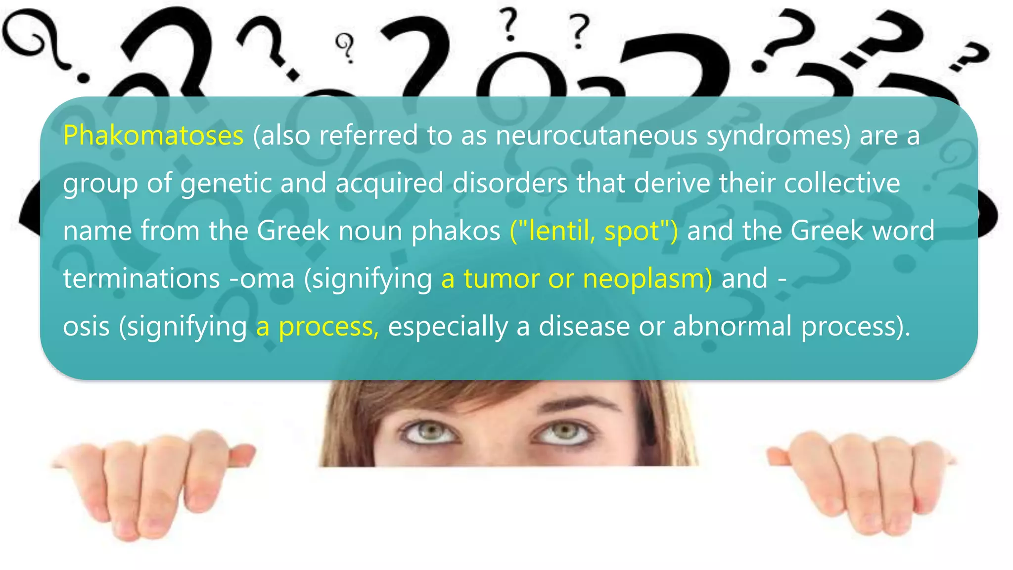
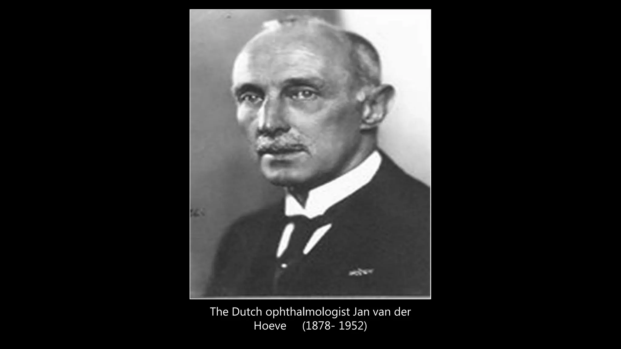
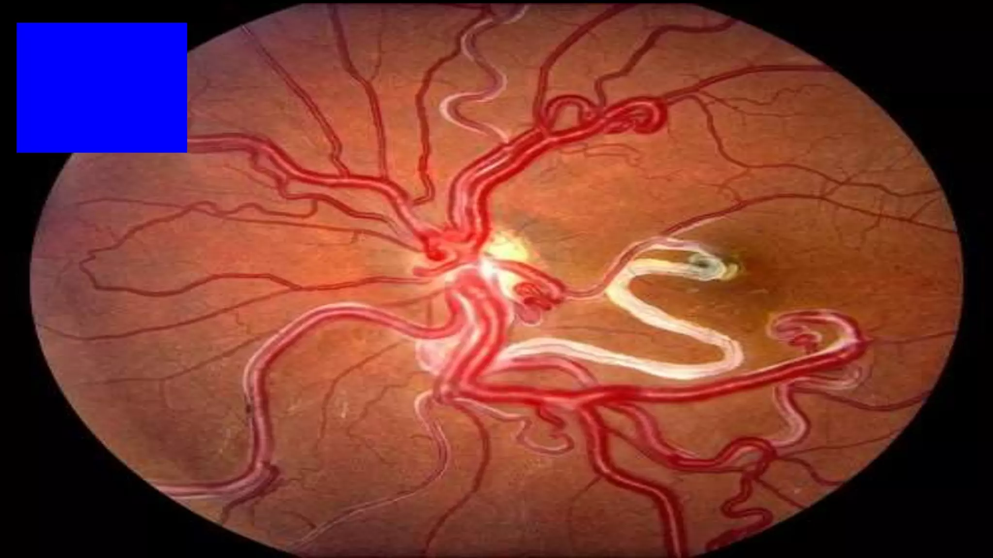
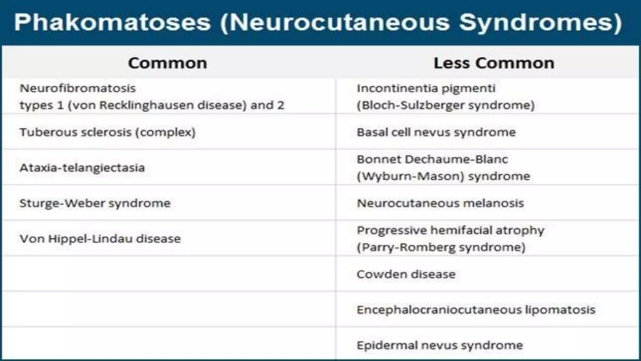
Call Girls Varanasi Just Call 9907093804 Top Class Call Girl Service Available
Ophthalmology Trivia - Feb 2020
1.
2.
3. Phakomatoses (also referred to as neurocutaneous syndromes) are a
group of genetic and acquired disorders that derive their collective
name from the Greek noun phakos ("lentil, spot") and the Greek word
terminations -oma (signifying a tumor or neoplasm) and -
osis (signifying a process, especially a disease or abnormal process).
5. The quest for Quasimodo the
“Hunchback” of Notre Dame de
Paris A further “renowned”, likely
NF1 sufferer, could have been
Quasimodo, the “Hunchback” of
Notre Dame de Paris, described by
the French poet, novelist and
dramatist of the Romantic
Movement, Victor Hugo (1802–
1885), in 19th century.
Phakomatoses are characterized by variable multisystem involvement. Typically, they affect the central nervous system (CNS), the eyes, and the skin, all of which derive from the same ectodermal origin.
A hamartoma is defined as a benign tumor or nodular growth that is composed of proliferating mature histologically normal cells that normally reside at the affected tissue. DDX?
Lisch nodules are dome-shaped gelatinous masses developing on the surface of the iris. Gold-tan to brown in color, they may grow up to 2 mm in diameter and attain variable sizes on the same iris. The presence of Lisch nodules is the most common clinical sign of NF1.
In 80% of eyes, Lisch nodules may be found in the inferior quadrants of the iris and this may be related to greater sun exposure, one of the postulated factors in the development of these benign tumefactions. Lisch nodules are almost always pathognomonic for NF1. However, they may occasionally be seen in some other rare conditions including Watson syndrome and neurofibromatosis type 2.
The differential diagnosis of Lisch nodules must include iris mamillations, irido-corneo-endothelial syndrome, Rieger's anomaly or syndrome, iris nevi, melanoma and inflammatory conditions like sarcoidosis, lepra, tuberculosis and syphilis.
6 OR MORE
1.5 CM
ON glioma
10-15 %
Mulbery retinal astrocytoma
A retinal astrocytoma (“mulberry lesion") adjacent to the optic nerve is typical of those found with tuberous sclerosis complex
calcified lesion 50%
Port wine stain
Trigeminal
Both macular SD-OCT findings showed multiple large inner retinal vessels that created a prominent shadowing artifact, retinal thickening, and speckling and heterogeneity of inner retinal layers.