SlideShare a Scribd company logo
Resident
Case Series
Sudden-onset visual
disturbances and headache in a
woman with a history of MS
leads to a visit to the ER
Bellows College
Presentation
2
• A 44-year-old Caucasian female presented to the ER complaining of sudden
onset of seeing a “thousand shades of gray, which later turned to black
patches” in her right eye associated with pain with eye movement and dull
headache. Her symptoms began 10 days prior and were progressively
worsening.
• Review of systems was notable for fevers, chills, night sweats, a recent
intentional 90-pound weight loss, tinnitus, nausea and vomiting.
• She denied any muscle, joint, dermatologic or genitourinary complaints
currently or in the past.
Bellows College
Medical History
3
• She was diagnosed with multiple sclerosis 20 years prior, after having
three episodes of “visual disturbances” in her right eye, presumably repeat
bouts of optic neuritis.
• After being treated with glatiramer acetate, discontinued 18 years ago,
she denied any new visual complaints.
• The remainder of her past medical history was remarkable for
hypertension, controlled with verapamil and clonidine.
Bellows College
Medical History
4
• She was a non-smoker and denied any recent travel or having pets.
• Two months prior she underwent a non-emergent laparoscopic
cholecystectomy with a complicated postoperative course,
resulting in an extended admission. Since then, she states she
just has not “felt right”.
Bellows College
Examination
• Vital signs were normal with a
BP of 126/82.
• VA uncorrected was 20/200 in
her right eye with no
improvement with pinhole and
20/20 in her left eye.
• Pupils were significant for a
RAPD in the right eye.
• The patient was able to identify only
the test plate in the right eye and
10/10 color plates in the left eye.
• Confrontation visual fields and
ocular motility were normal in both
eyes.
• IOP was 19 mmHg and 16 mmHg in
the right and left eyes, respectively.
5
Bellows College
Examination
• The anterior segment examination
of her right eye revealed:
1. Diffuse flat conjunctival
injection.
2. Fine KPs inferiorly.
3. 1+WBC in the AC.
4. 2+WBCs in the anterior
vitreous.
• There were no posterior
synechiae, iris nodules or visually
significant cataract.
• The left eye anterior segment
examination was unremarkable
with no signs of ocular
inflammation.
6
Bellows College 7
Bellows College 8
• The posterior exam of
the right eye revealed:
1. Significant vitreous cell.
2. Disc edema.
3. Peripapillary
hemorrhages.
4. Cotton wool spots.
5. Occlusive arteritis along
all four vascular arcades.
6. Macular edema.
7. The veins were slightly
engorged but did not
show any inflammatory
changes.
• Dilated funduscopic
exam of the left eye
was normal without
evidence of prior
inflammatory
episodes.
OD
Bellows College
The patient, a 44-year-old immunocompetent
Caucasian female with a diagnosis of MS secondary to
presumed episodes of right eye optic neuritis,
presented with 20/200 vision in the right eye and
clinical findings of a unilateral occlusive arteritis, disc
edema associated with peripapillary hemorrhages and
macular edema.
9
Bellows College 10
Bellows College
The differential of vasculitis is quite
extensive and is best broken down
according to conditions that affect
arteries, veins or both.
The differential of vasculitis is quite
extensive and is best broken down
according to conditions that affect
arteries, veins or both.
11
Conditions that affect veins include:
1. Sarcoidosis
2. Syphilis
3. Par planitis
4. Eales’ disease
5. Multiple sclerosis
Conditions that affect both arteries and
veins include:
1. (SLE)
2. Wegener’s granulomatosis
3. HLA-B27 associated disease
4. Behçet’s disease.
Conditions that affect arteries include:
1. Polyarteritis nodosa
2. Frosted-branch angiitis
3. Churg-Strauss syndrome
4. Acute retinal necrosis
5. Idiopathic retinal vasculitis, aneurysms
and neuroretinitis (IRVAN).
Bellows College
Differential Diagnosis?
• Despite the patient’s prior
diagnosis, MS was an unlikely
cause for several reasons. First,
MS causes a periphlebitis, not an
arteritis.1
• Approximately 11 % of MS
patients have venous sheathing
on routine color fundus
photography.2
• Second, according to the Optic
NeuritisTreatmentTrial, optic disc
edema associated with peripapillary
hemorrhages was never found to
lead to a diagnosis of MS.
• Finally, MS does not cause macular
edema.
• 1. RuckerWC. Sheathing of the retinal veins in Multiple Sclerosis. Mayo Clinc Proc
1972;47:335-40.
• 2.Young BR. Fluorescein angiography and retinal venous sheathing in multiple
sclerosis. Can J Ophthalmology 1976;11:31-6.
12
Bellows College
Laboratory workup
13
1. Anti-nuclear antibody (ANA)
2. Angiotensin converting enzyme (ACE)
3. ANCA, RPR/FTA
4. PPD
5. Bartonella antibodies
6. Rheumatoid factor
7. HIV antibodies
8. The ESR was 17 mm/hr and CRP was slightly elevated at 1.7 mg/L.
9. A chest X-ray was normal.
Bellows College
Workup
• While the laboratory tests were
pending, the patient was
admitted to the ophthalmology
inpatient service and started on
1g intravenous
methylprenisolone daily for
three days.
• Given the retinal vasculitis and
possible history of MS, MRI & MRA
of the brain were ordered.
• Imaging revealed non-specific
scattered subcortical and
periventricular white matter lesions
on FLAIR imaging and no signs of
cerebral vasculitis.
14
Bellows College 15
FFA (arterial-venous phase and late phase) showing areas of retinal non-
perfusion, corresponding to cotton-wool spots and leakage around disc and
arteries.
Bellows College 16
OCT of the right eye. Note vitritis, macular edema and subretinal fluid.
Bellows College
The absence of aneurysms on
FA helped ruled out IRVAN.
OCT
demonstrated
macular edema
and subretinal
fluid seen on
clinical exam
17
Fluorescein
angiography
helped confirm
the findings of
an occlusive
arteritis
Bellows College
Workup
18
• HLA typing revealed our patient was HLA-B51 positive.
• Dermatology was consulted to administer a pathergy test,
which was negative.
• In addition, a full exam of her skin and mucous
membranes did not show any evidence of oral or genital
sores.
Bellows College
Hulusi Behçet
Despite the lack
of additional
findings, the
leading diagnosis
was possible
Behçet’s disease
19
Bellows College 20A stamp (1980) in honor of Dr. Hulusi Behcet (1889-1948)
Bellows College 21
Behçet’s disease is a
chronic relapsing,
idiopathic, multisystem,
obliterative vasculitis
that can affect both
arteries and veins of all
calibers, characterized
by recurrent aphthous
oral ulcers, genital
ulceration and uveitis.
Vasculitis is a key
Pathogenetic
Component
Bellows College 22
The disease typically affects patients
fromTurkey, the Middle and Far East
(the ancient ‘Silk Road’ route), with a
lower prevalence in Europe and North
America.
Bellows College 23
Bellows College 24
Bellows College 25
Bellows College 26
Bellows College 27
Bellows College 28
Bellows College 29
Bellows College 30
Bellows College
Ocular features
• Ocular inflammation occurs in
about 70%, and tends to be
more severe in men; it is the
presenting manifestation in
about 10%.
• Signs are virtually always
bilateral eventually.
• Relapsing/remitting acute onset
panuveitis with retinal vasculitis and
often spontaneous resolution even
without treatment is the classical
pattern of eye involvement.
• Retinal vascular disease (vasculitis
and occlusion) is the main cause of
visual impairment.
31
Bellows College
Ocular features
• AAU, often bilateral, is typical. It is
not granulomatous.
• A transient mobile hypopyon in a
relatively white eye is characteristic.
• Vitritis may be severe; it is universal
in eyes with active posterior
segment disease.
• Retinitis: Transient superficial white
infiltrates that heal without scarring may
be seen during acute systemic disease.
• There may be deeper more diffuse retinitis
similar in appearance to viral inflammation.
• Exudative detachments can also occur.
• Inflammatory deposits analogous to KPs
may be seen on the inferior peripheral
retina.
32
Bellows College 33
Bellows College 34
Bellows College 35
Bellows College
Ocular features
• Retinal vasculitis :
• Arteritis as well as phlebitis, in
contrast to pure venous
involvement in sarcoidosis – can
manifest with sheathing,
perivascular haemorrhages and
occlusion.
• Vascular leakage may give rise to
diffuse retinal oedema and CMO.
• Optic disc hyperaemia and oedema.
Raised intracranial pressure can also
cause optic disc swelling and optic
atrophy in BD.
• Disc and retinal neovascularization
may be seen as a response to
inflammation and ischemia.
36
Bellows College 37
Bellows College 38
Bellows College
Ocular features
• Uncommon
manifestations:
conjunctivitis,
conjunctival ulcers,
episcleritis, scleritis and
ophthalmoplegia from
neurological
involvement.
• End-stage disease is
characterized by optic
atrophy, retinal atrophy
and gliosis, and sheathing,
attenuation and ghosting
of affected vessels ; the
vitreous tends to clear.
39
Bellows College 40
Bellows College 41
Bellows College 42
Bellows College
Ocular features
• Other complications
include posterior
synechiae, cataract,
glaucoma and,
uncommonly, retinal
detachment and
phthisis.
• Severe visual loss in males is
up to two-thirds of patients
at 10 years has been
reported, but is probably
much lower with aggressive
management; the rate in
women is about half that in
men.
43
Bellows College
Investigation
• HLA-B51.
• Pathergy test.
• Inflammatory markers (e.g.
ESR, CRP, complement levels,
WBCs) may be elevated.
• Thrombophilia screening is
appropriate in some patients
to exclude other causes of
thrombosis.
• FA delineates ischaemic areas
and aids detection of
posterior segment
inflammation and monitoring
of disease activity.
44
Bellows College 45
Bellows College 46
Bellows College
Treatment
• Topical steroids alone may be
adequate if – rarely – there is no
trace of posterior segment
involvement.
• Systemic steroids and
azathioprine (2.5 mg/kg/day) in
combination are recommended
for the initial management of
posterior uveitis in (EULAR)
2008 BD guidelines.
• Steroids should be tapered only
slowly.Topical and/or regional
steroids may also be used.
• Azathioprine may have a role in
prophylaxis.
47
Bellows College
Treatment
• Ciclosporin (2–5 mg/kg/day) or
infliximab, in combination with
azathioprine and systemic steroids, is
recommended by EULAR for severe
eye disease (> 2 lines reduction in visual
acuity and/or retinal vasculitis or
macular involvement).
• A recent study recommended a single
infliximab infusion as initial treatment
of posterior uveitis.
• Intravitreal administration is a novel
alternative route of administration for
infliximab.
• Infliximab or adalimumab should be
considered early for vision-threatening
Behçet disease (American Uveitis Society
recommendation).
48
Bellows College 49
Bellows College 50
Bellows College
Treatment
51
• Interferon- alfa (6 million IU per day subcutaneously
initially, gradually tapered) with or without steroids is a
EULAR-recommended alternative to the ciclosporin/
infliximab/azathioprine/steroid regimen for severe
disease; it should not be used in combination with
azathioprine (risk of myelosuppression).
• Anticoagulants are not recommended.
Bellows College 52
Bellows College 53
Bellows College
Follow up
54
• After three days of IV methylprednisolone, our patient was transitioned to
oral prednisone.
• Over the next month, the inflammation and macular edema resolved. Her
visual acuity returned to 20/30 with full recovery of her color vision.
• Rheumatology did not recommend steroid-sparing agents and continued
tapering her oral prednisone with no recurrence to date.
Bellows College 55
Bellows College 56
Bellows College 57

More Related Content

What's hot

UVEITIS
UVEITISUVEITIS
UVEITIS
Suraj Dhara
 
Multifocal Choroiditis
Multifocal ChoroiditisMultifocal Choroiditis
Multifocal Choroiditis
pinchasmd
 
Ocular sarcoidosis
Ocular sarcoidosisOcular sarcoidosis
Ocular sarcoidosis
Ahmed Eweidah
 
Scleritis & episcleritis
Scleritis & episcleritisScleritis & episcleritis
Scleritis & episcleritisPramod Sharma
 
Uveitis quiz (masquerade syndrome)
Uveitis quiz (masquerade syndrome)Uveitis quiz (masquerade syndrome)
Uveitis quiz (masquerade syndrome)
Panit Cherdchu
 
Uveitis (recent)
Uveitis (recent)Uveitis (recent)
Uveitis (recent)
Samhaa Mohammed
 
Childhood VKH
Childhood VKHChildhood VKH
Childhood VKH
shafique rahman
 
Uveitits classifications and Approach
Uveitits classifications and Approach Uveitits classifications and Approach
Uveitits classifications and Approach
AseelAlRashdi1
 
Ocular Manifestations of Inflammatory Bowel Disease
Ocular Manifestations of Inflammatory Bowel DiseaseOcular Manifestations of Inflammatory Bowel Disease
Ocular Manifestations of Inflammatory Bowel Disease
neurophq8
 
Apocrine Hidrocystoma
Apocrine HidrocystomaApocrine Hidrocystoma
Apocrine Hidrocystoma
pinchasmd
 
Eye in connective tissue disoreders
Eye in connective tissue disoredersEye in connective tissue disoreders
Eye in connective tissue disoreders
Amar Patil
 
Review of Uveitis
Review of UveitisReview of Uveitis
Review of Uveitis
Visionary Ophthamology
 
RE Anterior uveitis case presentation
RE Anterior uveitis case presentationRE Anterior uveitis case presentation
RE Anterior uveitis case presentation
Dr. Sudhanshu Shekhar
 
Uveitis investigations
Uveitis investigationsUveitis investigations
Uveitis investigations
pratik mohod
 
ocular Sarcoidosis
ocular Sarcoidosisocular Sarcoidosis
ocular Sarcoidosis
Sivateja Challa
 
Uveitic Glaucoma
Uveitic Glaucoma Uveitic Glaucoma
Uveitic Glaucoma
Visionary Ophthamology
 
Ocular leprosy by Dr. Iddi.pptx
Ocular leprosy by Dr. Iddi.pptxOcular leprosy by Dr. Iddi.pptx
Ocular leprosy by Dr. Iddi.pptx
Iddi Ndyabawe
 
Sympathetic ophthalmia
Sympathetic ophthalmiaSympathetic ophthalmia
Sympathetic ophthalmia
anudeep kannegolla
 

What's hot (20)

UVEITIS
UVEITISUVEITIS
UVEITIS
 
Multifocal Choroiditis
Multifocal ChoroiditisMultifocal Choroiditis
Multifocal Choroiditis
 
Heterochromia
HeterochromiaHeterochromia
Heterochromia
 
Ocular sarcoidosis
Ocular sarcoidosisOcular sarcoidosis
Ocular sarcoidosis
 
Scleritis & episcleritis
Scleritis & episcleritisScleritis & episcleritis
Scleritis & episcleritis
 
Uveitis quiz (masquerade syndrome)
Uveitis quiz (masquerade syndrome)Uveitis quiz (masquerade syndrome)
Uveitis quiz (masquerade syndrome)
 
Uveitis (recent)
Uveitis (recent)Uveitis (recent)
Uveitis (recent)
 
Childhood VKH
Childhood VKHChildhood VKH
Childhood VKH
 
Uveitits classifications and Approach
Uveitits classifications and Approach Uveitits classifications and Approach
Uveitits classifications and Approach
 
Ocular Manifestations of Inflammatory Bowel Disease
Ocular Manifestations of Inflammatory Bowel DiseaseOcular Manifestations of Inflammatory Bowel Disease
Ocular Manifestations of Inflammatory Bowel Disease
 
Apocrine Hidrocystoma
Apocrine HidrocystomaApocrine Hidrocystoma
Apocrine Hidrocystoma
 
Eye in connective tissue disoreders
Eye in connective tissue disoredersEye in connective tissue disoreders
Eye in connective tissue disoreders
 
Review of Uveitis
Review of UveitisReview of Uveitis
Review of Uveitis
 
RE Anterior uveitis case presentation
RE Anterior uveitis case presentationRE Anterior uveitis case presentation
RE Anterior uveitis case presentation
 
Uveitis investigations
Uveitis investigationsUveitis investigations
Uveitis investigations
 
ocular Sarcoidosis
ocular Sarcoidosisocular Sarcoidosis
ocular Sarcoidosis
 
Uveitic Glaucoma
Uveitic Glaucoma Uveitic Glaucoma
Uveitic Glaucoma
 
Ocular leprosy by Dr. Iddi.pptx
Ocular leprosy by Dr. Iddi.pptxOcular leprosy by Dr. Iddi.pptx
Ocular leprosy by Dr. Iddi.pptx
 
Glaucomatocyclitic Crisis
Glaucomatocyclitic CrisisGlaucomatocyclitic Crisis
Glaucomatocyclitic Crisis
 
Sympathetic ophthalmia
Sympathetic ophthalmiaSympathetic ophthalmia
Sympathetic ophthalmia
 

Similar to Behçet’s disease

Disorders of uveal tract
Disorders of uveal tractDisorders of uveal tract
Disorders of uveal tract
ManjeetKaur132
 
Post Dengue Choroiditis: Case Report
Post Dengue Choroiditis: Case ReportPost Dengue Choroiditis: Case Report
Post Dengue Choroiditis: Case Report
Dr. Jagannath Boramani
 
Ophthalmology revision for MD finals
Ophthalmology revision for  MD finalsOphthalmology revision for  MD finals
Ophthalmology revision for MD finals
Haitham Al Mahrouqi
 
Lecture 9 ON &R.pptx
Lecture 9 ON &R.pptxLecture 9 ON &R.pptx
Lecture 9 ON &R.pptx
HahLa2
 
OFTALMO -NOTES.pptx
OFTALMO -NOTES.pptxOFTALMO -NOTES.pptx
OFTALMO -NOTES.pptx
ScerbatiucCristina1
 
Retinal Vasculitis
Retinal VasculitisRetinal Vasculitis
Retinal Vasculitis
Shruti Laddha
 
case report.This is a 60 years old female patient, a known Glaucoma patient:pptx
case report.This is a 60 years old female patient, a known Glaucoma patient:pptxcase report.This is a 60 years old female patient, a known Glaucoma patient:pptx
case report.This is a 60 years old female patient, a known Glaucoma patient:pptx
fajrimohammed
 
Purtscher-Like Retinopathy
Purtscher-Like RetinopathyPurtscher-Like Retinopathy
Purtscher-Like Retinopathy
Dr. Jagannath Boramani
 
Retinopathy of prematurity
Retinopathy of prematurityRetinopathy of prematurity
Retinopathy of prematurity
sumit singh maharjan
 
OTHER RETINAL VASCULAR DISEASES by Iddi.pptx
OTHER RETINAL VASCULAR DISEASES by Iddi.pptxOTHER RETINAL VASCULAR DISEASES by Iddi.pptx
OTHER RETINAL VASCULAR DISEASES by Iddi.pptx
Iddi Ndyabawe
 
Eales disease
Eales diseaseEales disease
Eales disease
nrvdad
 
Leukocoria (White Pupillary Reflex)
Leukocoria (White Pupillary Reflex) Leukocoria (White Pupillary Reflex)
Leukocoria (White Pupillary Reflex)
Mohamed Abdelmongy
 
Iridocorneal endothelial syndrome
Iridocorneal endothelial syndromeIridocorneal endothelial syndrome
Iridocorneal endothelial syndrome
Iddi Ndyabawe
 
Sarcoidosis Complete
Sarcoidosis  CompleteSarcoidosis  Complete
Sarcoidosis Complete
navin mishra
 
GWR LE panuveitis.pptx
GWR LE panuveitis.pptxGWR LE panuveitis.pptx
GWR LE panuveitis.pptx
ssuser77a1e5
 
rvo.ppt retinal vein occlusion reti00nal
rvo.ppt retinal vein occlusion reti00nalrvo.ppt retinal vein occlusion reti00nal
rvo.ppt retinal vein occlusion reti00nal
ManuBansal32
 
Approach to Neurological causes of Vision loss.pptx
Approach to Neurological causes of Vision loss.pptxApproach to Neurological causes of Vision loss.pptx
Approach to Neurological causes of Vision loss.pptx
NeurologyKota
 
Uveitis
Uveitis Uveitis
830_Optic_nerve_final.ppt
830_Optic_nerve_final.ppt830_Optic_nerve_final.ppt
830_Optic_nerve_final.ppt
DarshuBoricha
 

Similar to Behçet’s disease (20)

Disorders of uveal tract
Disorders of uveal tractDisorders of uveal tract
Disorders of uveal tract
 
Post Dengue Choroiditis: Case Report
Post Dengue Choroiditis: Case ReportPost Dengue Choroiditis: Case Report
Post Dengue Choroiditis: Case Report
 
Ophthalmology revision for MD finals
Ophthalmology revision for  MD finalsOphthalmology revision for  MD finals
Ophthalmology revision for MD finals
 
Lecture 9 ON &R.pptx
Lecture 9 ON &R.pptxLecture 9 ON &R.pptx
Lecture 9 ON &R.pptx
 
OFTALMO -NOTES.pptx
OFTALMO -NOTES.pptxOFTALMO -NOTES.pptx
OFTALMO -NOTES.pptx
 
Retinal Vasculitis
Retinal VasculitisRetinal Vasculitis
Retinal Vasculitis
 
case report.This is a 60 years old female patient, a known Glaucoma patient:pptx
case report.This is a 60 years old female patient, a known Glaucoma patient:pptxcase report.This is a 60 years old female patient, a known Glaucoma patient:pptx
case report.This is a 60 years old female patient, a known Glaucoma patient:pptx
 
Purtscher-Like Retinopathy
Purtscher-Like RetinopathyPurtscher-Like Retinopathy
Purtscher-Like Retinopathy
 
Retinopathy of prematurity
Retinopathy of prematurityRetinopathy of prematurity
Retinopathy of prematurity
 
OTHER RETINAL VASCULAR DISEASES by Iddi.pptx
OTHER RETINAL VASCULAR DISEASES by Iddi.pptxOTHER RETINAL VASCULAR DISEASES by Iddi.pptx
OTHER RETINAL VASCULAR DISEASES by Iddi.pptx
 
Eales disease
Eales diseaseEales disease
Eales disease
 
Leukocoria (White Pupillary Reflex)
Leukocoria (White Pupillary Reflex) Leukocoria (White Pupillary Reflex)
Leukocoria (White Pupillary Reflex)
 
Iridocorneal endothelial syndrome
Iridocorneal endothelial syndromeIridocorneal endothelial syndrome
Iridocorneal endothelial syndrome
 
Sarcoidosis Complete
Sarcoidosis  CompleteSarcoidosis  Complete
Sarcoidosis Complete
 
GWR LE panuveitis.pptx
GWR LE panuveitis.pptxGWR LE panuveitis.pptx
GWR LE panuveitis.pptx
 
rvo.ppt retinal vein occlusion reti00nal
rvo.ppt retinal vein occlusion reti00nalrvo.ppt retinal vein occlusion reti00nal
rvo.ppt retinal vein occlusion reti00nal
 
Polypoidal Choroidal
Polypoidal Choroidal Polypoidal Choroidal
Polypoidal Choroidal
 
Approach to Neurological causes of Vision loss.pptx
Approach to Neurological causes of Vision loss.pptxApproach to Neurological causes of Vision loss.pptx
Approach to Neurological causes of Vision loss.pptx
 
Uveitis
Uveitis Uveitis
Uveitis
 
830_Optic_nerve_final.ppt
830_Optic_nerve_final.ppt830_Optic_nerve_final.ppt
830_Optic_nerve_final.ppt
 

More from Mohmmad Dmour , MD

Ophthalmology Trivia - Feb 2020
Ophthalmology Trivia - Feb 2020Ophthalmology Trivia - Feb 2020
Ophthalmology Trivia - Feb 2020
Mohmmad Dmour , MD
 
AMD
AMDAMD
Unilateral Optic disc swelling
Unilateral Optic disc swellingUnilateral Optic disc swelling
Unilateral Optic disc swelling
Mohmmad Dmour , MD
 
Facial palsy
Facial palsy Facial palsy
Facial palsy
Mohmmad Dmour , MD
 
Ophthalmic suturing 101
Ophthalmic suturing 101Ophthalmic suturing 101
Ophthalmic suturing 101
Mohmmad Dmour , MD
 
Areds
AredsAreds
Clinical cases - Cystoid Macular edema
Clinical cases - Cystoid Macular edema Clinical cases - Cystoid Macular edema
Clinical cases - Cystoid Macular edema
Mohmmad Dmour , MD
 
The case of the mysterious break
The case of the mysterious breakThe case of the mysterious break
The case of the mysterious break
Mohmmad Dmour , MD
 
Corneal Dystrophies
Corneal Dystrophies Corneal Dystrophies
Corneal Dystrophies
Mohmmad Dmour , MD
 
Red eye
Red eye Red eye
Ophthalmology High yield review
Ophthalmology High yield  review Ophthalmology High yield  review
Ophthalmology High yield review
Mohmmad Dmour , MD
 
Optics of ametropia
Optics of ametropiaOptics of ametropia
Optics of ametropia
Mohmmad Dmour , MD
 
Thyroid eye disease
Thyroid eye disease Thyroid eye disease
Thyroid eye disease
Mohmmad Dmour , MD
 
Aqueous humor dynamics
Aqueous humor dynamics Aqueous humor dynamics
Aqueous humor dynamics
Mohmmad Dmour , MD
 
The Orbit
The OrbitThe Orbit
Spherical lenses
Spherical lensesSpherical lenses
Spherical lenses
Mohmmad Dmour , MD
 
The limbus
The limbus The limbus
The limbus
Mohmmad Dmour , MD
 

More from Mohmmad Dmour , MD (17)

Ophthalmology Trivia - Feb 2020
Ophthalmology Trivia - Feb 2020Ophthalmology Trivia - Feb 2020
Ophthalmology Trivia - Feb 2020
 
AMD
AMDAMD
AMD
 
Unilateral Optic disc swelling
Unilateral Optic disc swellingUnilateral Optic disc swelling
Unilateral Optic disc swelling
 
Facial palsy
Facial palsy Facial palsy
Facial palsy
 
Ophthalmic suturing 101
Ophthalmic suturing 101Ophthalmic suturing 101
Ophthalmic suturing 101
 
Areds
AredsAreds
Areds
 
Clinical cases - Cystoid Macular edema
Clinical cases - Cystoid Macular edema Clinical cases - Cystoid Macular edema
Clinical cases - Cystoid Macular edema
 
The case of the mysterious break
The case of the mysterious breakThe case of the mysterious break
The case of the mysterious break
 
Corneal Dystrophies
Corneal Dystrophies Corneal Dystrophies
Corneal Dystrophies
 
Red eye
Red eye Red eye
Red eye
 
Ophthalmology High yield review
Ophthalmology High yield  review Ophthalmology High yield  review
Ophthalmology High yield review
 
Optics of ametropia
Optics of ametropiaOptics of ametropia
Optics of ametropia
 
Thyroid eye disease
Thyroid eye disease Thyroid eye disease
Thyroid eye disease
 
Aqueous humor dynamics
Aqueous humor dynamics Aqueous humor dynamics
Aqueous humor dynamics
 
The Orbit
The OrbitThe Orbit
The Orbit
 
Spherical lenses
Spherical lensesSpherical lenses
Spherical lenses
 
The limbus
The limbus The limbus
The limbus
 

Recently uploaded

Flu Vaccine Alert in Bangalore Karnataka
Flu Vaccine Alert in Bangalore KarnatakaFlu Vaccine Alert in Bangalore Karnataka
Flu Vaccine Alert in Bangalore Karnataka
addon Scans
 
micro teaching on communication m.sc nursing.pdf
micro teaching on communication m.sc nursing.pdfmicro teaching on communication m.sc nursing.pdf
micro teaching on communication m.sc nursing.pdf
Anurag Sharma
 
Local Advanced Lung Cancer: Artificial Intelligence, Synergetics, Complex Sys...
Local Advanced Lung Cancer: Artificial Intelligence, Synergetics, Complex Sys...Local Advanced Lung Cancer: Artificial Intelligence, Synergetics, Complex Sys...
Local Advanced Lung Cancer: Artificial Intelligence, Synergetics, Complex Sys...
Oleg Kshivets
 
Light House Retreats: Plant Medicine Retreat Europe
Light House Retreats: Plant Medicine Retreat EuropeLight House Retreats: Plant Medicine Retreat Europe
Light House Retreats: Plant Medicine Retreat Europe
Lighthouse Retreat
 
Superficial & Deep Fascia of the NECK.pptx
Superficial & Deep Fascia of the NECK.pptxSuperficial & Deep Fascia of the NECK.pptx
Superficial & Deep Fascia of the NECK.pptx
Dr. Rabia Inam Gandapore
 
Effective-Soaps-for-Fungal-Skin-Infections.pptx
Effective-Soaps-for-Fungal-Skin-Infections.pptxEffective-Soaps-for-Fungal-Skin-Infections.pptx
Effective-Soaps-for-Fungal-Skin-Infections.pptx
SwisschemDerma
 
Pharma Pcd Franchise in Jharkhand - Yodley Lifesciences
Pharma Pcd Franchise in Jharkhand - Yodley LifesciencesPharma Pcd Franchise in Jharkhand - Yodley Lifesciences
Pharma Pcd Franchise in Jharkhand - Yodley Lifesciences
Yodley Lifesciences
 
Vision-1.pptx, Eye structure, basics of optics
Vision-1.pptx, Eye structure, basics of opticsVision-1.pptx, Eye structure, basics of optics
Vision-1.pptx, Eye structure, basics of optics
Sai Sailesh Kumar Goothy
 
How STIs Influence the Development of Pelvic Inflammatory Disease.pptx
How STIs Influence the Development of Pelvic Inflammatory Disease.pptxHow STIs Influence the Development of Pelvic Inflammatory Disease.pptx
How STIs Influence the Development of Pelvic Inflammatory Disease.pptx
FFragrant
 
Novas diretrizes da OMS para os cuidados perinatais de mais qualidade
Novas diretrizes da OMS para os cuidados perinatais de mais qualidadeNovas diretrizes da OMS para os cuidados perinatais de mais qualidade
Novas diretrizes da OMS para os cuidados perinatais de mais qualidade
Prof. Marcus Renato de Carvalho
 
ARTHROLOGY PPT NCISM SYLLABUS AYURVEDA STUDENTS
ARTHROLOGY PPT NCISM SYLLABUS AYURVEDA STUDENTSARTHROLOGY PPT NCISM SYLLABUS AYURVEDA STUDENTS
ARTHROLOGY PPT NCISM SYLLABUS AYURVEDA STUDENTS
Dr. Vinay Pareek
 
ANATOMY AND PHYSIOLOGY OF URINARY SYSTEM.pptx
ANATOMY AND PHYSIOLOGY OF URINARY SYSTEM.pptxANATOMY AND PHYSIOLOGY OF URINARY SYSTEM.pptx
ANATOMY AND PHYSIOLOGY OF URINARY SYSTEM.pptx
Swetaba Besh
 
263778731218 Abortion Clinic /Pills In Harare ,
263778731218 Abortion Clinic /Pills In Harare ,263778731218 Abortion Clinic /Pills In Harare ,
263778731218 Abortion Clinic /Pills In Harare ,
sisternakatoto
 
Thyroid Gland- Gross Anatomy by Dr. Rabia Inam Gandapore.pptx
Thyroid Gland- Gross Anatomy by Dr. Rabia Inam Gandapore.pptxThyroid Gland- Gross Anatomy by Dr. Rabia Inam Gandapore.pptx
Thyroid Gland- Gross Anatomy by Dr. Rabia Inam Gandapore.pptx
Dr. Rabia Inam Gandapore
 
Pictures of Superficial & Deep Fascia.ppt.pdf
Pictures of Superficial & Deep Fascia.ppt.pdfPictures of Superficial & Deep Fascia.ppt.pdf
Pictures of Superficial & Deep Fascia.ppt.pdf
Dr. Rabia Inam Gandapore
 
Physiology of Special Chemical Sensation of Taste
Physiology of Special Chemical Sensation of TastePhysiology of Special Chemical Sensation of Taste
Physiology of Special Chemical Sensation of Taste
MedicoseAcademics
 
The Electrocardiogram - Physiologic Principles
The Electrocardiogram - Physiologic PrinciplesThe Electrocardiogram - Physiologic Principles
The Electrocardiogram - Physiologic Principles
MedicoseAcademics
 
Maxilla, Mandible & Hyoid Bone & Clinical Correlations by Dr. RIG.pptx
Maxilla, Mandible & Hyoid Bone & Clinical Correlations by Dr. RIG.pptxMaxilla, Mandible & Hyoid Bone & Clinical Correlations by Dr. RIG.pptx
Maxilla, Mandible & Hyoid Bone & Clinical Correlations by Dr. RIG.pptx
Dr. Rabia Inam Gandapore
 
Aortic Association CBL Pilot April 19 – 20 Bern
Aortic Association CBL Pilot April 19 – 20 BernAortic Association CBL Pilot April 19 – 20 Bern
Aortic Association CBL Pilot April 19 – 20 Bern
suvadeepdas911
 
Dehradun #ℂall #gIRLS Oyo Hotel 9719300533 #ℂall #gIRL in Dehradun
Dehradun #ℂall #gIRLS Oyo Hotel 9719300533 #ℂall #gIRL in DehradunDehradun #ℂall #gIRLS Oyo Hotel 9719300533 #ℂall #gIRL in Dehradun
Dehradun #ℂall #gIRLS Oyo Hotel 9719300533 #ℂall #gIRL in Dehradun
chandankumarsmartiso
 

Recently uploaded (20)

Flu Vaccine Alert in Bangalore Karnataka
Flu Vaccine Alert in Bangalore KarnatakaFlu Vaccine Alert in Bangalore Karnataka
Flu Vaccine Alert in Bangalore Karnataka
 
micro teaching on communication m.sc nursing.pdf
micro teaching on communication m.sc nursing.pdfmicro teaching on communication m.sc nursing.pdf
micro teaching on communication m.sc nursing.pdf
 
Local Advanced Lung Cancer: Artificial Intelligence, Synergetics, Complex Sys...
Local Advanced Lung Cancer: Artificial Intelligence, Synergetics, Complex Sys...Local Advanced Lung Cancer: Artificial Intelligence, Synergetics, Complex Sys...
Local Advanced Lung Cancer: Artificial Intelligence, Synergetics, Complex Sys...
 
Light House Retreats: Plant Medicine Retreat Europe
Light House Retreats: Plant Medicine Retreat EuropeLight House Retreats: Plant Medicine Retreat Europe
Light House Retreats: Plant Medicine Retreat Europe
 
Superficial & Deep Fascia of the NECK.pptx
Superficial & Deep Fascia of the NECK.pptxSuperficial & Deep Fascia of the NECK.pptx
Superficial & Deep Fascia of the NECK.pptx
 
Effective-Soaps-for-Fungal-Skin-Infections.pptx
Effective-Soaps-for-Fungal-Skin-Infections.pptxEffective-Soaps-for-Fungal-Skin-Infections.pptx
Effective-Soaps-for-Fungal-Skin-Infections.pptx
 
Pharma Pcd Franchise in Jharkhand - Yodley Lifesciences
Pharma Pcd Franchise in Jharkhand - Yodley LifesciencesPharma Pcd Franchise in Jharkhand - Yodley Lifesciences
Pharma Pcd Franchise in Jharkhand - Yodley Lifesciences
 
Vision-1.pptx, Eye structure, basics of optics
Vision-1.pptx, Eye structure, basics of opticsVision-1.pptx, Eye structure, basics of optics
Vision-1.pptx, Eye structure, basics of optics
 
How STIs Influence the Development of Pelvic Inflammatory Disease.pptx
How STIs Influence the Development of Pelvic Inflammatory Disease.pptxHow STIs Influence the Development of Pelvic Inflammatory Disease.pptx
How STIs Influence the Development of Pelvic Inflammatory Disease.pptx
 
Novas diretrizes da OMS para os cuidados perinatais de mais qualidade
Novas diretrizes da OMS para os cuidados perinatais de mais qualidadeNovas diretrizes da OMS para os cuidados perinatais de mais qualidade
Novas diretrizes da OMS para os cuidados perinatais de mais qualidade
 
ARTHROLOGY PPT NCISM SYLLABUS AYURVEDA STUDENTS
ARTHROLOGY PPT NCISM SYLLABUS AYURVEDA STUDENTSARTHROLOGY PPT NCISM SYLLABUS AYURVEDA STUDENTS
ARTHROLOGY PPT NCISM SYLLABUS AYURVEDA STUDENTS
 
ANATOMY AND PHYSIOLOGY OF URINARY SYSTEM.pptx
ANATOMY AND PHYSIOLOGY OF URINARY SYSTEM.pptxANATOMY AND PHYSIOLOGY OF URINARY SYSTEM.pptx
ANATOMY AND PHYSIOLOGY OF URINARY SYSTEM.pptx
 
263778731218 Abortion Clinic /Pills In Harare ,
263778731218 Abortion Clinic /Pills In Harare ,263778731218 Abortion Clinic /Pills In Harare ,
263778731218 Abortion Clinic /Pills In Harare ,
 
Thyroid Gland- Gross Anatomy by Dr. Rabia Inam Gandapore.pptx
Thyroid Gland- Gross Anatomy by Dr. Rabia Inam Gandapore.pptxThyroid Gland- Gross Anatomy by Dr. Rabia Inam Gandapore.pptx
Thyroid Gland- Gross Anatomy by Dr. Rabia Inam Gandapore.pptx
 
Pictures of Superficial & Deep Fascia.ppt.pdf
Pictures of Superficial & Deep Fascia.ppt.pdfPictures of Superficial & Deep Fascia.ppt.pdf
Pictures of Superficial & Deep Fascia.ppt.pdf
 
Physiology of Special Chemical Sensation of Taste
Physiology of Special Chemical Sensation of TastePhysiology of Special Chemical Sensation of Taste
Physiology of Special Chemical Sensation of Taste
 
The Electrocardiogram - Physiologic Principles
The Electrocardiogram - Physiologic PrinciplesThe Electrocardiogram - Physiologic Principles
The Electrocardiogram - Physiologic Principles
 
Maxilla, Mandible & Hyoid Bone & Clinical Correlations by Dr. RIG.pptx
Maxilla, Mandible & Hyoid Bone & Clinical Correlations by Dr. RIG.pptxMaxilla, Mandible & Hyoid Bone & Clinical Correlations by Dr. RIG.pptx
Maxilla, Mandible & Hyoid Bone & Clinical Correlations by Dr. RIG.pptx
 
Aortic Association CBL Pilot April 19 – 20 Bern
Aortic Association CBL Pilot April 19 – 20 BernAortic Association CBL Pilot April 19 – 20 Bern
Aortic Association CBL Pilot April 19 – 20 Bern
 
Dehradun #ℂall #gIRLS Oyo Hotel 9719300533 #ℂall #gIRL in Dehradun
Dehradun #ℂall #gIRLS Oyo Hotel 9719300533 #ℂall #gIRL in DehradunDehradun #ℂall #gIRLS Oyo Hotel 9719300533 #ℂall #gIRL in Dehradun
Dehradun #ℂall #gIRLS Oyo Hotel 9719300533 #ℂall #gIRL in Dehradun
 

Behçet’s disease

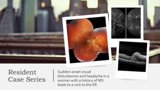
  • 1. Resident Case Series Sudden-onset visual disturbances and headache in a woman with a history of MS leads to a visit to the ER
  • 2. Bellows College Presentation 2 • A 44-year-old Caucasian female presented to the ER complaining of sudden onset of seeing a “thousand shades of gray, which later turned to black patches” in her right eye associated with pain with eye movement and dull headache. Her symptoms began 10 days prior and were progressively worsening. • Review of systems was notable for fevers, chills, night sweats, a recent intentional 90-pound weight loss, tinnitus, nausea and vomiting. • She denied any muscle, joint, dermatologic or genitourinary complaints currently or in the past.
  • 3. Bellows College Medical History 3 • She was diagnosed with multiple sclerosis 20 years prior, after having three episodes of “visual disturbances” in her right eye, presumably repeat bouts of optic neuritis. • After being treated with glatiramer acetate, discontinued 18 years ago, she denied any new visual complaints. • The remainder of her past medical history was remarkable for hypertension, controlled with verapamil and clonidine.
  • 4. Bellows College Medical History 4 • She was a non-smoker and denied any recent travel or having pets. • Two months prior she underwent a non-emergent laparoscopic cholecystectomy with a complicated postoperative course, resulting in an extended admission. Since then, she states she just has not “felt right”.
  • 5. Bellows College Examination • Vital signs were normal with a BP of 126/82. • VA uncorrected was 20/200 in her right eye with no improvement with pinhole and 20/20 in her left eye. • Pupils were significant for a RAPD in the right eye. • The patient was able to identify only the test plate in the right eye and 10/10 color plates in the left eye. • Confrontation visual fields and ocular motility were normal in both eyes. • IOP was 19 mmHg and 16 mmHg in the right and left eyes, respectively. 5
  • 6. Bellows College Examination • The anterior segment examination of her right eye revealed: 1. Diffuse flat conjunctival injection. 2. Fine KPs inferiorly. 3. 1+WBC in the AC. 4. 2+WBCs in the anterior vitreous. • There were no posterior synechiae, iris nodules or visually significant cataract. • The left eye anterior segment examination was unremarkable with no signs of ocular inflammation. 6
  • 8. Bellows College 8 • The posterior exam of the right eye revealed: 1. Significant vitreous cell. 2. Disc edema. 3. Peripapillary hemorrhages. 4. Cotton wool spots. 5. Occlusive arteritis along all four vascular arcades. 6. Macular edema. 7. The veins were slightly engorged but did not show any inflammatory changes. • Dilated funduscopic exam of the left eye was normal without evidence of prior inflammatory episodes. OD
  • 9. Bellows College The patient, a 44-year-old immunocompetent Caucasian female with a diagnosis of MS secondary to presumed episodes of right eye optic neuritis, presented with 20/200 vision in the right eye and clinical findings of a unilateral occlusive arteritis, disc edema associated with peripapillary hemorrhages and macular edema. 9
  • 11. Bellows College The differential of vasculitis is quite extensive and is best broken down according to conditions that affect arteries, veins or both. The differential of vasculitis is quite extensive and is best broken down according to conditions that affect arteries, veins or both. 11 Conditions that affect veins include: 1. Sarcoidosis 2. Syphilis 3. Par planitis 4. Eales’ disease 5. Multiple sclerosis Conditions that affect both arteries and veins include: 1. (SLE) 2. Wegener’s granulomatosis 3. HLA-B27 associated disease 4. Behçet’s disease. Conditions that affect arteries include: 1. Polyarteritis nodosa 2. Frosted-branch angiitis 3. Churg-Strauss syndrome 4. Acute retinal necrosis 5. Idiopathic retinal vasculitis, aneurysms and neuroretinitis (IRVAN).
  • 12. Bellows College Differential Diagnosis? • Despite the patient’s prior diagnosis, MS was an unlikely cause for several reasons. First, MS causes a periphlebitis, not an arteritis.1 • Approximately 11 % of MS patients have venous sheathing on routine color fundus photography.2 • Second, according to the Optic NeuritisTreatmentTrial, optic disc edema associated with peripapillary hemorrhages was never found to lead to a diagnosis of MS. • Finally, MS does not cause macular edema. • 1. RuckerWC. Sheathing of the retinal veins in Multiple Sclerosis. Mayo Clinc Proc 1972;47:335-40. • 2.Young BR. Fluorescein angiography and retinal venous sheathing in multiple sclerosis. Can J Ophthalmology 1976;11:31-6. 12
  • 13. Bellows College Laboratory workup 13 1. Anti-nuclear antibody (ANA) 2. Angiotensin converting enzyme (ACE) 3. ANCA, RPR/FTA 4. PPD 5. Bartonella antibodies 6. Rheumatoid factor 7. HIV antibodies 8. The ESR was 17 mm/hr and CRP was slightly elevated at 1.7 mg/L. 9. A chest X-ray was normal.
  • 14. Bellows College Workup • While the laboratory tests were pending, the patient was admitted to the ophthalmology inpatient service and started on 1g intravenous methylprenisolone daily for three days. • Given the retinal vasculitis and possible history of MS, MRI & MRA of the brain were ordered. • Imaging revealed non-specific scattered subcortical and periventricular white matter lesions on FLAIR imaging and no signs of cerebral vasculitis. 14
  • 15. Bellows College 15 FFA (arterial-venous phase and late phase) showing areas of retinal non- perfusion, corresponding to cotton-wool spots and leakage around disc and arteries.
  • 16. Bellows College 16 OCT of the right eye. Note vitritis, macular edema and subretinal fluid.
  • 17. Bellows College The absence of aneurysms on FA helped ruled out IRVAN. OCT demonstrated macular edema and subretinal fluid seen on clinical exam 17 Fluorescein angiography helped confirm the findings of an occlusive arteritis
  • 18. Bellows College Workup 18 • HLA typing revealed our patient was HLA-B51 positive. • Dermatology was consulted to administer a pathergy test, which was negative. • In addition, a full exam of her skin and mucous membranes did not show any evidence of oral or genital sores.
  • 19. Bellows College Hulusi Behçet Despite the lack of additional findings, the leading diagnosis was possible Behçet’s disease 19
  • 20. Bellows College 20A stamp (1980) in honor of Dr. Hulusi Behcet (1889-1948)
  • 21. Bellows College 21 Behçet’s disease is a chronic relapsing, idiopathic, multisystem, obliterative vasculitis that can affect both arteries and veins of all calibers, characterized by recurrent aphthous oral ulcers, genital ulceration and uveitis. Vasculitis is a key Pathogenetic Component
  • 22. Bellows College 22 The disease typically affects patients fromTurkey, the Middle and Far East (the ancient ‘Silk Road’ route), with a lower prevalence in Europe and North America.
  • 31. Bellows College Ocular features • Ocular inflammation occurs in about 70%, and tends to be more severe in men; it is the presenting manifestation in about 10%. • Signs are virtually always bilateral eventually. • Relapsing/remitting acute onset panuveitis with retinal vasculitis and often spontaneous resolution even without treatment is the classical pattern of eye involvement. • Retinal vascular disease (vasculitis and occlusion) is the main cause of visual impairment. 31
  • 32. Bellows College Ocular features • AAU, often bilateral, is typical. It is not granulomatous. • A transient mobile hypopyon in a relatively white eye is characteristic. • Vitritis may be severe; it is universal in eyes with active posterior segment disease. • Retinitis: Transient superficial white infiltrates that heal without scarring may be seen during acute systemic disease. • There may be deeper more diffuse retinitis similar in appearance to viral inflammation. • Exudative detachments can also occur. • Inflammatory deposits analogous to KPs may be seen on the inferior peripheral retina. 32
  • 36. Bellows College Ocular features • Retinal vasculitis : • Arteritis as well as phlebitis, in contrast to pure venous involvement in sarcoidosis – can manifest with sheathing, perivascular haemorrhages and occlusion. • Vascular leakage may give rise to diffuse retinal oedema and CMO. • Optic disc hyperaemia and oedema. Raised intracranial pressure can also cause optic disc swelling and optic atrophy in BD. • Disc and retinal neovascularization may be seen as a response to inflammation and ischemia. 36
  • 39. Bellows College Ocular features • Uncommon manifestations: conjunctivitis, conjunctival ulcers, episcleritis, scleritis and ophthalmoplegia from neurological involvement. • End-stage disease is characterized by optic atrophy, retinal atrophy and gliosis, and sheathing, attenuation and ghosting of affected vessels ; the vitreous tends to clear. 39
  • 43. Bellows College Ocular features • Other complications include posterior synechiae, cataract, glaucoma and, uncommonly, retinal detachment and phthisis. • Severe visual loss in males is up to two-thirds of patients at 10 years has been reported, but is probably much lower with aggressive management; the rate in women is about half that in men. 43
  • 44. Bellows College Investigation • HLA-B51. • Pathergy test. • Inflammatory markers (e.g. ESR, CRP, complement levels, WBCs) may be elevated. • Thrombophilia screening is appropriate in some patients to exclude other causes of thrombosis. • FA delineates ischaemic areas and aids detection of posterior segment inflammation and monitoring of disease activity. 44
  • 47. Bellows College Treatment • Topical steroids alone may be adequate if – rarely – there is no trace of posterior segment involvement. • Systemic steroids and azathioprine (2.5 mg/kg/day) in combination are recommended for the initial management of posterior uveitis in (EULAR) 2008 BD guidelines. • Steroids should be tapered only slowly.Topical and/or regional steroids may also be used. • Azathioprine may have a role in prophylaxis. 47
  • 48. Bellows College Treatment • Ciclosporin (2–5 mg/kg/day) or infliximab, in combination with azathioprine and systemic steroids, is recommended by EULAR for severe eye disease (> 2 lines reduction in visual acuity and/or retinal vasculitis or macular involvement). • A recent study recommended a single infliximab infusion as initial treatment of posterior uveitis. • Intravitreal administration is a novel alternative route of administration for infliximab. • Infliximab or adalimumab should be considered early for vision-threatening Behçet disease (American Uveitis Society recommendation). 48
  • 51. Bellows College Treatment 51 • Interferon- alfa (6 million IU per day subcutaneously initially, gradually tapered) with or without steroids is a EULAR-recommended alternative to the ciclosporin/ infliximab/azathioprine/steroid regimen for severe disease; it should not be used in combination with azathioprine (risk of myelosuppression). • Anticoagulants are not recommended.
  • 54. Bellows College Follow up 54 • After three days of IV methylprednisolone, our patient was transitioned to oral prednisone. • Over the next month, the inflammation and macular edema resolved. Her visual acuity returned to 20/30 with full recovery of her color vision. • Rheumatology did not recommend steroid-sparing agents and continued tapering her oral prednisone with no recurrence to date.

Editor's Notes

  1. Color fundus photo of right eye. Note the peripapillary hemorrhages, disc edema, cotton wool spots and arterial sheathing.
  2. Frosted-branch angiitis OPTIC NEURTIS IN MS BEHCEBT DISEASE IRVAN ACUTE RETINAL NECROSIS
  3. the Turkish dermatologist and scientist who first recognized the three main symptoms of the syndrome in one of his patients in 1924 and reported his research on the disease in Journal of Skin and Venereal Diseases in 1936.
  4. Vasculitis is a key pathogenetic component and may involve small, medium and large veins and arteries
  5. The U.S. prevalence is 0.4 per 100,000 with a female predominance
  6. It is strongly associated with HLA-B51; the ethnic groups with a higher prevalence of BD also have a higher rate of HLA-B51 positivity. The peak age of onset is the third decade; reported gender prevalence varies with ethnicity. Theories exist that Behçet’s disease is an autoimmune condition triggered by an infectious agent in a genetically predisposed individual.. Mortality is around 5% at 5–10 years, typically due to cardiovascular or CNS complications.
  7. Diagnostic criteria of the Behçet's Disease Research Committee of Japan (2003 revision). The criteria were developed by the Behçet’s Research Committee of Japan in 1974 and were later revised in 2003 to give greater weight to the presence of ocular inflammation.3 Our patient only satisfied one major criterion. recurrent oral ulceration : characterized by oral ulcers at least three times in a 12-month period, plus at least two of genital ulceration, ocular inflammation, characteristic skin lesions (erythema nodosum , pseudofolliculitis, acneiform nodules, papulopustular lesions) Vascular lesions. Aneurysms, including pulmonary and coronary, and venous thrombosis/thrombophlebitis Arthritis occurs in 30%, though arthralgia is more common. Dermatographia , similar to the pathergy reaction, indicates skin hypersensitivity and consists of the formation of erythematous lines following stroking or scratching. Neurological manifestations (5%) such as meningoencephalitis of the brainstem, dural sinus thrombosis and cerebral aneurysms. Gastrointestinal inflammation, especially ileocaecal. Hepatic and renal lesions are relatively uncommon.
  8. (B) superficial thrombophlebitis; (C) dermatographia
  9. Ocular involvement occurs in approximately 70 to 90 percent of cases and tends to affect both anterior and posterior structures.
  10. associated with a hypopyon, which shifts with gravity and head position. Posterior segment inflammation occurs in up to 93 percent of patients with ocular disease and portends a poor prognosis.
  11. (B) retinal infiltrates; Recurrent exudative retinal detachment
  12. Vascular occlusive episodes are a feared complication and if it involves the macula, visual acuity is reduced. Optic nerve involvement occurs in about 25 percent of patients.
  13. occlusive vasculitis;
  14. Inflammatory retinal vein occlusion with associated vitritis and retinal vasculitis before (A) and after (B) treatment with high dose oral steroid.
  15. (D) end-stage disease
  16. (A) Acute branch retinal vein occlusion. (B) Total vascular obliteration and optic atrophy secondary to recurrent vascular occlusion
  17. Intracranial venous thrombosis. (A) Male patient presents with headaches and disc oedema. (B) Magnetic resonance imaging and (C) magnetic resonance angiography demonstrating superior sagittal sinus thrombosis.
  18. HLA-B51 has a high association with Behçet’s, but is not part of the diagnostic criteria. 
  19. a pathergy reaction: pustule 24–48 hours after a sterile needle prick (>95% specific, but often negative in European and North American patients). Other tests that can help support a diagnosis of Behçet’s disease, but are not pathognomonic, are the pathergy test (skin prick) and HLA-B51 testing. During the pathergy test the forearm is pricked with a sterile needle and observed for two days. A positive test occurs if a pustule forms at the site of skin trauma. Only a minority of patients demonstrate this finding. The reaction is not common in the United States, while about half of all patients in Japan and Middle Eastern countries have this phenomenon.
  20. right retinal oedema with relative cilioretinal sparing, mild papillary hyperaemia and venous congestion. Multiple dot–blot haemorrhages were not present initially but developed within a few days Fundus photography and fluorescein angiography obtained 3 days after presentation. Note grossly impaired perfusion, retinal whitening and relative cilioretinal sparing.
  21. Immunosuppressants are the mainstay of treatment; availability and expense may limit therapeutic options in many regions. The initial goal of therapy is swift control of intraocular inflammation, typically accomplished using a combination of high-dose intravenous, oral, periocular, intravitreal or topical corticosteroids. Equally important is preventing additional inflammatory episodes. Long-term treatment with steroids should be avoided in cases of significant posterior uveitis, as chronic steroid use has not been found to reduce recurrences or improve the visual prognosis.6 Many steroid-sparing agents currently exist to prevent recurrence of inflammation. Some of the most commonly employed steroid-sparing medications include infliximab, azathioprine, cyclosporine-A, cyclophosphamide, tacrolimus and methotrexate.
  22. Hypertension, nephrotoxicity and neurotoxicity are concerns with ciclosporin, which should be avoided in patients with CNS involvement unless it is determined that severe eye disease warrants the risk. Infliximab may lead to activation of tuberculosis, and screening-positive patients should receive prophylactic treatment (e.g. isoniazid).
  23. This 20 year old woman had Behcet's disease with retinitis that responded rapidly to oral prednisone and subcutaneous injection of adalimumab. A - Initial fundus appearance of retinitis. The yellow arrow indicates white paravascular retinitis. Subretinal fluid is present but is difficult to appreciate in this photograph (see OCT, panel C). B - One month after beginning oral prednisone 40 mg/day and after one injection of subcutaneous adalimumab, the retinitis has regressed, as has the subretinal fluid. The blue arrow indicates residual retinal pigment epithelial metaplastic changes. C - An optical coherence tomograph line scan showing the retinal edema and subretinal fluid (orange arrow) at the initial presentation. D - One month after treatment, the edema and subretinal fluid have resolved. The retinal pigment epithelial metaplasia is indicated by the red arrow.
  24. Active Behçet uveitis showing vitritis and retinal infiltrates, located close to the fovea (A) and in the retinal periphery (B). HD-OCT of a patient with active Behçet uveitis showing vitritis and cystoid macular edema with a small serous foveal detachment (C); (D) complete resolution of the vitritis and the macular edema 5 weeks after starting Golimumab therapy
  25. Fundus photography and fluorescein angiography of one patient (Case 4) with Behçet uveitis. (A) Before vascular occlusion (at the 4-month follow-up): retinal vasculitis of the left eye. Fluorescein angiography (FA) shows diffuse leakage of fluorescein dye from the whole retinal vasculature and the optic disc. (B) Retinal vascular occlusion (before interferon alfa-2a treatment, at the 8-month follow-up): branch retinal vein occlusion with intraretinal edema of the left eye. FA shows blocked vascular filling at the inferotemporal venous branch. (C) After interferon alfa-2a treatment (at the 14-month follow-up): tortuous retinal vessels at the inferotemporal area, with improved retinal hemorrhage and edema. FA shows reperfusion; however, narrow inferotemporal venous branch with wide peripheral non-perfusion areas are also noted.
  26. . Color fundus photo of right eye. Note resolved disc and macular edema. Intraretinal hemorrhages still present.
  27. OCT of the right eye. Note resolved macular edema and subretinal fluid. Hard exudates are present.