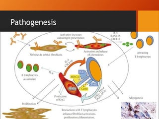
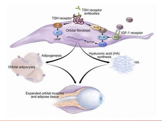
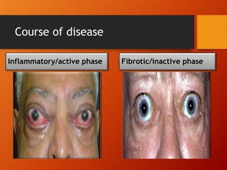
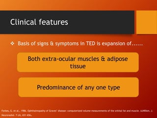
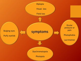
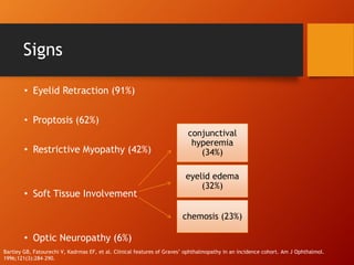
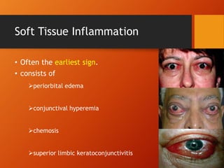
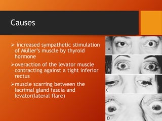
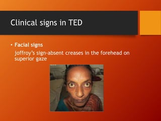
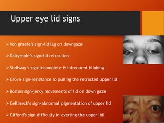
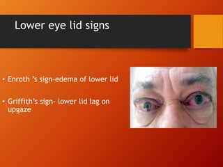
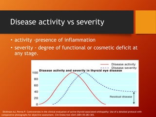
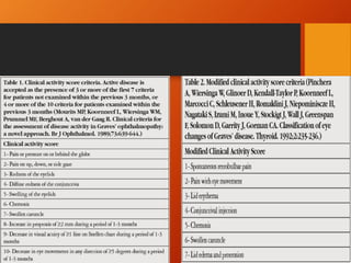
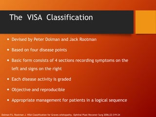
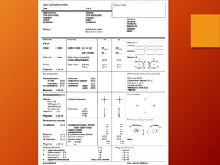
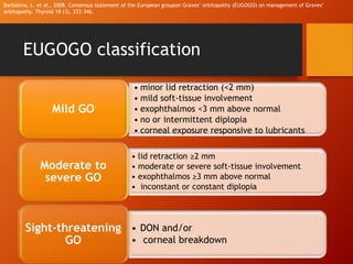
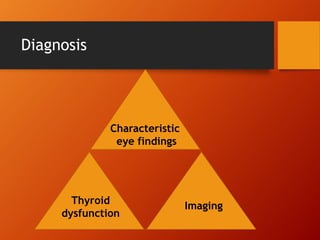
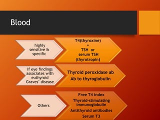
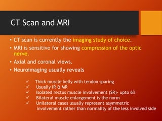
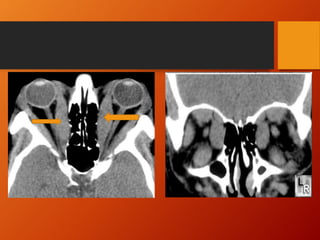
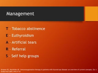
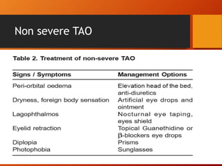
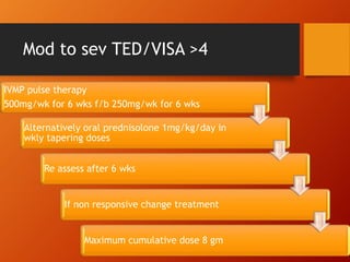
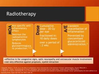
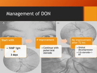
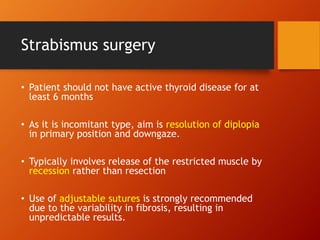
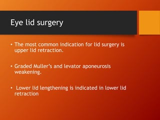
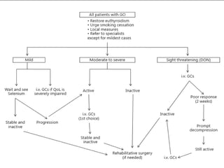
Thyroid eye disease, also known as Graves' ophthalmopathy, is an autoimmune condition causing inflammation and accumulation of fluid in the orbit, raising pressure. It produces symptoms related to proptosis, extraocular muscle involvement, and soft tissue changes. Management involves treatment of thyroid dysfunction, artificial tears, tobacco cessation, immunosuppression with steroids for moderate to severe disease, and orbital decompression or strabismus surgery for complications like dysthyroid optic neuropathy or diplopia. The disease progresses through inflammatory and fibrotic phases, and different classification systems evaluate severity and activity to guide treatment.
Optic neuritis is inflammation of the optic nerve that can be caused by demyelination, infection, or other autoimmune processes. It is classified based on location of inflammation (retrobulbar, papillitis, neuroretinitis) and etiology. Common symptoms include sudden unilateral vision loss, eye pain on movement, and color vision changes. Examination may reveal reduced acuity, afferent pupillary defect, and optic disc swelling or atrophy. Brain MRI and lumbar puncture help diagnose underlying causes like multiple sclerosis. For demyelinating optic neuritis, high-dose intravenous steroids can hasten recovery of vision lost.
1. Differential diagnosis of disc edema includes conditions like papilledema, optic neuritis, ischemic optic neuropathy, diabetic papillopathy, and hypertensive retinopathy.
2. Papilledema is caused by increased intracranial pressure and presents with bilateral disc swelling and normal vision, while optic neuritis typically causes unilateral vision loss and eye pain.
3. Diabetic papillopathy presents as transient unilateral or bilateral disc edema that resolves within months without vision loss, while malignant hypertension can lead to bilateral disc edema and vision changes as part of hypertensive retinopathy.
The document summarizes key points from a conference on diabetes and insulin. It discusses the various ocular manifestations of diabetes, including retinopathy, neuropathy, glaucoma and cataracts. It notes that up to 95% of those with type 1 diabetes and 60% of those with type 2 diabetes develop retinopathy. Risk factors include blood sugar levels, blood pressure, duration of diabetes, blood lipids, ethnicity and pregnancy. Screening guidelines are provided based on diabetes type and risk level. Imaging techniques like OCT and treatments like anti-VEGF injections plus laser therapy are reviewed. Novel research areas are also highlighted.
This document provides an overview of different types of corneal dystrophies, including their classification, clinical features, histopathology, and management. It discusses epithelial and subepithelial dystrophies, corneal dystrophies of Bowman's layer, stromal corneal dystrophies, and Descemet membrane and endothelial dystrophies. The key points are that corneal dystrophies are inherited, bilateral, and slowly progressive disorders that begin early in life and are characterized by corneal opacification without relationship to environmental factors. Diagnosis involves classification based on the anatomical layer affected and treatment typically involves managing symptoms although surgery may be needed if vision is impaired.
This document provides information on rhegmatogenous retinal detachment including:
- The pathogenesis which involves vitreoretinal traction from liquefaction of the vitreous gel creating a hole that allows fluid into the subretinal space.
- Symptoms include photopsia, floaters, and visual field defects.
- Signs include Marcus Gunn pupil, low intraocular pressure, retinal breaks, and mild iritis.
- Treatment options depend on factors like location and size of retinal break, state of vitreous gel, and include laser photocoagulation, cryotherapy, scleral buckling, pneumatic retinopexy, and drainage of subretinal fluid in
This document discusses thyroid diseases and their ocular manifestations. It focuses on Graves' disease, which causes Graves' ophthalmopathy - an autoimmune inflammatory disorder affecting the eye area. Symptoms include eyelid retraction, swelling, redness, and bulging eyes. Risk factors include female sex, smoking, and preexisting thyroid conditions. The document outlines pathogenesis, clinical features classified by severity, diagnostic tests, and treatment options including medications, surgery, and radiation therapy.
Choroidal melanomas are the most common primary intraocular malignancies in adults. They arise from melanocytes within the choroid and can be pigmented or amelanotic. Risk factors include light iris color and increased sun exposure. Diagnosis is based on clinical appearance, ultrasound, and fluorescein angiography. Prognosis depends on tumor size, cell type, genetic factors, and presence of extrascleral extension. The liver is the most common site of metastasis.
Age-related macular degeneration (AMD) is a leading cause of blindness that results from thickening of Bruch's membrane with age, interfering with metabolism in the retinal pigment epithelium (RPE) and photoreceptors. This causes deposits called drusen that damage the overlying RPE and underlying tissue. There are two forms: dry AMD involves gradual RPE and photoreceptor atrophy, while wet AMD features choroidal neovascularization that leaks fluid and blood, causing rapid vision loss if untreated. Risk factors include age, genetics, smoking, and diet.
Macular hole is a defect in the macula involving its full thickness. It was first described in 1869. Idiopathic macular holes are the most common type and affect people over age 55. Staging of macular holes ranges from stage 1 to 4 based on size and pathology. Symptoms include decreased vision and metamorphopsia. Diagnosis involves examination, OCT, and sometimes FA. Treatment is usually vitrectomy surgery for stages 2-4 to relieve traction on the macula. Prognosis depends on pre-op vision and hole size/duration, with most patients gaining vision after surgery.
This document discusses various types of corneal dystrophies presented by Dr. Puneet Sharma. It defines corneal dystrophies as a group of progressive, genetically determined disorders that cause non-inflammatory opacification of the cornea. It then describes several specific corneal dystrophies categorized by the layer of the cornea they affect (epithelial, stromal, endothelial). For each dystrophy, it covers inheritance pattern, histology, onset, signs/symptoms, and treatment. The document provides detailed information on Cogan dystrophy, Meesmann dystrophy, Reis-Bucklers dystrophy, granular dystrophy types 1 and 2, lattice dystrophy types 1 and 2, and Fuchs endothelial
Pseudoexfoliation syndrome is a systemic condition characterized by grey-white fibrillar deposits that can lead to open-angle glaucoma. It involves the trabecular meshwork, lens, ciliary body and other ocular tissues, and is a major risk factor for glaucoma. Treatment involves managing elevated intraocular pressure through medications, laser trabeculoplasty, trabeculectomy or cataract surgery due to the increased risk of complications from zonular weakness.
This document discusses retinal vascular occlusions, including their classification, presentation, investigations, and management. It begins by introducing retinal arterial and venous occlusions as significant causes of blindness. It then covers the anatomy of retinal blood supply and classifies occlusions by anatomical site. The remainder of the document details the characteristics, risk factors, clinical features, investigations, and treatment approaches for various types of retinal arterial occlusions (central retinal artery occlusion, branch retinal artery occlusion, cilioretinal artery occlusion) and venous occlusions (central retinal vein occlusion, branch retinal vein occlusion, hemiretinal vein occlusion).
This document discusses central retinal artery occlusion (CRAO), an ophthalmic emergency caused by obstruction of the central retinal artery. It affects around 1 in 100,000 people and often leads to severe vision loss. CRAO can be caused by emboli or thrombi and is associated with atherosclerosis and other vascular risk factors. The diagnosis involves examining the eye for signs like a cherry red spot at the macula. Acute management focuses on restoring retinal blood flow through measures like ocular massage or vasodilators. Long term management aims to prevent further vascular events through treating underlying risk factors.
Congenital Glaucoma is one of the most common causes of irreversible childhood blindness. This presentation covers this topic in detail that can aid physicians in effective patient care.
PS: The slides in the preview look skewed, download the presentation to view the font used in Office 2012 and upwards.
Duane's retraction syndrome involves congenital miswiring of the medial and lateral rectus muscles, causing limited eye movement. There are typically four types based on the pattern of limited adduction and/or abduction. Treatment may involve glasses, prisms, botulinum toxin injections, or surgery such as recession of the medial or lateral rectus muscles to improve eye alignment and positioning. Brown syndrome similarly involves a congenital or acquired restriction of eye elevation in adduction, believed to be caused by an abnormality of the superior oblique tendon. It is characterized by limited elevation in adduction and downshoot, and may cause a vertical eye misalignment.
The document discusses the anatomy and function of the macula and various tests used to evaluate macular function. The macula is located at the center of the retina and contains the highest concentration of photoreceptors, enabling sharp, central vision and color perception. Several tests are described that can help diagnose and monitor macular diseases by assessing visual acuity, contrast sensitivity, retinal imaging, and potential remaining vision in the context of opaque ocular media.
- The document discusses the evaluation of proptosis, which is the abnormal forward protrusion of the eyeball. It defines different types of orbital abnormalities and provides the approach to examining a patient with proptosis, including taking a thorough history, performing a local and systemic examination, ordering appropriate imaging and lab tests, and considering histopathological studies if needed. The causes of proptosis can be divided into categories such as inflammatory, mass effect, vascular changes, and infiltrative processes. Key aspects of the evaluation are to determine if the proptosis is unilateral or bilateral and whether there are associated signs and symptoms to suggest an underlying cause.
Vitreous hemorrhage occurs when blood leaks into the vitreous humor of the eye. It can be nondispersed, allowing some retinal view, or dispersed throughout the vitreous. Common causes include diabetic retinopathy, retinal vein occlusion, trauma, and posterior vitreous detachment. Patients experience painless vision loss and floaters. Examination may show a red eye or obscured retina. Treatment focuses on the underlying cause through laser, surgery, or observation depending on severity and risk factors.
The document discusses paralytic strabismus, including:
1) Hering's law of equal innervation and Sherington's law of reciprocal innervation which are important in diagnosing paralytic strabismus.
2) The sequelae of ocular muscle palsy including overactions and underactions of muscles.
3) Methods for investigating incomitant strabismus including cover tests, motility examination, and Hess screen plots to identify the affected muscle.
Thyroid eye disease (TED), also known as Graves' orbitopathy, is a self-limiting autoimmune condition that affects the eye muscles and fatty tissues behind the eyes. It is commonly associated with Graves' disease and hyperthyroidism. Treatment depends on the severity and includes corticosteroids to reduce inflammation, radiation therapy, orbital decompression surgery, and eyelid or muscle surgery to correct motility disorders or abnormalities. While TED has no cure currently, treatment aims to manage active inflammation and correct residual issues once the disease becomes inactive.
This document discusses different types of esotropia (inward eye turning), including accommodative esotropia caused by uncorrected refractive errors, convergence excess esotropia caused by a high AC/A ratio, and early onset esotropia appearing before age 6 months. Accommodative esotropia is treated initially with corrective lenses, while convergence excess may require bifocals or surgery. Early onset esotropia often requires surgery by age 1-2 years to align the eyes, with risks of undercorrection, amblyopia, or development of dissociated vertical deviation. The document provides details on diagnostic criteria and management approaches for various forms of esotropia.
Most retinal surgeons are trained to create formal retinal drawings of the fundus.
Retinal drawings are useful to document pathology, although more and more people now prefer fundus photographs.
Can be used for serial follow up of patients to document changes in the pathology.
This document provides information about ectopia lentis, or displacement of the crystalline lens. It begins with classifications of ectopia lentis based on location and etiology. Congenital causes like Marfan syndrome and homocystinuria are described. Presentation, examination findings, complications, workup, and management approaches are outlined. Surgical techniques depend on degree and location of lens subluxation/dislocation and may involve lens removal with or without intraocular lens implantation. Management of subluxated lenses in children poses additional challenges due to risk of complications with contact lenses or suture-fixated intraocular lenses.
Diabetic retinopathy is damage to the blood vessels of the retina due to diabetes. A study compared the standard treatment of pan-retinal photocoagulation (PRP) to initial treatment with intravitreal anti-VEGF injections (ranibizumab) to delay or prevent the need for PRP. At 2 years, vision was maintained or improved in the ranibizumab group while vision remained unchanged in the PRP group. The study suggests initial treatment of proliferative diabetic retinopathy with anti-VEGF injections like ranibizumab may be as effective as immediate PRP treatment and could help delay or reduce the need for destructive PRP therapy.
Coats' disease is a rare eye condition characterized by abnormal blood vessels in the retina. It most commonly affects young boys under 10 years old. The document discusses the history, signs and symptoms, classification, diagnosis, differential diagnosis, and management of Coats' disease. Treatment options depend on the stage of the disease and aim to obliterate abnormal retinal vessels using laser photocoagulation, cryotherapy, surgery, or newer adjunct treatments like intravitreal anti-VEGF agents. Indian studies found Coats' disease patients often present with more severe vision loss and extensive retinal involvement compared to other populations.
This document provides information on the anatomy and diseases of the vitreous humor. It discusses that the vitreous humor is a jelly-like structure that fills the back of the eye and provides support. Common diseases include vitreous liquefaction, detachment, hemorrhage, and opacities. Vitreous liquefaction is the most common degenerative change and causes floaters. Posterior vitreous detachment often occurs in older individuals and may lead to retinal tears or breaks. Vitreous opacities can result from inflammatory cells, aggregates, tumors or hemorrhages. Vitreous hemorrhage usually stems from retinal vessels and can cause vision loss.
A systematic approach with practical tips to diagnose and manage optic disc pallor. Disc pallor is often encountered in the routine clinical practice and remains a diagnostic enigma for most ophthalmologist. I illustrate the relevant practical points to be looked out for to deal with disc pallor.
Neovascular glaucoma is a severe form of secondary glaucoma characterized by fibrovascular proliferation in the anterior chamber angle caused by chronic retinal ischemia. The most common causes are diabetic retinopathy, central retinal vein occlusion, and ocular ischemic disease. The proliferation of new blood vessels leads to the formation of a membrane that can cause open or closed angle glaucoma with very high intraocular pressure. Treatment involves controlling the underlying cause, lowering intraocular pressure through medications, laser treatment or surgery like trabeculectomy with anti-metabolites or tube shunts, and preventing further neovascularization through panretinal photocoagulation. Early diagnosis and aggressive treatment is important but successful management is challenging and
Thyroid ophthalmopathy is an autoimmune condition that can cause mild to severe and potentially sight-threatening eye disease. Graves' disease is the most common associated thyroid abnormality. Symptoms include eyelid retraction, proptosis, and restrictive myopathy. Signs include eyelid retraction in 91% of patients, proptosis in 62%, and restrictive myopathy in 42%. The condition is classified in several ways, including the Wernicke, CAS, VISA, and EUGOGO systems, which grade severity based on symptoms and signs.
This document provides information about Thyroid Eye Disease (TED). It discusses the epidemiology, etiology, pathogenesis, clinical features, grading systems, evaluation, and management of TED. TED is an autoimmune inflammatory disorder characterized by eye lid retraction, proptosis, and restrictive extraocular muscle involvement. It commonly affects women more than men and is associated with Graves' hyperthyroidism. The document outlines various clinical signs, grading systems including NO SPECS and Mourits classifications, evaluation approach, and stepwise management strategies based on disease severity from mild to refractory disease.
Thyroid eye disease (TED) is an autoimmune inflammatory disorder affecting the eye muscles and surrounding tissues. It is commonly associated with Graves' disease. Symptoms include eye pain, swelling of eyelids, and issues with eye movement. Examination may reveal eyelid retraction, proptosis, and restrictive myopathy. Management involves medications like steroids to reduce inflammation during active phases, with surgery to correct eye muscle issues and proptosis during inactive phases. The goal is to improve symptoms, eye health, and appearance.
This document provides an overview of dysthyroid ophthalmopathy (also known as Graves' ophthalmopathy). It discusses the history, incidence, signs and symptoms, classification systems, pathogenesis, and clinical course. Key points include that it is an autoimmune condition associated with thyroid dysfunction, characterized by inflammation and swelling of extraocular muscles and orbital tissues leading to proptosis, diplopia, and optic nerve compression in severe cases. Classification systems evaluate activity, severity and organ involvement. The condition follows a self-limiting clinical course typically lasting 1-5 years and divided into active inflammatory and quiescent fibrotic phases.
Thyroid eye disease (TED), also known as Graves' ophthalmopathy, is an autoimmune disorder affecting the eyes that is commonly associated with Graves' disease and hyperthyroidism. It causes inflammation and swelling of the muscles and fatty tissues behind the eyes. The document discusses the epidemiology, risk factors, pathogenesis, clinical features including proptosis, lid retraction, optic neuropathy, and restrictive myopathy, as well as treatments such as steroids, radiation, orbital decompression surgery, and eyelid surgery. Management involves treatment of both the eye symptoms and any underlying thyroid abnormalities.
Thyroid ophthalmopathy (TED) is an autoimmune disorder where antibodies against the TSH receptor cause inflammation and swelling of the extraocular muscles and orbital tissues. The classic signs include eyelid retraction, proptosis, diplopia, and issues with eye movement. The symptoms result from infiltration of lymphocytes, plasma cells and fibrosis of the extraocular muscles leading to enlargement. Risk factors include Graves' disease, smoking, radiation exposure, and other autoimmune disorders. Diagnosis involves evaluating for these clinical signs and ruling out other potential causes of eye swelling.
Thyroid eye disease (TED), also known as thyroid-associated orbitopathy or Graves' ophthalmopathy, is the most common cause of both unilateral and bilateral proptosis in adults. It is associated with Graves' disease, the most common form of hyperthyroidism. TED causes soft tissue inflammation and swelling around the eyes, which can lead to proptosis, lid retraction, diplopia, and optic neuropathy. Treatment depends on severity and includes lubricants, steroids, radiotherapy, and in severe cases, surgery. Left untreated, TED can cause vision loss due to optic nerve compression.
This document provides information about proptosis (forward protrusion of the eyeball). It discusses the types of proptosis, differential diagnosis, investigations, and classifications. Proptosis can be caused by space-occupying lesions in the orbit or endocrinological disorders. Measurement of proptosis and examination involves assessing factors like duration, progression, and pulsations. Key differentials discussed are thyroid eye disease and pseudotumor. Imaging tests like CT scan and MRI are important investigations. Management depends on severity and involves medications, radiation, surgery.
This document discusses various eye conditions and how they relate to dentistry. It begins by explaining how to test visual acuity using Snellen charts and describes common causes of decreased vision like cataracts, age-related macular degeneration, glaucoma, and diabetic retinopathy. Specific eye conditions that can be related to dental issues are then outlined, such as Marcus Gunn jaw winking, giant cell arteritis, and rare ophthalmic complications from dental anesthesia. The document concludes by mentioning acute red eye conditions and dry eye disorders like Sjogren's syndrome that are less connected to dentistry.
Thyroid eye disease (TED), also known as Graves' ophthalmopathy, occurs in 25-50% of patients with Graves' disease. It is an autoimmune disorder caused by antibodies that lead to inflammation and swelling of the extraocular muscles and orbital tissues. Common symptoms include bulging eyes, dry eyes, double vision, and eye pain. Treatment involves controlling the hyperthyroidism medically or with radioactive iodine, using steroids to reduce inflammation, and potentially orbital decompression surgery for severe proptosis or optic nerve compression. Smoking worsens the condition so patients are advised to quit.
This document discusses neurocutaneous syndromes, which are disorders characterized by abnormalities of the skin and central nervous system. Some key syndromes mentioned include neurofibromatosis, tuberous sclerosis, Sturge-Weber syndrome, and Von Hippel-Lindau syndrome. Neurofibromatosis type 1 is described in detail, outlining its diagnostic criteria involving cafe-au-lait spots, freckling, and tumors. Tuberous sclerosis is also summarized, noting its diagnostic criteria involve tumors in multiple organ systems. Sturge-Weber syndrome links a port-wine stain on the face with leptomeningeal angiomas in the brain.
This document discusses Graves' disease and its management. Some key points:
- Graves' disease most commonly affects females ages 40-44 and males ages 45-49. Symptoms include eye protrusion, double vision, and eye pain or pressure.
- Assessment involves measuring eye protrusion, eye muscle function and signs of inflammation. Disease severity is classified as mild, moderate or severe.
- Treatment depends on severity but may include anti-thyroid medications, steroids, radiation therapy, surgery to decompress the orbit or correct eye muscle imbalances, and eyelid surgery.
- Management aims to control thyroid function, reduce inflammation and symptoms, and correct structural issues like eye protrusion and double vision. Care must
This document discusses optic disc swelling (papilledema) caused by increased intracranial pressure. It presents a case of a 35-year-old woman with severe headaches and vision issues. Examination found bilateral disc edema. CT scan revealed a brain tumor causing pressure. The patient was diagnosed with papilledema from the tumor and underwent surgery. The document then discusses the causes, presentation, stages, histopathology, and treatment of papilledema, emphasizing the importance of an eye exam for patients with headaches to identify potential intracranial issues.
Swollen optic nerve_presentation_last_revision 103112 disregard all othersArash Eslami
This document discusses the evaluation and differential diagnosis of a patient presenting with a swollen optic disc. The 58-year-old male patient woke up with spots in his vision in the left eye and was found to have unilateral disc swelling upon examination. Key points addressed include ruling out giant cell arteritis with urgent bloodwork and evaluating for signs of increased intracranial pressure. The differential diagnosis discussion centers on anterior ischemic optic neuropathy versus papilledema. Factors supporting NAION include the patient's risk factors of diabetes and hypertension as well as the clinical features matching NAION such as unilateral involvement and visual field defects. Close monitoring over subsequent months tracked improvement consistent with NAION.
This document discusses thyroid eye disease (TED), also known as Graves' ophthalmopathy. It covers the pathology, signs and symptoms, investigations, and treatment approaches for TED. Key points include that TED is an autoimmune disorder characterized by infiltrative orbitopathy, common signs include lid retraction, proptosis, restrictive myopathy, and optic neuropathy. Investigations include serological tests, CT or MRI imaging, and visual field testing. Treatment focuses on managing acute congestive orbitopathy, compressive optic neuropathy, motility disorders, and eyelid abnormalities, and may include corticosteroids, radiation therapy, or decompression surgery.
The ocular presentation of systemic diseasesRuwida Alorfy
The document summarizes various ocular manifestations of systemic diseases. It discusses how conditions like diabetes, hypertension, sarcoidosis, thyroid disease, and infections can present with issues like uveitis, cataracts, retinal vessel abnormalities, proptosis, ptosis, and abnormal eye movements. It provides details on characteristic signs, potential etiologies, and implications for underlying systemic illnesses for each type of ocular presentation.
Title: Sense of Smell
Presenter: Dr. Faiza, Assistant Professor of Physiology
Qualifications:
MBBS (Best Graduate, AIMC Lahore)
FCPS Physiology
ICMT, CHPE, DHPE (STMU)
MPH (GC University, Faisalabad)
MBA (Virtual University of Pakistan)
Learning Objectives:
Describe the primary categories of smells and the concept of odor blindness.
Explain the structure and location of the olfactory membrane and mucosa, including the types and roles of cells involved in olfaction.
Describe the pathway and mechanisms of olfactory signal transmission from the olfactory receptors to the brain.
Illustrate the biochemical cascade triggered by odorant binding to olfactory receptors, including the role of G-proteins and second messengers in generating an action potential.
Identify different types of olfactory disorders such as anosmia, hyposmia, hyperosmia, and dysosmia, including their potential causes.
Key Topics:
Olfactory Genes:
3% of the human genome accounts for olfactory genes.
400 genes for odorant receptors.
Olfactory Membrane:
Located in the superior part of the nasal cavity.
Medially: Folds downward along the superior septum.
Laterally: Folds over the superior turbinate and upper surface of the middle turbinate.
Total surface area: 5-10 square centimeters.
Olfactory Mucosa:
Olfactory Cells: Bipolar nerve cells derived from the CNS (100 million), with 4-25 olfactory cilia per cell.
Sustentacular Cells: Produce mucus and maintain ionic and molecular environment.
Basal Cells: Replace worn-out olfactory cells with an average lifespan of 1-2 months.
Bowman’s Gland: Secretes mucus.
Stimulation of Olfactory Cells:
Odorant dissolves in mucus and attaches to receptors on olfactory cilia.
Involves a cascade effect through G-proteins and second messengers, leading to depolarization and action potential generation in the olfactory nerve.
Quality of a Good Odorant:
Small (3-20 Carbon atoms), volatile, water-soluble, and lipid-soluble.
Facilitated by odorant-binding proteins in mucus.
Membrane Potential and Action Potential:
Resting membrane potential: -55mV.
Action potential frequency in the olfactory nerve increases with odorant strength.
Adaptation Towards the Sense of Smell:
Rapid adaptation within the first second, with further slow adaptation.
Psychological adaptation greater than receptor adaptation, involving feedback inhibition from the central nervous system.
Primary Sensations of Smell:
Camphoraceous, Musky, Floral, Pepperminty, Ethereal, Pungent, Putrid.
Odor Detection Threshold:
Examples: Hydrogen sulfide (0.0005 ppm), Methyl-mercaptan (0.002 ppm).
Some toxic substances are odorless at lethal concentrations.
Characteristics of Smell:
Odor blindness for single substances due to lack of appropriate receptor protein.
Behavioral and emotional influences of smell.
Transmission of Olfactory Signals:
From olfactory cells to glomeruli in the olfactory bulb, involving lateral inhibition.
Primitive, less old, and new olfactory systems with different path
micro teaching on communication m.sc nursing.pdfAnurag Sharma
Microteaching is a unique model of practice teaching. It is a viable instrument for the. desired change in the teaching behavior or the behavior potential which, in specified types of real. classroom situations, tends to facilitate the achievement of specified types of objectives.
share - Lions, tigers, AI and health misinformation, oh my!.pptxTina Purnat
• Pitfalls and pivots needed to use AI effectively in public health
• Evidence-based strategies to address health misinformation effectively
• Building trust with communities online and offline
• Equipping health professionals to address questions, concerns and health misinformation
• Assessing risk and mitigating harm from adverse health narratives in communities, health workforce and health system
Ozempic: Preoperative Management of Patients on GLP-1 Receptor Agonists Saeid Safari
Preoperative Management of Patients on GLP-1 Receptor Agonists like Ozempic and Semiglutide
ASA GUIDELINE
NYSORA Guideline
2 Case Reports of Gastric Ultrasound
These lecture slides, by Dr Sidra Arshad, offer a quick overview of the physiological basis of a normal electrocardiogram.
Learning objectives:
1. Define an electrocardiogram (ECG) and electrocardiography
2. Describe how dipoles generated by the heart produce the waveforms of the ECG
3. Describe the components of a normal electrocardiogram of a typical bipolar lead (limb II)
4. Differentiate between intervals and segments
5. Enlist some common indications for obtaining an ECG
6. Describe the flow of current around the heart during the cardiac cycle
7. Discuss the placement and polarity of the leads of electrocardiograph
8. Describe the normal electrocardiograms recorded from the limb leads and explain the physiological basis of the different records that are obtained
9. Define mean electrical vector (axis) of the heart and give the normal range
10. Define the mean QRS vector
11. Describe the axes of leads (hexagonal reference system)
12. Comprehend the vectorial analysis of the normal ECG
13. Determine the mean electrical axis of the ventricular QRS and appreciate the mean axis deviation
14. Explain the concepts of current of injury, J point, and their significance
Study Resources:
1. Chapter 11, Guyton and Hall Textbook of Medical Physiology, 14th edition
2. Chapter 9, Human Physiology - From Cells to Systems, Lauralee Sherwood, 9th edition
3. Chapter 29, Ganong’s Review of Medical Physiology, 26th edition
4. Electrocardiogram, StatPearls - https://www.ncbi.nlm.nih.gov/books/NBK549803/
5. ECG in Medical Practice by ABM Abdullah, 4th edition
6. Chapter 3, Cardiology Explained, https://www.ncbi.nlm.nih.gov/books/NBK2214/
7. ECG Basics, http://www.nataliescasebook.com/tag/e-c-g-basics
TEST BANK For Community Health Nursing A Canadian Perspective, 5th Edition by...Donc Test
TEST BANK For Community Health Nursing A Canadian Perspective, 5th Edition by Stamler, Verified Chapters 1 - 33, Complete Newest Version Community Health Nursing A Canadian Perspective, 5th Edition by Stamler, Verified Chapters 1 - 33, Complete Newest Version Community Health Nursing A Canadian Perspective, 5th Edition by Stamler Community Health Nursing A Canadian Perspective, 5th Edition TEST BANK by Stamler Test Bank For Community Health Nursing A Canadian Perspective, 5th Edition Pdf Chapters Download Test Bank For Community Health Nursing A Canadian Perspective, 5th Edition Pdf Download Stuvia Test Bank For Community Health Nursing A Canadian Perspective, 5th Edition Study Guide Test Bank For Community Health Nursing A Canadian Perspective, 5th Edition Ebook Download Stuvia Test Bank For Community Health Nursing A Canadian Perspective, 5th Edition Questions and Answers Quizlet Test Bank For Community Health Nursing A Canadian Perspective, 5th Edition Studocu Test Bank For Community Health Nursing A Canadian Perspective, 5th Edition Quizlet Test Bank For Community Health Nursing A Canadian Perspective, 5th Edition Stuvia Community Health Nursing A Canadian Perspective, 5th Edition Pdf Chapters Download Community Health Nursing A Canadian Perspective, 5th Edition Pdf Download Course Hero Community Health Nursing A Canadian Perspective, 5th Edition Answers Quizlet Community Health Nursing A Canadian Perspective, 5th Edition Ebook Download Course hero Community Health Nursing A Canadian Perspective, 5th Edition Questions and Answers Community Health Nursing A Canadian Perspective, 5th Edition Studocu Community Health Nursing A Canadian Perspective, 5th Edition Quizlet Community Health Nursing A Canadian Perspective, 5th Edition Stuvia Community Health Nursing A Canadian Perspective, 5th Edition Test Bank Pdf Chapters Download Community Health Nursing A Canadian Perspective, 5th Edition Test Bank Pdf Download Stuvia Community Health Nursing A Canadian Perspective, 5th Edition Test Bank Study Guide Questions and Answers Community Health Nursing A Canadian Perspective, 5th Edition Test Bank Ebook Download Stuvia Community Health Nursing A Canadian Perspective, 5th Edition Test Bank Questions Quizlet Community Health Nursing A Canadian Perspective, 5th Edition Test Bank Studocu Community Health Nursing A Canadian Perspective, 5th Edition Test Bank Quizlet Community Health Nursing A Canadian Perspective, 5th Edition Test Bank Stuvia
Knee anatomy and clinical tests 2024.pdfvimalpl1234
This includes all relevant anatomy and clinical tests compiled from standard textbooks, Campbell,netter etc..It is comprehensive and best suited for orthopaedicians and orthopaedic residents.
Basavarajeeyam is an important text for ayurvedic physician belonging to andhra pradehs. It is a popular compendium in various parts of our country as well as in andhra pradesh. The content of the text was presented in sanskrit and telugu language (Bilingual). One of the most famous book in ayurvedic pharmaceutics and therapeutics. This book contains 25 chapters called as prakaranas. Many rasaoushadis were explained, pioneer of dhatu druti, nadi pareeksha, mutra pareeksha etc. Belongs to the period of 15-16 century. New diseases like upadamsha, phiranga rogas are explained.
2. Lets start..
• Thyroid eye disease is an autoimmune disease
producing symptoms related to inflammation,
accumulation of fluid in the orbit and also to
adipogenesis raising intra-orbital pressure.
3. Girl you are in grave danger!!
• Per 100,000
person year
3
16
• Bartley GB, Fatourechi V, Kadrmas EF, Jacobsen SJ, Ilstrup DM, Garrity JA, et al. The incidence of Graves’ ophthalmopathy in Olmsted
County, Minnesota. Am J Ophthalmol 1995;120:511-7.
5. Asians (7.7%)Europeans (42%)
Tellez M, Cooper J, Edmonds C. Graves’ ophthalmopathy in relation to cigarette smoking and ethnic origin. Clin Endocrinol (Oxf) 1992;36:291-4.
7. Etiology
• Graves’ hyperthyroidism (90%)
• Hypothyroid Hashimoto’s thyroiditis
• Euthyroid subjects with no current or past
evidence of thyroid hyper or hypofunction (so-
called euthyroid Graves’ disease).1
1.Burch HB, Wartofsky L. Graves’ ophthalmopathy: current concepts regarding pathogenesis and management. Endocr Rev.1993;14(6):747–793.
8. Risk factors
• Smoking (strongest modifiable
risk factor)
• Family history
• Monozygotic twins
11. Course of disease
Inflammatory/active phase
• red and painful
• one to two years (range 6
months to 5 years).
• Active inflammation recurs
in about 1% of patients
after months to years of
inactivity
Fibrotic/inactive phase
• permanent structural
changes.
• no reliable test or sign.
• Stable clinical findings for
6 months suggest that the
patient has passed from
the active to the inactive
phase.
12. Clinical features
Forbes, G. et al., 1986. Ophthalmopathy of Graves’ disease: computerized volume measurements of the orbital fat and muscle. AJNRAm. J.
Neuroradiol. 7 (4), 651–656.
Both extra-ocular muscles & adipose
tissue
Predominance of any one type
Basis of signs & symptoms in TED is expansion of......
14. Signs
• Eyelid Retraction (91%)
• Proptosis (62%)
• Restrictive Myopathy (42%)
• Soft Tissue Involvement
• Optic Neuropathy (6%)
conjunctival
hyperemia
(34%)
eyelid edema
(32%)
chemosis (23%)
Bartley GB, Fatourechi V, Kadrmas EF, et al. Clinical features of Graves’ ophthalmopathy in an incidence cohort. Am J Ophthalmol.
1996;121(3):284–290.
15. Soft Tissue Inflammation
• Often the earliest sign.
• consists of
periorbital edema
conjunctival hyperemia
chemosis
superior limbic keratoconjunctivitis
16. Eyelid retraction
• Also called Dalrymple’s sign.
Normally, upper eyelid- 2mm below limbus
lower eyelid-inferior limbus
When retraction occurs, the sclera (white) can
be seen
• Lateral flare (almost pathognomic)
17. Causes
increased sympathetic stimulation
of Müller’s muscle by thyroid
hormone
overaction of the levator muscle
contracting against a tight inferior
rectus
muscle scarring between the
lacrimal gland fascia and
levator(lateral flare)
18. Proptosis
• Usually (90%) bilateral
• TAO is the most common cause of unilateral and
bilateral proptosis in adults
• Axial
• Resulting from enlargement of the extraocular
muscles and adipose tissue, as well as orbital fat
deposits and the infiltration of orbital tissues by
GAGs and leukocytes
19. • It does not respond to
hyperthyroidism treatment,
and is permanent in 70% of
cases.
• Severe proptosis prevents
adequate lid closure, and may
lead to severe exposure
keratopathy and corneal
ulceration.
20. Proptosis measurement
• Hertel exophthalmometer
• Distance between the lateral
orbital rim and the anterior
surface of the cornea.
• The upper limit of normal
whites -18 mm
blacks -21 mm
Asians -12-18 mm.
• A difference in reading of more
than 2 mm between the two
eyes is suggestive of proptosis.
22. Restrictive Myopathy
• Eye movements are restricted due to edema in the
extraocular muscles during the infiltrative stage and the
subsequent fibrosis.
• Despite expansion of the extraocular muscles in TAO, the
muscle fibers themselves are normal.
• IR>MR>SR>LR1
• Pressure exerted by a fibrotic inferior rectus muscle on the
globe may cause a spike in intraocular pressure during
upgaze.2
1.Bartley GB, Fatourechi V, Kadrmas EF, Jacobsen SJ, Ilstrup DM, Garrity JA, Gorman CA. Clinical features of Graves’ ophthalmopathy in an
incidence cohort. Am J Ophthalmol. 1996;121:284-290.
2.Konuk O, Onaran Z, Ozhan Oktar S, Yucel C, Unal M. Intraocular pressure and superior ophthalmic vein blood flow velocity in Graves’
orbitopathy: relation with the clinical features. Graefes Arch Clin Exp Ophthalmol. 2009;247:1555-1559
23. • Thyroid ophthalmopathy
+
exotropia
• Convergence insufficiency
• Inferior rectus muscle
restriction may mimic double
elevator palsy
• Forced ductions or elevated
intraocular pressure with eye
movement for confirmation.
} Concurrent MG
24. Dysthyroid Optic Neuropathy
(DON)
• Pressure from enlarged muscles on the optic nerve or
the vessels that supply it.
• This occurs when the enlarged EOM expand to
compress the optic nerve instead of producing
exophthalmos.
• Gradual decline in visual acuity, color vision
disturbance, development of an afferent pupillary
defect and central or paracentral scotomas
• Bilateral, simultaneous optic neuropathy can occur
which would eliminate a relative afferent pupillary
defect
McKeag D, Lane C, Lazarus JH, Baldeschi L, Boboridis K, Dickinson AJ, Hullo AI, Kahaly G, Krassas G, Marcocci C, Marinò M, Mourits MP, Nardi M, Neoh C,
Orgiazzi J, Perros P, Pinchera A, Pitz S, Prummel MF, Sartini MS, Wiersinga WM; European Group on Graves’ Orbitopathy (EUGOGO). Clinical features of
dysthyroid optic neuropathy: a European Group on Graves’Orbitopathy (EUGOGO) survey. Br J Ophthalmol. 2007;91:455-458.
25. Clinical signs in TED
• Facial signs
joffroy’s sign-absent creases in the forehead on
superior gaze
26. Eyelid signs
Kocher’s sign-staring appearance
Vigouroux sign-eyelid fullness
Rosenbach’s sign-tremors of eyelids
Riesman’s sign-Bruit over the eyelids
27. Upper eye lid signs
Von graefe’s sign-lid lag on downgaze
Dalrymple’s sign-lid retraction
Stellwag’s sign-incomplete & infrequent blinking
Grove sign-resistance to pulling the retracted upper lid
Boston sign-jerky movements of lid on down gaze
Gellineck’s sign-abnormal pigmentation of upper lid
Gifford’s sign-difficulty in everting the upper lid
28. Lower eye lid signs
• Enroth ’s sign-edema of lower lid
• Griffith’s sign- lower lid lag on
upgaze
30. Extraocular movement signs
Moebius sign-unable to converge eyes
Ballet’s sign-restriction of one or more EOM
Suker’s sign-poor fixation on abduction
Jendrassik’s sign-paralysis of all EOM
32. Disease activity vs severity
• activity -presence of inflammation
• severity - degree of functional or cosmetic deficit at
any stage.
Dickinson AJ, Perros P. Controversies in the clinical evaluation of active thyroid-associated orbitopathy: Use of a detailed protocol with
comparative photographs for objective assessment. Clin Endocrinol (Oxf) 2001;55:283-303.
34. WERNER´S CLASSIFICATION - NOSPECS
• Class 0: No signs or symptoms
• Class 1: Only signs (lid retraction, stare ± lid lag)
• Class 2: Soft tissue involvement
• Class 3: Proptosis
• Class 4: Extraocular muscle involvement
• Class 5: Corneal involvement
• Class 6: Sight loss (optic nerve involvement)
Werner SC. Classification of the eye changes of Graves’ disease. Am J Ophthalmol 1969;68:646-8.
35. Limitations
• Does not necessarily show sequential
involvement
• Inability to assess disease activity
• No prognostic or clinical implication
36. Clinical activity score
• In 1989, Mourits et al.developed the Clinical
Activity Score (CAS) for evaluating
ophthalmopathy activity.
• Found to be of value in predicting the outcome
of immunosuppressive treatment and
immunotherapy because of its high specificity
and high positive predictive value.
• CAS ≥4 were more responsive to treatment as
compared to patients with CAS<4
Mourits MP, Koornneef L, Wiersinga WM, Prummel MF, Berghout A, van der Gaag R. Clinical criteria for the assessment of disease activity in Graves’
ophthalmopathy: A novel approach. Br J Ophthalmol 1989;73:639-44.
37.
38. Clinical Activity Score
1. Pain on or behind the globe
2. Pain on eye movement
3. Redness of the eyelids
4. Redness of the conjunctiva
5. Swelling of the eyelids
6. Chemosis
7. Swollen caruncle
8. Increase of proptosis
9. Decreased eye movement
10. Decreased visual acuity
Pain
Redness
Swelling
Function
39. The VISA Classification
Devised by Peter Dolman and Jack Rootman
Based on four disease points
Basic form consists of 4 sections recording symptoms on the
left and signs on the right
Each disease activity is graded
Objective and reproducible
Appropriate management for patients in a logical sequence
Dolman PJ, Rootman J. VISA Classification for Graves orbitopathy. Ophthal Plast Reconstr Surg 2006;22:319-24
40.
41. EUGOGO classification
• minor lid retraction (<2 mm)
• mild soft-tissue involvement
• exophthalmos <3 mm above normal
• no or intermittent diplopia
• corneal exposure responsive to lubricants
Mild GO
• lid retraction ≥2 mm
• moderate or severe soft-tissue involvement
• exophthalmos ≥3 mm above normal
• inconstant or constant diplopia
Moderate to
severe GO
• DON and/or
• corneal breakdown
Sight-threatening
GO
Bartalena, L. et al., 2008. Consensus statement of the European groupon Graves’ orbitopathy (EUGOGO) on management of Graves’
orbitopathy. Thyroid 18 (3), 333–346.
45. CT Scan and MRI
• CT scan is currently the imaging study of choice.
• MRI is sensitive for showing compression of the optic
nerve.
• Axial and coronal views.
• Neuroimaging usually reveals
Thick muscle belly with tendon sparing
Usually IR & MR
Isolated rectus muscle involvement (SR)- upto 6%
Bilateral muscle enlargement is the norm
Unilateral cases usually represent asymmetric
involvement rather than normality of the less involved side
46.
47. Management
Krassas GE, Heufelder AE. Immunosuppressive therapy in patients with thyroid eye disease: an overview of current concepts. Eur J
Endocrinol 2001;144:311-8
T – Tobacco abstinence
E – Euthyroidism
A – Artificial tears
R – Referral
S – Self help groups
50. Mod to sev TED/VISA >4
IVMP pulse therapy
500mg/wk for 6 wks f/b 250mg/wk for 6 wks
Alternatively oral prednisolone 1mg/kg/day in
wkly tapering doses
Re assess after 6 wks
If non responsive change treatment
Maximum cumulative dose 8 gm
51. Radiotherapy
1.Kung AVC, Michon J, Tai KS, et al. the effect of somatostatin versus corticosteroids in the treatment of Graves’ ophthalmopathy. Thyroid 1996;6:381-4.
2. Bartalena L, Tanda L. Immunotherapy for Graves’ orbitopathy: easy enthusiasm but let’s keep trying. J Endocrinol Invest 2006;29:1012-6.
3. Nielsen Ch, El Fassi D, Hasselbalch HC, et al. B-cell depletion with rituximab in the treatment of autoimmune diseases. Graves’ ophthalmopathy the latest addition to an
expanding family. Expert Opin Biol Ther 2007;7:1061-78.
MOA
Non specific anti-
inflammatory
effect
Destroys the
radiosensitive
lymphocytes
Reduces
glycosaminoglyca
ns production.1,2,3
Dose
Cumulative
dose : 20 Gy
per eye
fractioned in:
10 daily doses
over a period of
: 2 wks
A/E
transient
exacerbation of
inflammation
Prevented by
concomitant
glucocorticoid
administration.3
•effective in for congestive signs, optic neuropathy and extraocular muscle involvement
•not very effective against proptosis, eyelid retraction
52. Management of DON
Start with
• IVMP 1gm
If improvement
• Continue with
pulse/oral
steroids
No improvement
after 72 hrs
• Orbital
decompression
• f/b steroids+/-
RT3 days
54. Types of surgery
• Orbital decompression
• Strabismus surgery
• Eyelid surgery
55. Orbital decompression
• Indications
compressive optic neuropathy
severe exposure keratopathy
• Transantral approach is the most preferred
• A/E are vision loss, diplopia ,periorbital
numbness, sinusitis, oroantral fistula, facial
neuralgia,, globe malposition
56. Strabismus surgery
• Patient should not have active thyroid disease for at
least 6 months
• As it is incomitant type, aim is resolution of diplopia
in primary position and downgaze.
• Typically involves release of the restricted muscle by
recession rather than resection
• Use of adjustable sutures is strongly recommended
due to the variability in fibrosis, resulting in
unpredictable results.
57. Eye lid surgery
• The most common indication for lid surgery is
upper lid retraction.
• Graded Muller’s and levator aponeurosis
weakening.
• Lower lid lengthening is indicated in lower lid
retraction