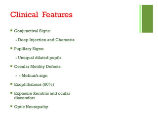
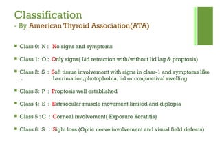
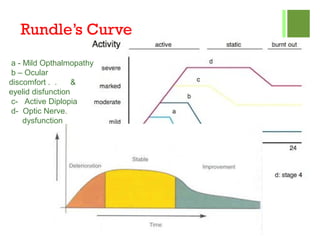
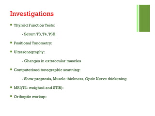
Thyroid Diseases
Ocular Manifestations
JeevanShrestha
Presented by: Jeevan Shrestha
KMCTH
 I have no conflict of interest or disclosure in relation to
this presentation.
Thyroid Disorders
 Grave’s Disease
 Hashimoto's Thyroiditis
 Thyroid Carcinoma
 Primary Hyperthyroidism
 Neck Irradiation
JeevanShrestha
Grave’s Ophthalmopathy
 Is an autoimmune inflammatory disorder affecting the orbit
around the eye, characterized by upper eyelid retraction, lid lag,
swelling, redness, conjunctivitis, and bulging eyes.
 Various Names:
 Thyroid Eye Disease(TED)
 Thyroid-associated Ophthalmopathy (TAO)
 Dysthyroid Ophthalmopathy
 Thyroid Orbitopathy
 Endocrine Ophthalmopathy
 Sex: More common in female than male 4:1
 Smoking
 Middle age
 Autoimmune thyroid disease
 HLA-DR3 and HLA-B8
 TED associated with Hyperthyroidism(90%), Hypothyroidism(4%),
Euthyroidism(6%)
Risk factors
Onset
 20% of TED is diagnosed same time as hyperthyroidism
 60% of eye disease occur after 1 year of thyroid disease
 Only 30% of hyperthyroidism  TED
Pathogenesis
 Inflammatory targets:
 Primary: Orbital fibroblast
 Secondary: Extraocular muscles
 Activated T-cells act and stimulate
adipogenesis, fibroblast proliferation
and glycosaminoglycan synthesis.
 Enlargement of extraocular muscles
due to edema and infiltration
 Orbital soft tissue infiltrated with
lymphocytes, macrophages and mast
cells
Pathogenesis
 Autoimmune disorder(IgG mediated)
 Enlargement of Extraocular Muscles
- By Increase in Glycosaminoglycans
 Cellular Infiltration of Interstitial Tissues
- With lymohocytes, plasma cells and macrophages, mast cell
- Fibrosis
 Proliferation of Orbital fat, Connective tissue and Lacrimal Gland
- With retention of fluid and GAG
Grave’s Ophthalmopathy
Axial CT
Extraocular Muscle Enlargement
(Fusiform Appearance)
Clinical Features
 LID SIGNS
- Retraction of Upper Lids
(Dalrymple’s Sign) in 90%
- Lid Lag in 50% (von Graefe’s Sign)
- Fullness of Eyelids(Enroth’s sign)
- Difficulty in Eversion of Upper Lid
(Gifford’s Sign)
- Infrequent Blinking( Stellwag’s Sign)
Clinical Features
 Conjunctival Signs:
- Deep Injection and Chemosis
 Pupillary Signs:
- Unequal dilated pupils
 Occular Motility Defects:
- - Mobius’s sign
 Exophthalmos (60%)
 Exposure Keratitis and ocular
discomfort
 Optic Neuropathy
Classification
- By American Thyroid Association(ATA)
 Class 0: N : No signs and symptoms
 Class 1: O : Only signs( Lid retraction with/without lid lag & proptosis)
 Class 2: S : Soft tissue involvement with signs in class-1 and symptoms like
. Lacrimation,photophobia, lid or conjunctival swelling
 Class 3: P : Proptosis well established
 Class 4: E : Extraocular muscle movement limited and diplopia
 Class 5 : C : Corneal involvement( Exposure Keratitis)
 Class 6: S : Sight loss (Optic nerve involvement and visual field defects)
Rundle’s Curve
a - Mild Opthalmopathy
b – Ocular
discomfort . . &
eyelid disfunction
c- Active Diplopia
d- Optic Nerve.
dysfunction
Diagnosis
 Grave’s Ophthalmopathy:
 10-20% Precede hyperthyroid
 40% Concurrent
 30% < 6 months after diagnosis
 10-20% > 6 moths after Dx
Investigations
 Thyroid Function Tests:
- Serum T3,T4,TSH
 Positional Tonometry:
 Ultrasonography:
- Changes in extraocular muscles
 Computerised tonographic scanning:
- Show proptosis, Muscle thickness, Optic Nerve thickening
 MRI(T2- weighed and STIR):
 Orthoptic workup:
Treatment
A) Non- surgical Management:
- Smoking Cessation
- Head elevation at night & cold compressors in morning(Reduce periorbital
edema)
- Lubricating artificial tear drops
- Eyelid taping
- Guanethidine 5% eyedrop ( Decrease lid retraction)
- Prisms
- Systemic steroids
- Radiotherapy
- Combined therapy : ( Low dose steroids + Azathioprine + Irradiation)
Treatment
(B) Surgical Management:
i)Orbital Decompression:
- Two wall Decompression (Orbital floor and medial wall removed)
- Three wall Decompression (Floor, medial and lateral wall removed)
- Four wall Decompression ( Three wall removal plus lateral half of roof and. .
. large portion of
sphenoid at apex)
ii) Extraocular muscle surgery:
- Always done after orbital decompression
- To achieve binocular single vision in reading position
Treatment
iii) Eyelid Surgery :
- Mullerotomy
- Levator recession/disinsertion
- Scleral grafts
- Recession of lower eyelid retractors
- Blepheroplasty
 Dalrymple’s sign: Lid retraction
 • von Graefe’s sign: Upper lid lag on downward
Gaze
 • Kocher’s sign: Increased lid retraction with visual fixation
 • Ballet’s sign: Palsy of one or more extraocular muscles
 • Suker’s sign:Weakness of fixation on lateral gaze
 • Cowen’s sign: Jerky papillary contraction to consensual light
 • Knies’ sign: Unequal dilatation of the pupils
 • Jeffrey’s sign: Absence of forehead wrinkling on upward gaze
 Griffith’s sign: Lower lid lag on downward gaze
 • Stellwag’s sign: Infrequent blinking
 • Enroth’s sign: Puffy swelling of the lids
 • Mobius’ sign:Weakness of convergence
mobius
THANK YOU
Marty
Feldman
Presented By :
Jeevan Shrestha

THYROID EYE DISEASE

  • 1.
  • 2.
     I haveno conflict of interest or disclosure in relation to this presentation.
  • 3.
    Thyroid Disorders  Grave’sDisease  Hashimoto's Thyroiditis  Thyroid Carcinoma  Primary Hyperthyroidism  Neck Irradiation JeevanShrestha
  • 4.
    Grave’s Ophthalmopathy  Isan autoimmune inflammatory disorder affecting the orbit around the eye, characterized by upper eyelid retraction, lid lag, swelling, redness, conjunctivitis, and bulging eyes.  Various Names:  Thyroid Eye Disease(TED)  Thyroid-associated Ophthalmopathy (TAO)  Dysthyroid Ophthalmopathy  Thyroid Orbitopathy  Endocrine Ophthalmopathy
  • 5.
     Sex: Morecommon in female than male 4:1  Smoking  Middle age  Autoimmune thyroid disease  HLA-DR3 and HLA-B8  TED associated with Hyperthyroidism(90%), Hypothyroidism(4%), Euthyroidism(6%) Risk factors
  • 6.
    Onset  20% ofTED is diagnosed same time as hyperthyroidism  60% of eye disease occur after 1 year of thyroid disease  Only 30% of hyperthyroidism  TED
  • 7.
    Pathogenesis  Inflammatory targets: Primary: Orbital fibroblast  Secondary: Extraocular muscles  Activated T-cells act and stimulate adipogenesis, fibroblast proliferation and glycosaminoglycan synthesis.  Enlargement of extraocular muscles due to edema and infiltration  Orbital soft tissue infiltrated with lymphocytes, macrophages and mast cells
  • 8.
    Pathogenesis  Autoimmune disorder(IgGmediated)  Enlargement of Extraocular Muscles - By Increase in Glycosaminoglycans  Cellular Infiltration of Interstitial Tissues - With lymohocytes, plasma cells and macrophages, mast cell - Fibrosis  Proliferation of Orbital fat, Connective tissue and Lacrimal Gland - With retention of fluid and GAG
  • 9.
    Grave’s Ophthalmopathy Axial CT ExtraocularMuscle Enlargement (Fusiform Appearance)
  • 10.
    Clinical Features  LIDSIGNS - Retraction of Upper Lids (Dalrymple’s Sign) in 90% - Lid Lag in 50% (von Graefe’s Sign) - Fullness of Eyelids(Enroth’s sign) - Difficulty in Eversion of Upper Lid (Gifford’s Sign) - Infrequent Blinking( Stellwag’s Sign)
  • 11.
    Clinical Features  ConjunctivalSigns: - Deep Injection and Chemosis  Pupillary Signs: - Unequal dilated pupils  Occular Motility Defects: - - Mobius’s sign  Exophthalmos (60%)  Exposure Keratitis and ocular discomfort  Optic Neuropathy
  • 12.
    Classification - By AmericanThyroid Association(ATA)  Class 0: N : No signs and symptoms  Class 1: O : Only signs( Lid retraction with/without lid lag & proptosis)  Class 2: S : Soft tissue involvement with signs in class-1 and symptoms like . Lacrimation,photophobia, lid or conjunctival swelling  Class 3: P : Proptosis well established  Class 4: E : Extraocular muscle movement limited and diplopia  Class 5 : C : Corneal involvement( Exposure Keratitis)  Class 6: S : Sight loss (Optic nerve involvement and visual field defects)
  • 13.
    Rundle’s Curve a -Mild Opthalmopathy b – Ocular discomfort . . & eyelid disfunction c- Active Diplopia d- Optic Nerve. dysfunction
  • 14.
    Diagnosis  Grave’s Ophthalmopathy: 10-20% Precede hyperthyroid  40% Concurrent  30% < 6 months after diagnosis  10-20% > 6 moths after Dx
  • 15.
    Investigations  Thyroid FunctionTests: - Serum T3,T4,TSH  Positional Tonometry:  Ultrasonography: - Changes in extraocular muscles  Computerised tonographic scanning: - Show proptosis, Muscle thickness, Optic Nerve thickening  MRI(T2- weighed and STIR):  Orthoptic workup:
  • 16.
    Treatment A) Non- surgicalManagement: - Smoking Cessation - Head elevation at night & cold compressors in morning(Reduce periorbital edema) - Lubricating artificial tear drops - Eyelid taping - Guanethidine 5% eyedrop ( Decrease lid retraction) - Prisms - Systemic steroids - Radiotherapy - Combined therapy : ( Low dose steroids + Azathioprine + Irradiation)
  • 17.
    Treatment (B) Surgical Management: i)OrbitalDecompression: - Two wall Decompression (Orbital floor and medial wall removed) - Three wall Decompression (Floor, medial and lateral wall removed) - Four wall Decompression ( Three wall removal plus lateral half of roof and. . . large portion of sphenoid at apex) ii) Extraocular muscle surgery: - Always done after orbital decompression - To achieve binocular single vision in reading position
  • 18.
    Treatment iii) Eyelid Surgery: - Mullerotomy - Levator recession/disinsertion - Scleral grafts - Recession of lower eyelid retractors - Blepheroplasty
  • 21.
     Dalrymple’s sign:Lid retraction  • von Graefe’s sign: Upper lid lag on downward Gaze  • Kocher’s sign: Increased lid retraction with visual fixation
  • 22.
     • Ballet’ssign: Palsy of one or more extraocular muscles  • Suker’s sign:Weakness of fixation on lateral gaze  • Cowen’s sign: Jerky papillary contraction to consensual light  • Knies’ sign: Unequal dilatation of the pupils  • Jeffrey’s sign: Absence of forehead wrinkling on upward gaze  Griffith’s sign: Lower lid lag on downward gaze  • Stellwag’s sign: Infrequent blinking  • Enroth’s sign: Puffy swelling of the lids  • Mobius’ sign:Weakness of convergence mobius
  • 23.