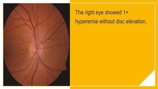
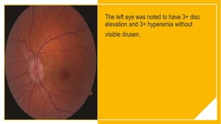
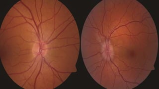
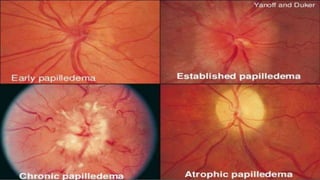
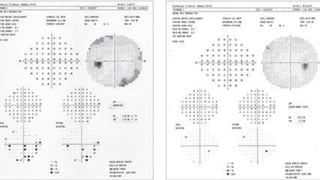
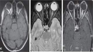
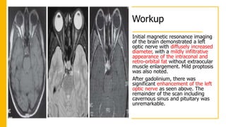
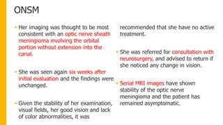
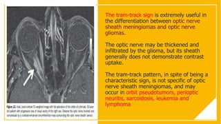
Case
Presentation
A middle-aged woman seeks a
LASIK consult, but unilateral disc
edema leads the examination on a
new course.
PRESENTATION:
 A 44-year-old woman with a medical
history of depression and ocular
history of mild myopia presented for
LASIK evaluation.
 Her best-corrected visual acuity was
20/20 in both eyes, but examination
revealed a swollen optic disc in the
left eye.
 LASIK surgery was declined and she
was referred for evaluation of the
unilateral optic nerve head swelling.
She recalled minor head trauma three
months earlier.
 Systemic review of symptoms was
negative except for a subjective
sensation of tingling of left side of
her head and earlobe. She denied
pain and decreased vision.
Medical History
 She denied any history of
vision loss, eye pain or
systemic neurologic symptoms
(weakness, numbness,
paralysis).
 She smoked one pack of
cigarettes per day for 30 years,
described minor social alcohol
use and no other drug use.
 Medications included only
Escitalopram (Lexapro) 10 mg
daily.
 She had no family history of
systemic problems.
On examination…
• Examination showed a best corrected
visual acuity of 20/20 in each eye.
• Pupil examination noted 1 mm of
anisocoria that was the same in light
and dark with no afferent pupillary
defect.
• Applanation intraocular pressures
were 18 mmHg and 15 mmHg.
• She had full ocular motility, full
confrontational fields and color plate
testing was full.
On examination…
• Hertel measurements were 15
mm OD and 16 mm OS with a
base of 105 mm.
• She denied red desaturation.
• Anterior segment examination
revealed normal findings
The right eye showed 1+
hyperemia without disc elevation.
The left eye was noted to have 3+ disc
elevation and 3+ hyperemia without
visible drusen.
What is your
differential
diagnosis?
What further
workup would you
pursue?
A broad differential diagnosis can be generated
for unilateral optic disc edema.
 Traumatic
 Demyelinating/autoimmune (multiple
sclerosis, neuromyelitis optica, lupus
erythematosis, thyroid eye disease)
 Infectious conditions (tuberculosis,
lyme disease, syphilis, herpetic,
bartonella)
 Infiltrative/inflammatory (neoplasm,
sarcoidosis, drusen)
 Congestive (orbital
pseudotumor/cellulitis)
 Vascular (diabetic papillopathy, giant
cell arteritis, nonarteritic anterior
ischemic optic neuropathy).
Workup
B-scan ultrasonography
revealed no optic disc
drusen.
Humphrey visual field
testing showed
peripheral relative and
absolute scotomas
Workup
Initial magnetic resonance imaging
of the brain demonstrated a left
optic nerve with diffusely increased
diameter, with a mildly infiltrative
appearance of the intraconal and
retro-orbital fat without extraocular
muscle enlargement. Mild proptosis
was also noted.
After gadolinium, there was
significant enhancement of the left
optic nerve as seen above. The
remainder of the scan including
cavernous sinus and pituitary was
unremarkable.
Diagnosis
• Based on her clinical findings,
her differential diagnosis was
narrowed to:
1. Optic nerve meningioma
2. Optic nerve drusen
3. Resolving optic neuritis
4. Optic nerve glioma.
ONSM
ONSM
 Her imaging was thought to be most
consistent with an optic nerve sheath
meningioma involving the orbital
portion without extension into the
canal.
 She was seen again six weeks after
initial evaluation and the findings were
unchanged.
 Given the stability of her examination,
visual fields, her good vision and lack
of color abnormalities, it was
recommended that she have no active
treatment.
 She was referred for consultation with
neurosurgery, and advised to return if
she noticed any change in vision.
 Serial MRI images have shown
stability of the optic nerve
meningioma and the patient has
remained asymptomatic.
Discussion
 Optic nerve sheath meningiomas are
benign neoplasms of the optic nerve
sheath.
 Although rare, ONSM is the second-
most common primary optic nerve
tumor and represents 1-2% of all
meningioma
 Show positive immunoreactivity for
vimentin and may contain psammoma
bodies.
 Malignant transformation is
exceedingly rare.
 For unknown reasons, there is a
higher incidence of optic nerve sheath
meningiomas in middle-aged women.
 There is also an association with
neurofibromatosis type 2.
A classic triad of an ONSM:
Progressive visual loss
Optic nerve atrophy
Presence of optociliary collateral vessels
Discussion
 The tumor typically tracks along the
optic nerve sheath and can grow to
surround and compress the optic
nerve, central retinal artery or the
central retinal vein.
 Vision loss is usually caused by a
compressive optic neuropathy, but
patients may live without symptoms
for decades.
 The mass can also grow back towards
the chiasm and cause contralateral
field deficits.
 Patients may demonstrate the classic,
pathognomonic clinical triad for
ONSM, consisting of painless, slowly
progressive vision loss, optic atrophy,
and optociliary shunt vessels.
 Axial proptosis, optic disc edema,
color vision abnormalities and
extraocular muscle movement
abnormalities may also be noted.
The tram-track sign is extremely useful in
the differentiation between optic nerve
sheath meningiomas and optic nerve
gliomas.
The optic nerve may be thickened and
infiltrated by the glioma, but its sheath
generally does not demonstrate contrast
uptake.
The tram-track pattern, in spite of being a
characteristic sign, is not specific of optic
nerve sheath meningiomas, and may
occur in orbit pseudotumors, perioptic
neuritis, sarcoidosis, leukemia and
lymphoma
Discussion
 MRI will demonstrate diffuse, tubular
thickening of the optic nerve sheath
encasing the optic nerve, often
producing a characteristic "tram
track" sign on axial cuts or a
"doughnut" sign on coronal cuts. The
tumor enhances with contrast
infusion.
 In asymptomatic patients, observation
with exams every six to 12 months
including vision, pupils, color vision,
visual fields as well as periodic MRI
imaging is indicated.
 Many patients do have progressive
visual loss over time, but there are
currently no tests available that can
predict the aggressiveness of these
benign neoplasms.
 If there is progression of a patient’s
symptoms, the optic nerve can be
approached with radiotherapy or
gamma knife surgery.
Unilateral Optic disc swelling

Unilateral Optic disc swelling

  • 1.
    Case Presentation A middle-aged womanseeks a LASIK consult, but unilateral disc edema leads the examination on a new course.
  • 2.
    PRESENTATION:  A 44-year-oldwoman with a medical history of depression and ocular history of mild myopia presented for LASIK evaluation.  Her best-corrected visual acuity was 20/20 in both eyes, but examination revealed a swollen optic disc in the left eye.  LASIK surgery was declined and she was referred for evaluation of the unilateral optic nerve head swelling. She recalled minor head trauma three months earlier.  Systemic review of symptoms was negative except for a subjective sensation of tingling of left side of her head and earlobe. She denied pain and decreased vision.
  • 3.
    Medical History  Shedenied any history of vision loss, eye pain or systemic neurologic symptoms (weakness, numbness, paralysis).  She smoked one pack of cigarettes per day for 30 years, described minor social alcohol use and no other drug use.  Medications included only Escitalopram (Lexapro) 10 mg daily.  She had no family history of systemic problems.
  • 4.
    On examination… • Examinationshowed a best corrected visual acuity of 20/20 in each eye. • Pupil examination noted 1 mm of anisocoria that was the same in light and dark with no afferent pupillary defect. • Applanation intraocular pressures were 18 mmHg and 15 mmHg. • She had full ocular motility, full confrontational fields and color plate testing was full.
  • 5.
    On examination… • Hertelmeasurements were 15 mm OD and 16 mm OS with a base of 105 mm. • She denied red desaturation. • Anterior segment examination revealed normal findings
  • 6.
    The right eyeshowed 1+ hyperemia without disc elevation.
  • 7.
    The left eyewas noted to have 3+ disc elevation and 3+ hyperemia without visible drusen.
  • 10.
    What is your differential diagnosis? Whatfurther workup would you pursue?
  • 11.
    A broad differentialdiagnosis can be generated for unilateral optic disc edema.  Traumatic  Demyelinating/autoimmune (multiple sclerosis, neuromyelitis optica, lupus erythematosis, thyroid eye disease)  Infectious conditions (tuberculosis, lyme disease, syphilis, herpetic, bartonella)  Infiltrative/inflammatory (neoplasm, sarcoidosis, drusen)  Congestive (orbital pseudotumor/cellulitis)  Vascular (diabetic papillopathy, giant cell arteritis, nonarteritic anterior ischemic optic neuropathy).
  • 12.
    Workup B-scan ultrasonography revealed nooptic disc drusen. Humphrey visual field testing showed peripheral relative and absolute scotomas
  • 16.
    Workup Initial magnetic resonanceimaging of the brain demonstrated a left optic nerve with diffusely increased diameter, with a mildly infiltrative appearance of the intraconal and retro-orbital fat without extraocular muscle enlargement. Mild proptosis was also noted. After gadolinium, there was significant enhancement of the left optic nerve as seen above. The remainder of the scan including cavernous sinus and pituitary was unremarkable.
  • 17.
    Diagnosis • Based onher clinical findings, her differential diagnosis was narrowed to: 1. Optic nerve meningioma 2. Optic nerve drusen 3. Resolving optic neuritis 4. Optic nerve glioma.
  • 18.
  • 19.
    ONSM  Her imagingwas thought to be most consistent with an optic nerve sheath meningioma involving the orbital portion without extension into the canal.  She was seen again six weeks after initial evaluation and the findings were unchanged.  Given the stability of her examination, visual fields, her good vision and lack of color abnormalities, it was recommended that she have no active treatment.  She was referred for consultation with neurosurgery, and advised to return if she noticed any change in vision.  Serial MRI images have shown stability of the optic nerve meningioma and the patient has remained asymptomatic.
  • 20.
    Discussion  Optic nervesheath meningiomas are benign neoplasms of the optic nerve sheath.  Although rare, ONSM is the second- most common primary optic nerve tumor and represents 1-2% of all meningioma  Show positive immunoreactivity for vimentin and may contain psammoma bodies.  Malignant transformation is exceedingly rare.  For unknown reasons, there is a higher incidence of optic nerve sheath meningiomas in middle-aged women.  There is also an association with neurofibromatosis type 2.
  • 21.
    A classic triadof an ONSM: Progressive visual loss Optic nerve atrophy Presence of optociliary collateral vessels
  • 23.
    Discussion  The tumortypically tracks along the optic nerve sheath and can grow to surround and compress the optic nerve, central retinal artery or the central retinal vein.  Vision loss is usually caused by a compressive optic neuropathy, but patients may live without symptoms for decades.  The mass can also grow back towards the chiasm and cause contralateral field deficits.  Patients may demonstrate the classic, pathognomonic clinical triad for ONSM, consisting of painless, slowly progressive vision loss, optic atrophy, and optociliary shunt vessels.  Axial proptosis, optic disc edema, color vision abnormalities and extraocular muscle movement abnormalities may also be noted.
  • 24.
    The tram-track signis extremely useful in the differentiation between optic nerve sheath meningiomas and optic nerve gliomas. The optic nerve may be thickened and infiltrated by the glioma, but its sheath generally does not demonstrate contrast uptake. The tram-track pattern, in spite of being a characteristic sign, is not specific of optic nerve sheath meningiomas, and may occur in orbit pseudotumors, perioptic neuritis, sarcoidosis, leukemia and lymphoma
  • 25.
    Discussion  MRI willdemonstrate diffuse, tubular thickening of the optic nerve sheath encasing the optic nerve, often producing a characteristic "tram track" sign on axial cuts or a "doughnut" sign on coronal cuts. The tumor enhances with contrast infusion.  In asymptomatic patients, observation with exams every six to 12 months including vision, pupils, color vision, visual fields as well as periodic MRI imaging is indicated.  Many patients do have progressive visual loss over time, but there are currently no tests available that can predict the aggressiveness of these benign neoplasms.  If there is progression of a patient’s symptoms, the optic nerve can be approached with radiotherapy or gamma knife surgery.

Editor's Notes

  • #4 is an antidepressant of the selective serotonin reuptake inhibitor (SSRI) class
  • #7 You can use this type of slide for text, images, shapes and tables to help add information in a different way. Duplicate this slide to add additional images of important location in your visit.
  • #8 You can use this type of slide for text, images, shapes and tables to help add information in a different way. Duplicate this slide to add additional images of important location in your visit.
  • #9 but both eyes were otherwise within normal limits (See Figure 1). Her blood pressure was 122/66.
  • #14 Figure 2. Humphrey visual field testing showed peripheral relative and absolute scotomas.
  • #21 The most common primary tumour of the optic nerve is the benign glioma. 
  • #22 known as Hoyt-Spencer triad, are
  • #25 You can use this type of slide for text, images, shapes and tables to help add information in a different way. Duplicate this slide to add additional images of important location in your visit.
  • #26 Biopsy of the tumor is not necessary for diagnosis and carries a high risk of damage to the optic nerve.