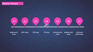
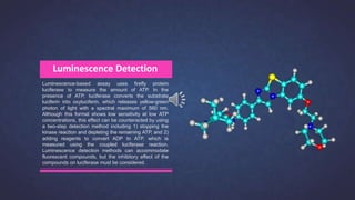
哎呀小小草 POWERPOINT
Kinases are arguably one of the most important drug targets due
to the roles their dysregulation, mutations, and overexpression
play in all major pathologies, including metabolic, cardiovascular,
degenerative, inflammatory, and infectious diseases. Finding the
best method for kinase screening and profiling can be a challenging
task and will depend on your research objective, the type of kinase,
availability of an antibody, the size of the kinase substrate and the
analytical instruments available.
Creative Bioarray provides a comprehensive set of assay
technologies to solve all of your research objectives - from
target discovery to compound screening, profiling and hit
confirmation. Our breadth of expertise and experience in
kinase research enables our scientists to support you
every step of the way through the drug discovery process.
Kinase Assays
Creative Bioarray
https://dda.creative-bioarray.com/
1-631-626-9181
Email: info@creative-bioarray.com
Kinase Assays
01 02 03 04 05 06 07
Radiometric
Assay
FRET Assay TRF Assay FP Assay Luminescence
Assay
Mobility Shift
Assay
Cell-based
ELISA Assay
Radiometric kinase assay using 32P-labelled ATP or
33P-labelled ATP is one of the oldest techniques used
to study protein phosphorylation. They rely on the
transfer of radioactive phosphate groups from ATP to
the substrate by a kinase, allowing detection of
activity. The radiolabeled phosphate incorporated into
the substrate is directly proportional to the kinase
activity.
Radiometric Assay
Advantages
Radiometric assay formats are the gold
standard for kinase screening, which can
produce reproducible, high-quality data
and directly measure enzyme activity.
Limitations
They are labor intensive and require safe
disposal of radioactive materials, so they
are usually not suitable for high-throughput
screening.
Fluorescence resonance energy
transfer (FRET) involves the
transfer of non-radiative energy
from a donor fluorophore to a
close-proximity acceptor
fluorophore. When the donor
and acceptor are spatially close
to each other, the acceptor
molecule will quench any
fluorescence emission from the
donor molecule.
Fluorescence Resonance Energy Transfer (FRET) Assay
The first step of the FRET kinase assay is to
incubate the FRET labeled substrate with ATP and
kinase, resulting in the transfer of γ-phosphate to
the substrate peptide. In the second reaction step,
a site-specific protease is added to the mixture. If
the protease cleaves a non-phosphorylated peptide,
the fluorescent donor and acceptor will be
sufficiently separated to disrupt the FRET, thus
generating a fluorescent signal that can be
measured as a ratio of donor emission to acceptor
emission.
Advantages of FRET
HTS friendly
Homogenous reaction
Limitations of FRET
Uses synthetic peptide substrate
Requires counter screening against coupling enzyme
Demonstrates a high false-positive rate for fluorescent compounds
Substrate requires modification for fluorophore labeling
Time-resolved fluorescence (TRF) uses fluorophores
with a long decay time, and monitors the fluorescence
as a function of time after excitation by a flash of light.
Lanthanide ions, such as europium, samarium, and
terbium, are often used in this technology due to their
longer emission lifetimes (hundreds of microseconds
compared to several nanoseconds for conventional
organic fluorophores).
Time Resolved Fluorescence (TRF)
Advantages &
Limitations
The advantage of this technology is that contaminating
fluorescent molecules which contribute to background
noise have a shorter decay time than lanthanide ions,
so the signal is much cleaner. There are several
commercially available TRF assays, mainly based either
on ELISA assay or combined with FRET assay.
Fluorescence polarization (FP), a technique that monitors
molecular movement and rotation, is a widely used
detection method for kinase inhibitors in HTS. The principle
of this assay is that smaller molecules rotate faster than
larger molecules. A fluorescently labeled peptide substrate
in the solution rotates quickly and so will have a low
fluorescence polarization. When the phosphorylation
reaction is initiated by addition of a kinase and ATP, any
phosphorylated peptide substrates will be recognized by
the phospho-antibody. The binding of the antibody will slow
down the rotation of the molecule (high fluorescence
polarization), and this change in speed can be measured.
Fluorescence Polarization (FP) Assay
Advantages &
Limitations
Since this is a one-step assay without additional
washing steps, it is very suitable for screening a large
number of kinase inhibitor candidates. However, one
of the major drawbacks for this and other
fluorescence-based assays, is that unused fluorescent
compounds and labeled substrates may produce high
background signals and false positives.
Luminescence-based assay uses firefly protein
luciferase to measure the amount of ATP. In the
presence of ATP, luciferase converts the substrate
luciferin into oxyluciferin, which releases yellow-green
photon of light with a spectral maximum of 560 nm.
Although this format shows low sensitivity at low ATP
concentrations, this effect can be counteracted by using
a two-step detection method including 1) stopping the
kinase reaction and depleting the remaining ATP, and 2)
adding reagents to convert ADP to ATP, which is
measured using the coupled luciferase reaction.
Luminescence detection methods can accommodate
fluorescent compounds, but the inhibitory effect of the
compounds on luciferase must be considered.
Luminescence Detection
The mobility shift assay takes advantage of the fact
that a phosphorylated peptide substrate is more
negatively charged than the same substrate in the
unphosphorylated state. Therefore, when a mixture of
these peptides is subjected to gel electrophoresis, the
phosphorylated and unphosphorylated substrates will
have different mobility.
Mobility Shift Assay
Advantages & Limitations
Since the difference in charge between the peptide
and the substrate is key, the result of mobility shift
assay is greatly affected by the size and sequence of
the substrate. Therefore, this assay is not suitable for
the analysis of larger peptides or whole protein, and
works best when the peptide substrate contains only
specific phosphorylation site sequences.
Cell-based functional kinase screening provides a
reliable, quantitative mean for determining the functional
inhibitory effects of drug candidates on kinase targets of
interest in a physiologically relevant cellular environment.
This allows for a more predictive characterization of the
organism’s physiological response to the drug, and
greatly reduces the chances of false positive/negative
data.
Cell-based ELISA Assay
Enzyme-linked immunosorbent assay (ELISA) can be
used to quantitate kinase levels and assess their activity.
In this format, the substrate is captured by the membrane
and detected by a specific anti-phosphorylated substrate
antibody. The detection can also be very sensitive when
using antibodies labeled with highly fluorescent dyes.
Advantages & Limitations
ELISA is popular because it is generally fast,
inexpensive, easy to accommodate multiple samples,
and does not require highly specialized equipment.
However, the requirement of separation with multiple
washing steps limits the use of ELISA in HTS. In
addition, the development of high-quality phospho-
specific antibodies presents another challenge.
A large number of commercially available kinase assays have been developed in recent years based
on the technologies described above. Each technology has its strengths and weaknesses, and the
most appropriate assay will depend on the aim of the project. For high-throughput screening drug
discovery projects, methods that can be easily automated and miniaturized, such as the
fluorescence-based assays, are the most suitable. For projects that require a more in-depth profile of
the kinase function, a more robust assay such as radiometric assay will provide more reliable results.
Conclusion

Technologies to study kinases

  • 2.
    哎呀小小草 POWERPOINT Kinases arearguably one of the most important drug targets due to the roles their dysregulation, mutations, and overexpression play in all major pathologies, including metabolic, cardiovascular, degenerative, inflammatory, and infectious diseases. Finding the best method for kinase screening and profiling can be a challenging task and will depend on your research objective, the type of kinase, availability of an antibody, the size of the kinase substrate and the analytical instruments available.
  • 3.
    Creative Bioarray providesa comprehensive set of assay technologies to solve all of your research objectives - from target discovery to compound screening, profiling and hit confirmation. Our breadth of expertise and experience in kinase research enables our scientists to support you every step of the way through the drug discovery process. Kinase Assays Creative Bioarray https://dda.creative-bioarray.com/ 1-631-626-9181 Email: info@creative-bioarray.com
  • 4.
    Kinase Assays 01 0203 04 05 06 07 Radiometric Assay FRET Assay TRF Assay FP Assay Luminescence Assay Mobility Shift Assay Cell-based ELISA Assay
  • 6.
    Radiometric kinase assayusing 32P-labelled ATP or 33P-labelled ATP is one of the oldest techniques used to study protein phosphorylation. They rely on the transfer of radioactive phosphate groups from ATP to the substrate by a kinase, allowing detection of activity. The radiolabeled phosphate incorporated into the substrate is directly proportional to the kinase activity. Radiometric Assay
  • 7.
    Advantages Radiometric assay formatsare the gold standard for kinase screening, which can produce reproducible, high-quality data and directly measure enzyme activity. Limitations They are labor intensive and require safe disposal of radioactive materials, so they are usually not suitable for high-throughput screening.
  • 9.
    Fluorescence resonance energy transfer(FRET) involves the transfer of non-radiative energy from a donor fluorophore to a close-proximity acceptor fluorophore. When the donor and acceptor are spatially close to each other, the acceptor molecule will quench any fluorescence emission from the donor molecule. Fluorescence Resonance Energy Transfer (FRET) Assay The first step of the FRET kinase assay is to incubate the FRET labeled substrate with ATP and kinase, resulting in the transfer of γ-phosphate to the substrate peptide. In the second reaction step, a site-specific protease is added to the mixture. If the protease cleaves a non-phosphorylated peptide, the fluorescent donor and acceptor will be sufficiently separated to disrupt the FRET, thus generating a fluorescent signal that can be measured as a ratio of donor emission to acceptor emission.
  • 10.
    Advantages of FRET HTSfriendly Homogenous reaction Limitations of FRET Uses synthetic peptide substrate Requires counter screening against coupling enzyme Demonstrates a high false-positive rate for fluorescent compounds Substrate requires modification for fluorophore labeling
  • 12.
    Time-resolved fluorescence (TRF)uses fluorophores with a long decay time, and monitors the fluorescence as a function of time after excitation by a flash of light. Lanthanide ions, such as europium, samarium, and terbium, are often used in this technology due to their longer emission lifetimes (hundreds of microseconds compared to several nanoseconds for conventional organic fluorophores). Time Resolved Fluorescence (TRF)
  • 13.
    Advantages & Limitations The advantageof this technology is that contaminating fluorescent molecules which contribute to background noise have a shorter decay time than lanthanide ions, so the signal is much cleaner. There are several commercially available TRF assays, mainly based either on ELISA assay or combined with FRET assay.
  • 15.
    Fluorescence polarization (FP),a technique that monitors molecular movement and rotation, is a widely used detection method for kinase inhibitors in HTS. The principle of this assay is that smaller molecules rotate faster than larger molecules. A fluorescently labeled peptide substrate in the solution rotates quickly and so will have a low fluorescence polarization. When the phosphorylation reaction is initiated by addition of a kinase and ATP, any phosphorylated peptide substrates will be recognized by the phospho-antibody. The binding of the antibody will slow down the rotation of the molecule (high fluorescence polarization), and this change in speed can be measured. Fluorescence Polarization (FP) Assay
  • 16.
    Advantages & Limitations Since thisis a one-step assay without additional washing steps, it is very suitable for screening a large number of kinase inhibitor candidates. However, one of the major drawbacks for this and other fluorescence-based assays, is that unused fluorescent compounds and labeled substrates may produce high background signals and false positives.
  • 18.
    Luminescence-based assay usesfirefly protein luciferase to measure the amount of ATP. In the presence of ATP, luciferase converts the substrate luciferin into oxyluciferin, which releases yellow-green photon of light with a spectral maximum of 560 nm. Although this format shows low sensitivity at low ATP concentrations, this effect can be counteracted by using a two-step detection method including 1) stopping the kinase reaction and depleting the remaining ATP, and 2) adding reagents to convert ADP to ATP, which is measured using the coupled luciferase reaction. Luminescence detection methods can accommodate fluorescent compounds, but the inhibitory effect of the compounds on luciferase must be considered. Luminescence Detection
  • 20.
    The mobility shiftassay takes advantage of the fact that a phosphorylated peptide substrate is more negatively charged than the same substrate in the unphosphorylated state. Therefore, when a mixture of these peptides is subjected to gel electrophoresis, the phosphorylated and unphosphorylated substrates will have different mobility. Mobility Shift Assay
  • 21.
    Advantages & Limitations Sincethe difference in charge between the peptide and the substrate is key, the result of mobility shift assay is greatly affected by the size and sequence of the substrate. Therefore, this assay is not suitable for the analysis of larger peptides or whole protein, and works best when the peptide substrate contains only specific phosphorylation site sequences.
  • 23.
    Cell-based functional kinasescreening provides a reliable, quantitative mean for determining the functional inhibitory effects of drug candidates on kinase targets of interest in a physiologically relevant cellular environment. This allows for a more predictive characterization of the organism’s physiological response to the drug, and greatly reduces the chances of false positive/negative data. Cell-based ELISA Assay Enzyme-linked immunosorbent assay (ELISA) can be used to quantitate kinase levels and assess their activity. In this format, the substrate is captured by the membrane and detected by a specific anti-phosphorylated substrate antibody. The detection can also be very sensitive when using antibodies labeled with highly fluorescent dyes.
  • 24.
    Advantages & Limitations ELISAis popular because it is generally fast, inexpensive, easy to accommodate multiple samples, and does not require highly specialized equipment. However, the requirement of separation with multiple washing steps limits the use of ELISA in HTS. In addition, the development of high-quality phospho- specific antibodies presents another challenge.
  • 25.
    A large numberof commercially available kinase assays have been developed in recent years based on the technologies described above. Each technology has its strengths and weaknesses, and the most appropriate assay will depend on the aim of the project. For high-throughput screening drug discovery projects, methods that can be easily automated and miniaturized, such as the fluorescence-based assays, are the most suitable. For projects that require a more in-depth profile of the kinase function, a more robust assay such as radiometric assay will provide more reliable results. Conclusion