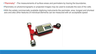
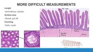
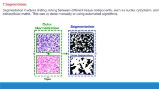
Morphometry involves obtaining quantitative information about biological tissue structures through measurements of their size, shape, and other metrics. It provides objective criteria that can improve reproducibility in histopathology. Key applications include tumor classification, diagnostic accuracy, and distinguishing conditions like hyperplasia vs carcinoma. Techniques include planimetry, point counting, pixel counting in digital images. Morphometry of features like nuclear size and shape correlates with biological behaviors and can aid diagnosis of conditions like endometrial hyperplasia vs cancer or malignant mesothelioma. Next-generation techniques continue advancing this quantitative approach.
Cytologic assessment of bronchopulmonary lesionsAseem Jain
This document provides an overview of cytologic assessment of bronchopulmonary lesions. It discusses the normal histology of the respiratory system and various cytologic sampling techniques used to evaluate the lungs, such as sputum samples, bronchial brushings, washings and lavage. The cytology of normal respiratory cells and endogenous material is described. A variety of benign pulmonary conditions and infectious processes are outlined. Specific lung diseases like tuberculosis, sarcoidosis and nocardiosis are discussed through their characteristic cytologic findings.
Cytogenetic Analysis in Hematological Malignanciesspa718
The document discusses the importance of cytogenetic analysis in hematological malignancies. Some key points:
- Many hematological malignancies have clonal chromosomal abnormalities that can aid in diagnosis, classification, and risk stratification.
- Certain recurrent abnormalities are specific to certain tumor subtypes and can predict treatment response and clinical outcome.
- Genes at breakpoints of recurrent abnormalities play a role in tumorigenesis and can be treatment targets.
- Cytogenetic analysis is useful for diagnosis, risk stratification, treatment selection, and monitoring treatment response in hematological cancers like CML, AML, ALL, lymphoma, MDS, MM, and CLL.
This document discusses Perls stain, which is used to identify iron deposits in tissue samples. It provides background on pigments in living tissue, including endogenous pigments like hemosiderin and hematogenous pigments. The history of Prussian blue and its use as Perls stain is described. The principle of the stain is that hydrochloric acid releases ferric ions from hemosiderin, which then react with potassium ferrocyanide to form insoluble Prussian blue pigment. Staining protocols, quality control, and clinical applications for identifying iron deposits in organs are covered.
This document discusses tissue microarrays (TMAs), which allow analysis of hundreds of tissue samples on a single slide. It describes how TMAs are constructed by taking small tissue cores from donor blocks and embedding them in a recipient block. The advantages of TMAs include high throughput analysis and relatively low cost. Various types of TMAs are used for applications like immunohistochemistry, in situ hybridization, and analyzing protein/DNA expression. The document outlines the steps to construct a TMA, including defining the research question, selecting cases, determining core size and number, making a map, and embedding the cores. Quality controls and limitations are also discussed.
Flow cytometry for cell componenet analysisRAJA GOPAL
Flow cytometry is a technique that uses lasers and fluorescence to analyze physical and chemical characteristics of cells as they flow in a fluid stream. It allows simultaneous analysis of thousands of cells per second based on parameters like cell size, granularity, and detection of cell surface antigens using specific antibodies labeled with fluorochromes of different colors. Specimens suitable for analysis include blood, bone marrow, body fluids, and cell suspensions generated from tissues. Flow cytometry has various applications like immunophenotyping, DNA analysis, diagnosis of conditions like PNH, reticulated cell counting, and blood bank testing.
This document provides information on various cytochemical staining techniques used in hematology, including myeloperoxidase, esterase, alkaline phosphatase, acid phosphatase, Sudan black B, periodic acid Schiff, and Toluidine blue staining. It describes the principle, reagents, procedure, and interpretation for each stain. These stains are used to classify and diagnose different types of leukemia by identifying cellular enzymes and components in blood and bone marrow samples.
Immunohistochemistry is a technique used to identify antigens in tissue samples using antigen-antibody interactions. It has made a large impact in disease diagnosis since its development in the 1940s-1970s. The technique involves using labeled antibodies that specifically bind to antigens in tissue sections. This binding is then visualized using markers like enzymes or fluorescent dyes. Several methods have been developed to increase the signal and reduce background noise, including indirect labeling techniques and polymer-based methods. Proper tissue processing and antibody selection are important for obtaining high quality results with immunohistochemistry.
This document discusses the role of immunohistochemistry (IHC) in diagnosing soft tissue tumours. It begins by defining soft tissue and the WHO classification of soft tissue tumours. IHC is an important ancillary technique that can be used to identify discrete tissue components using antigen-antibody binding. The document outlines the IHC protocol and discusses various markers that can help diagnose different types of soft tissue tumours, including markers for fibroblastic, adipocytic, vascular, neural, osseous and cartilaginous tumours. Specific markers and the tumours they are useful for identifying are provided. The document emphasizes that IHC should be used along with other techniques as markers sometimes show cross-reactivity.
Pitfalls in diagnosis of soft tissue tumors of childhoodSonic V S
The document discusses several potential pitfalls in the diagnosis of soft tissue tumors in children. It covers:
1) Misclassification of specific sarcomas like rhabdomyosarcoma subtypes and non-rhabdomyosarcoma soft tissue sarcomas.
2) Benign lesions that can be misdiagnosed as sarcomas, and sarcomas that can be misdiagnosed as benign.
3) Misgrading the aggressiveness of sarcomas.
4) Non-soft tissue tumors that are sometimes misdiagnosed as soft tissue sarcomas. Careful histology, immunohistochemistry, cytogenetics and molecular analysis are needed to arrive at
This document discusses pathology informatics and describes several key concepts:
- Pathology informatics involves using information technology to facilitate pathology practices like patient care, disease understanding, and education. It requires integrating diverse data sources and presenting data to support decisions.
- Key components of a pathology information system include managing specimens, acquiring and processing data, and generating integrated reports that can be accessed across hospital systems.
- Standards like SNOMED, ICD-10, and LOINC are important for representing and communicating information between different medical systems.
- A laboratory information system is a comprehensive software that manages the pathology laboratory workflow and reporting.
This document discusses automation in hematology. It begins by outlining the necessity for automation in cell counting, diagnosing various blood conditions, and performing multiple tests on a single platform. The document then covers the advantages and disadvantages of automation, including increased speed and accuracy versus high costs. It describes the various principles used in automated hematology analyzers, such as electrical impedance, optical light scattering, and flow cytometry. Finally, it provides details on specific analyzers like the Pentra ES 60 and Pentra DF Nexus.
This document discusses quality control procedures for automated hematology analyzers. It describes the use of histograms, flags, and quality control measures to ensure accurate results. Key points:
- Histograms provide graphical representations of cell populations to analyze size, distribution, and detect abnormalities. Flags signal when abnormalities are detected to mandate blood smear examination.
- Internal quality control involves continuous validation of test reliability using control samples processed like patient samples. External quality control evaluates comparability to peer laboratories.
- Accuracy measures closeness to true values while precision measures reproducibility. Controls contain same constituents as patient samples to monitor analyzer performance. Results are plotted on Levey-Jennings charts against manufacturer ranges.
- Westgard
This document discusses fungal infections in histopathology. It provides advantages of histopathology for fungal identification, describes special staining techniques used to identify fungi in different tissues, outlines major fungal forms seen, and lists true and opportunistic fungal pathogens. It also describes 8 cases presenting with fungal infections and provides the diagnoses for each case. The document serves as a reference for histopathologists to identify fungi in tissue samples.
The document discusses cryostats, which are devices used to cut thin frozen sections of tissues for examination under a microscope. Cryostats contain a microtome inside a freezer unit that can rapidly freeze tissue samples and cut sections as thin as 1 micrometer at temperatures below freezing. The cryostat process allows for quick diagnosis by freezing and sectioning tissues within minutes rather than having to dehydrate, embed in paraffin, and section as with traditional microtomes.
This document provides information on cryostat frozen section procedures. It discusses the types of cryostats, handling of specimens, the microtome, freezing methods, artifact avoidance, quality assurance, maintenance and more. Cryostats are used to cut thin frozen sections of tissues for histological examination. Rapid freezing is important to minimize artifacts from ice crystal formation. Sections are cut very thin, typically 1-100 micrometers, for examination. Proper handling, freezing, sectioning and disinfection are required to obtain high quality samples.
Chromogenic in situ hybridization (CISH) is a technique that combines in situ hybridization with immunohistochemistry to detect specific DNA sequences within tissue samples using brightfield microscopy. CISH uses nucleic acid probes labeled with enzymes that produce colored precipitates when exposed to chromogenic substrates, allowing visualization of gene amplification or rearrangements. CISH has advantages over fluorescence in situ hybridization (FISH) in being cheaper, more stable, and easier to use, though it is less sensitive than FISH. CISH is commonly used to assess HER2 gene amplification in breast cancer and detect genetic fusions in cancers.
Honing and stropping are processes used to sharpen knives. Honing removes nicks and irregularities from the cutting edge to make it straight and sharp. It is done using a hone, which must be kept clean and lubricated. The process involves pushing and pulling the knife across the hone in strokes until the edge is smooth. Stropping further polishes the edge after honing. It straightens and aligns the microscopic teeth at the edge without removing material, resulting in an even sharper, mirror-like finish.
Squash cytology of cns paediatric tumoursSumanth Deva
This document provides an overview of squash cytology techniques for diagnosing pediatric central nervous system tumors, describing the smear patterns and key cytological features of common tumor types like medulloblastoma, atypical teratoid/rhabdoid tumors, choroid plexus papilloma, and choroid plexus carcinoma. Squash cytology allows rapid examination of biopsy samples during neurosurgery to aid diagnosis and surgical decision making.
This document discusses several staining methods used for staining nerve tissue, including H&E staining, Nissl staining, Luxol-fast blue staining, Kluver–Barrera staining, Bodian silver staining, Holzer staining, and Gallyas–Braak staining. Each method selectively stains different components of nerve tissue such as cell nuclei, myelin sheaths, nerve fibers, and abnormal protein deposits. The staining methods allow clear visualization of neuronal structures and identification of lesions or abnormalities in nervous system tissue.
The document discusses various automated techniques used in histopathology. It describes automated tissue processors that use either tissue transfer systems or self-contained fluid exchange to process tissue through a series of solutions. Microwave and rapid processors are also discussed as alternatives that can significantly reduce processing time. The document also mentions automated tools for tasks like tissue microarrays, embedding, sectioning with microtomes and cryostats. Overall, the key benefits of automation include improved efficiency, customized schedules, reduced processing time and safer handling of chemicals.
Immunohistochemistry in diagnosis of soft tissue tumours seminarPannaga Kumar
This document discusses immunohistochemistry in the diagnosis of soft tissue tumors. It begins by introducing soft tissue and the classification of soft tissue tumors. It then discusses various ancillary techniques used, focusing on immunohistochemistry. It provides details on common markers used to identify muscle, neural, melanocytic, endothelial and other types of differentiation. It discusses the applications and diagnostic utility of various markers for different tumor types. In summary, the document is a comprehensive overview of immunohistochemistry techniques and markers useful in the diagnosis and classification of soft tissue tumors.
This presentation in mainly focused of understanding of automation and its utility in cytopathology. It will be very usefull for postgraduate in pathology, cytopathologist and cytotechnicians.
Lymph nodes are bean-shaped organs found throughout the body that filter lymph and house immune cells. A lymph node contains a fibrous capsule enclosing compartments of connective tissue and lymphocytes. The parenchyma is divided into an outer cortex and inner medulla. A normal lymph node contains mature lymphocytes, plasma cells, centrocytes, centroblasts, and immunoblasts. Lymphadenopathy refers to enlarged lymph nodes, which can be caused by infection, inflammation, autoimmune disease, or cancer metastasis. Physical examination of lymph nodes considers location, number, size, consistency, tenderness, and mobility to evaluate causes of lymphadenopathy.
The document discusses effusion cytology. It begins by describing the anatomy of serous cavities and membranes that line them, producing serous fluid. Any excess fluid is an effusion, indicating a pathological process. Effusions can be classified as hydrostatic, infectious, inflammatory, or malignant. Samples are collected and prepared as smears for staining.
Normal components in effusions include mesothelial cells, histiocytes, lymphocytes, and other inflammatory cells. Reactive mesothelial cells can appear atypical but maintain a uniform appearance. Malignant effusions result from direct extension or metastasis of cancers. Identifying malignant cells involves comparing size, shape and number to determine the primary tumor type and origin. The most
Quality assurance in pathology is important to minimize errors and ensure accurate diagnoses. External quality assessment programs help laboratories evaluate performance and harmonize results. A quality assurance program involves establishing standards and protocols, monitoring compliance, conducting regular reviews, and participating in proficiency testing. The goal is to maintain accuracy, precision and reliability throughout the pre-analytical, analytical and post-analytical phases to optimize patient care.
GRADE CATEGORIZATION OF TUMOUR CELLS WITH STANDARD AND REFERENTIAL FRONTIER A...pharmaindexing
This document summarizes a research paper that proposes a new method for classifying brain tumor grades using image processing techniques. The method involves preprocessing MRI images to isolate the tumor region using thresholding and image subtraction. The tumor area is then segmented into four quadrants. Standard points mark the initial tumor location, while growth points registered in later images indicate tumor expansion over time. Comparing growth point changes across patient images at different stages allows calculating the tumor growth rate, aiding pathologists in diagnosis and treatment recommendations.
Fractal Parameters of Tumour Microscopic Images as Prognostic Indicators of C...cscpconf
This document summarizes a study that analyzed fractal parameters of tumor microscopic images as prognostic indicators for clinical outcomes in early breast cancer. The study analyzed 92 breast cancer patients without systemic treatment. It calculated fractal dimension and lacunarity from digital images of hematoxylin and eosin stained tumor sections. Higher fractal dimension, indicating greater structural complexity, associated with higher risk of distant metastasis. Lower lacunarity, indicating less heterogeneity, also associated with higher metastasis risk. The fractal parameters provided prognostic value comparable to standard clinicopathological factors and indicated potential for use in clinical prognosis to complement molecular approaches.
FRACTAL PARAMETERS OF TUMOUR MICROSCOPIC IMAGES AS PROGNOSTIC INDICATORS OF C...csandit
Research in the field of breast cancer outcome prognosis has been focused on molecular biomarkers, while neglecting the discovery of novel tumour histology structural clues. We thus
aimed to improve breast cancer prognosis by fractal analysis of tumour histomorphology. This study included 92 breast cancer patients without systemic treatment. Fractal parametersfractal dimension and lacunarity of the breast tumour microscopic histology possess prognostic value comparable to the major clinicopathological prognostic parameters. Fractal analysis was performed for the first time on routinely produced archived pan-tissue stained primary breast tumour sections, indicating its potential for clinical use as a simple and cost-effective prognostic indicator of distant metastasis risk to complement the molecular approaches for
cancer risk prognosis.
This document presents a genetic algorithm-based classification method for classifying different types of lung cancer in needle biopsy images. It first segments cell nuclei from biopsy images and extracts color, texture, and shape features from the nuclei. A dictionary learning approach is used to build discriminative subdictionaries for each feature type. In testing, features from an image are classified at the cell level and then fused at the image level via majority voting. The method achieves higher accuracy than using single features or existing classification methods, demonstrating its effectiveness in classifying lung cancer types in biopsy images.
Pathomics Based Biomarkers, Tools, and Methodsimgcommcall
This document discusses pathomics-based biomarkers, tools, and methods for multi-scale integrative analysis in biomedical informatics. It summarizes several projects involving extracting quantitative features from pathology and radiology images using image segmentation and analysis techniques. These features are then linked to molecular data and clinical outcomes using statistical and machine learning methods to develop biomarkers. The tools and methods described aim to standardize and optimize feature extraction while accounting for uncertainties.
Human health is the real wealth for a society. Consequently prevention of health from complex diseases like cancer needs the diagnosis of these entire viruses at an early stage. Colon cancer, the most common one, reached the highest rate among all the other types recently. Colorectal cancer gets developed either in colon or in the rectum inside the large intestine, due to the abnormal growth of the cells. Computer-aided decision support system has become one of the major research topics in medical imaging field during the past two decades to detect cancers. Detecting and screening of colorectal cancers are done by a Computed Tomography. The implemented algorithm determines the locations and features of glands which are affected by cancer tissues and save this
information for the subsequent diagnosis. The proposed algorithm carries out the diagnosis with two modules:
One known as the gland detection and the other one referred as the nuclei detection. Gland detection is performed in the proposed algorithm using color segmentation either through HSV or LAB transformation. Noise removal and erosion of the input image is performed for enhancing the selection of the affected tissues. The boundary detection and connection is established through Markov Chain model to identify the affected tissues with proper threshold. The first module detects the glands where the possibly of miss detection is more. Hence to remove the miss detected glands the algorithm proceed for the second module referred as nuclei detection. The most well
known region growing methodology is slightly modified to increase the speed and reduce the memory size To provide the execution in low-end clients, the whole image is cracked into smaller tiles and after the processing of each individual tiles , the results are to be merged to get back the original size. After nuclei detection if the number of nucleus is more that glands are miss detected glands and they are removed.
Brain Tumor Area Calculation in CT-scan image using Morphological Operationsiosrjce
IOSR Journal of Computer Engineering (IOSR-JCE) is a double blind peer reviewed International Journal that provides rapid publication (within a month) of articles in all areas of computer engineering and its applications. The journal welcomes publications of high quality papers on theoretical developments and practical applications in computer technology. Original research papers, state-of-the-art reviews, and high quality technical notes are invited for publications.
This document presents a method for calculating the area of brain tumors in CT scan images using morphological operations. The proposed method involves 10 steps: 1) inputting a CT scan image, 2) cropping the image, 3) converting to grayscale, 4) applying morphological gradient filtering, 5) histogram equalization, 6) selecting the region of interest, 7) subtracting and thresholding, 8) morphological closing, and 9) calculating the tumor area. The method is tested on different tumor images and more accurately calculates tumor area compared to radiologists who assume tumor shapes. The algorithm provides automated tumor highlighting and area calculation to assist physicians.
Bridging the STEM gender gap through cultural inclusion and educational opportunity, this opportunity was granted to a selected set of women from UB to showcase their research.
An overview of automatic brain tumor detection frommagnetic resonance imagesMangesh Lingampalle
The document discusses several techniques for automatically detecting brain tumors from magnetic resonance (MR) images. It begins with an overview of MR imaging and challenges of manual tumor detection. Several existing techniques are then summarized, including thresholding-based methods, fuzzy classification with deformable models, using wavelets and statistics to segment tissues, feature extraction with Adaboost classification, and color-converted k-means clustering. The document proposes a technique using undecimated wavelet transform (UDWT) and Gabor filters for preprocessing, followed by morphological operations and parameter analysis to detect tumors. Automatic detection techniques could help address limitations of manual detection and improve diagnosis of brain tumors.
Iaetsd classification of lung tumour usingIaetsd Iaetsd
This document describes a study that aims to classify lung tumors using geometric and texture features extracted from chest x-ray images. The study uses 75 chest x-ray images (25 from small-cell lung cancer, 25 from non-small cell lung cancer, and 25 from tuberculosis) to extract geometric features like area, shape, and distance from texture features calculated using gray level co-occurrence matrices. Active shape models are used to segment the lung fields for feature extraction. The extracted features are then analyzed to determine the optimal features for classifying different types of lung abnormalities.
Clinical Radiotherapy Planning basics for beginnersDina Barakat
1) External beam radiotherapy involves delivering high-energy x-ray beams from outside the patient's body to treat tumors. The ICRU recommends doses be within ±7-5% of the prescribed dose to the target.
2) Treatment planning involves acquiring patient images like CT scans, outlining the tumor and organs at risk, determining beam geometry, and calculating dose distributions.
3) Virtual simulation uses digitally reconstructed radiographs from CT images to plan beam placement, replacing conventional simulation using x-rays. This allows direct use of patient anatomy in planning.
A Review of Super Resolution and Tumor Detection Techniques in Medical Imagingijtsrd
Images with high resolution are desirable in many applications such as medical imaging, video surveillance, astronomy etc. In medical imaging, images are obtained for medical investigative purposes and for providing information about the anatomy, the physiologic and metabolic activities of the volume below the skin. Medical imaging is an important diagnosis instrument to determine the presence of certain diseases. Therefore increasing the image resolution should significantly improve the diagnosis ability for corrective treatment. Brain tumor detection is used for identifying the tumor present in the Brain. MRI images help the doctors for identifying the Brain tumor size and shape of the tumor. The purpose of this report to provide a survey of research related super resolution and tumor detection methods. Fathimath Safana C. K | Sherin Mary Kuriakose ""A Review of Super Resolution and Tumor Detection Techniques in Medical Imaging"" Published in International Journal of Trend in Scientific Research and Development (ijtsrd), ISSN: 2456-6470, Volume-3 | Issue-3 , April 2019, URL: https://www.ijtsrd.com/papers/ijtsrd23525.pdf
Paper URL: https://www.ijtsrd.com/engineering/electronics-and-communication-engineering/23525/a-review-of-super-resolution-and-tumor-detection-techniques-in-medical-imaging/fathimath-safana-c-k
Digital Pathology, FDA Approval and Precision MedicineJoel Saltz
Digital pathology platforms combined with machine learning can improve the consistency and quality of clinical decision making by precisely scoring known criteria from pathology images and predicting treatment outcomes and cancer types. Researchers are developing tools to extract features from pathology images, link these features to molecular data and clinical outcomes, and use these integrated datasets to gain new insights into cancer and select the best interventions. The SEER Virtual Tissue Repository aims to enable population-level cancer research by creating a linked collection of de-identified clinical data and whole slide images from pathology samples that can be analyzed using computational methods.
Pathomics Based Biomarkers and Precision MedicineJoel Saltz
Role of Digital Pathology Data Science (Pathomics) in precision medicine. Features from billions or trillions of objects segmented from digital Pathology data can be employed to predict patient outcome and steer treatment.
Presentation at Imaging 2020, Jackson Hole, WY September 2016
Brain Tumor Segmentation and Volume Estimation from T1-Contrasted and T2 MRIsCSCJournals
This document presents a novel, fully automatic method for brain tumor segmentation and volume estimation using T1-contrasted and T2 MRI scans. The method involves 5 main steps: 1) preprocessing images using anisotropic diffusion filtering, 2) segmenting tumor regions using k-means clustering, 3) combining segmented regions using logical and morphological operations, 4) applying temporal smoothing for error checking, and 5) measuring the tumor volume. The method was tested on brain volumes from a public dataset and compared to 5 other state-of-the-art algorithms, outperforming them in accuracy and closeness to actual tumor volumes.
This document discusses quantitative analysis of radiologic images using techniques like image segmentation, registration, and statistical atlases. It notes that computers can help address human limitations in measurement, detection of subtle abnormalities, and evaluation of complex patterns. Key techniques include using statistical atlases to capture normal anatomical variation, registering images to integrate data from multiple individuals, and performing pattern classification using machine learning to aid diagnosis. The goal is for computers to complement humans by providing increased reproducibility, quantification, and analysis of non-focal or complex spatio-temporal changes.
30 SLIDE SHOW JOURNAL Mimickers Melanoma in Situ Dt. 20.11.2023.pptxSarithaRani4
This study examines expression of the PRAME marker in mimickers of melanoma in situ (MIS), including Paget's disease, extramammary Paget's disease, and pagetoid squamous cell carcinoma in situ. The study finds that PRAME expression is variable in these mimickers and therefore is not a reliable marker to differentiate them from MIS. PRAME staining was seen in some cases of each entity, with weak or moderate intensity in most cases. The study concludes that while PRAME may suggest a non-squamous lineage, other immunostains are needed to confirm diagnoses of pagetoid lesions.
The document provides information about embedding and section cutting techniques in histopathology. It discusses the aims of embedding tissue, such as providing support and preventing distortion. Common embedding mediums include paraffin wax, resin, agar, and gelatin. Paraffin wax is the most popular due to being inexpensive, non-toxic, and allowing long term tissue storage. The document outlines the tissue embedding process and describes equipment used, including embedding molds and the Tissue-Tek system. Tissue orientation in the block is important for visualizing the desired morphology.
This document provides an overview of amyloidosis, including:
- Amyloidosis is caused by the deposition of abnormal protein fibrils in tissues, which can damage organs.
- There are different types of amyloid proteins including light chains, serum amyloid A, and beta-amyloid.
- Amyloidosis can be primary (associated with plasma cell disorders), secondary (associated with chronic inflammation), hereditary, or localized.
- Organs commonly involved include the kidneys, heart, liver, spleen, and tongue. Deposits appear pink with Congo red staining and apple-green under polarized light.
A semen analysis measures the amount and quality of semen and sperm. It is one of the first tests done to evaluate male fertility issues. It determines semen volume, sperm concentration, motility, morphology, pH and presence of white blood cells or other abnormalities. The sample is collected through masturbation and analyzed under a microscope to measure these parameters. Normal ranges have been established by the WHO. Abnormal results could indicate issues that reduce fertility. The analysis provides important information about male reproductive health and ability to conceive.
This document provides an overview of apoptosis, or programmed cell death. It begins with an introduction defining apoptosis and its importance in development, aging, and disease. It then describes the main types and mechanisms of apoptosis, including the extrinsic and intrinsic pathways. Key features of apoptosis like morphology, assays to detect it, and differences from necrosis are summarized. The document concludes by discussing other forms of programmed cell death and clinical applications of understanding apoptosis.
Bone marrow examination involves bone marrow aspiration and trephine biopsy. Bone marrow aspiration uses a specialized needle to obtain a bone marrow tissue sample, while trephine biopsy removes a small core of bone marrow. These procedures are used to diagnose hematological disorders and cancers, stage lymphomas and solid tumors, and detect infections. The iliac crest is a common site for bone marrow aspiration and biopsy. Nurses prepare equipment, assist during the procedure, monitor the patient, and ensure follow-up care after the procedure.
This document discusses bone marrow examination, including its purpose, types (aspiration and biopsy), indications, contraindications, procedure details, instruments used, complications, and findings in conditions like iron deficiency anemia and megaloblastic anemia. Bone marrow examination assesses hematopoiesis through aspiration or biopsy of the medullary cavity in bones. Aspiration is used to evaluate cellularity and differentials while biopsy allows evaluation of morphology, architecture, and focal lesions. Common sites include the iliac crest and sternum.
This document discusses various central nervous system pathologies including hydrocephalus, meningitis, and encephalitis. It defines hydrocephalus as increased cerebrospinal fluid in the skull and describes the circulation and types. Meningitis is defined as inflammation of the meninges and its types include acute pyogenic, viral, and chronic forms. Encephalitis is defined as brain parenchyma infection and its causes include bacterial, viral, and fungal forms. Key pathological features of each condition are summarized.
The document discusses meningitis and encephalitis. It defines meningitis as an inflammation of the protective membranes covering the brain and spinal cord, known as the meninges. There are various causes of meningitis including bacterial, viral, parasitic and non-infectious. Common symptoms include headache, fever and neck stiffness. Diagnosis involves spinal fluid analysis and imaging. Treatment depends on the cause but may include antibiotics, antivirals and steroids. Encephalitis additionally involves inflammation of the brain tissue and can be caused by viruses, bacteria, fungi or parasites. It presents with fever and neurological symptoms. Treatment focuses on treating the underlying infection and managing complications.
30 SLIDE SHOW JOURNAL Mimickers Melanoma in Situ Dt. 20.09.2023.pptxSarithaRani4
This document discusses the expression of PRAME in mimickers of melanoma in situ (MIS), including Paget's disease, extramammary Paget's disease, and pagetoid squamous cell carcinoma in situ. The study examines 31 cases using PRAME immunohistochemistry. It finds that PRAME expression is not reliable for differentiating these mimickers from MIS, as some cases of each showed variable, weak PRAME positivity. PRAME may be slightly better at distinguishing pagetoid squamous cell carcinoma, but is not specific enough on its own. The conclusion is that while PRAME can be used in MIS diagnosis, other stains are needed to confirm diagnoses of pagetoid lesions.
This document discusses 6 cases involving cell death and different types of biopsies. Case 1 involves a child with cervical lymph node enlargement where a lymph node biopsy was performed. Case 2 describes a woman with axillary lymph node enlargement, fever, weight loss and core biopsy of the lymph node. Case 3 is about an elderly man with bloody stools, abdominal pain and ulcers found on colonoscopy who had a stem cell transplant for multiple myeloma. Case 4 presents a woman with jaundice, fatigue and abnormal liver enzymes who developed liver failure. Case 5 involves a woman with ulceration and swelling of the hard palate. Case 6 describes a man with nasal obstruction and discharge who had an ulcero-pro
This document discusses targeted cancer therapy, which aims to interfere with specific molecular targets involved in carcinogenesis and tumor growth. It describes various types of targeted therapies, including hormonal therapy, signal transduction inhibitors like tyrosine kinase inhibitors, gene expression modulators, apoptosis inducers, angiogenesis inhibitors, and immunotherapies. While offering benefits over chemotherapy, targeted therapies also face challenges such as drug resistance and high costs. Researchers continue developing new targeted drugs and methods to improve cancer treatment.
How to Manage Your Lost Opportunities in Odoo 17 CRMCeline George
Odoo 17 CRM allows us to track why we lose sales opportunities with "Lost Reasons." This helps analyze our sales process and identify areas for improvement. Here's how to configure lost reasons in Odoo 17 CRM
A review of the growth of the Israel Genealogy Research Association Database Collection for the last 12 months. Our collection is now passed the 3 million mark and still growing. See which archives have contributed the most. See the different types of records we have, and which years have had records added. You can also see what we have for the future.
हिंदी वर्णमाला पीपीटी, hindi alphabet PPT presentation, hindi varnamala PPT, Hindi Varnamala pdf, हिंदी स्वर, हिंदी व्यंजन, sikhiye hindi varnmala, dr. mulla adam ali, hindi language and literature, hindi alphabet with drawing, hindi alphabet pdf, hindi varnamala for childrens, hindi language, hindi varnamala practice for kids, https://www.drmullaadamali.com
This presentation includes basic of PCOS their pathology and treatment and also Ayurveda correlation of PCOS and Ayurvedic line of treatment mentioned in classics.
Strategies for Effective Upskilling is a presentation by Chinwendu Peace in a Your Skill Boost Masterclass organisation by the Excellence Foundation for South Sudan on 08th and 09th June 2024 from 1 PM to 3 PM on each day.
ISO/IEC 27001, ISO/IEC 42001, and GDPR: Best Practices for Implementation and...PECB
Denis is a dynamic and results-driven Chief Information Officer (CIO) with a distinguished career spanning information systems analysis and technical project management. With a proven track record of spearheading the design and delivery of cutting-edge Information Management solutions, he has consistently elevated business operations, streamlined reporting functions, and maximized process efficiency.
Certified as an ISO/IEC 27001: Information Security Management Systems (ISMS) Lead Implementer, Data Protection Officer, and Cyber Risks Analyst, Denis brings a heightened focus on data security, privacy, and cyber resilience to every endeavor.
His expertise extends across a diverse spectrum of reporting, database, and web development applications, underpinned by an exceptional grasp of data storage and virtualization technologies. His proficiency in application testing, database administration, and data cleansing ensures seamless execution of complex projects.
What sets Denis apart is his comprehensive understanding of Business and Systems Analysis technologies, honed through involvement in all phases of the Software Development Lifecycle (SDLC). From meticulous requirements gathering to precise analysis, innovative design, rigorous development, thorough testing, and successful implementation, he has consistently delivered exceptional results.
Throughout his career, he has taken on multifaceted roles, from leading technical project management teams to owning solutions that drive operational excellence. His conscientious and proactive approach is unwavering, whether he is working independently or collaboratively within a team. His ability to connect with colleagues on a personal level underscores his commitment to fostering a harmonious and productive workplace environment.
Date: May 29, 2024
Tags: Information Security, ISO/IEC 27001, ISO/IEC 42001, Artificial Intelligence, GDPR
-------------------------------------------------------------------------------
Find out more about ISO training and certification services
Training: ISO/IEC 27001 Information Security Management System - EN | PECB
ISO/IEC 42001 Artificial Intelligence Management System - EN | PECB
General Data Protection Regulation (GDPR) - Training Courses - EN | PECB
Webinars: https://pecb.com/webinars
Article: https://pecb.com/article
-------------------------------------------------------------------------------
For more information about PECB:
Website: https://pecb.com/
LinkedIn: https://www.linkedin.com/company/pecb/
Facebook: https://www.facebook.com/PECBInternational/
Slideshare: http://www.slideshare.net/PECBCERTIFICATION
Exploiting Artificial Intelligence for Empowering Researchers and Faculty, In...Dr. Vinod Kumar Kanvaria
Exploiting Artificial Intelligence for Empowering Researchers and Faculty,
International FDP on Fundamentals of Research in Social Sciences
at Integral University, Lucknow, 06.06.2024
By Dr. Vinod Kumar Kanvaria
This slide is special for master students (MIBS & MIFB) in UUM. Also useful for readers who are interested in the topic of contemporary Islamic banking.
Film vocab for eal 3 students: Australia the movie
morphometry.pptx
1. ROLE OF MORPHOMETRY IN
HISTOPATHOLOGY
PRESENTOR-DR SANDHYA
MODERATOR-DR PRAVEEN SIR
2. INTRODUCTION
Histopathology is crucial for diagnostic and therapeutic decisions in many diseases.
The created classifications, however, have not always allowed explicit recognition of
diagnostic categories.
So, considerable intra- and interobserver variations are possible under diagnostic
circumstances.
Better reproducibility can be reached by applying traditional methods like morphometry
as well as special stains, electron microscopy and immunohistochemistry
3. What is Morphometry?
A body of methods for obtaining information about shape and size of a structure in terms
of quantities such as:
Volume
Surface area
Relative amounts of each component
Orientation, Interconnections
Distribution of substructures
4. Morphometric approach includes item classification and grading, point counting and
intersection counting methods, and the use of various semiautomatic or automatic
instruments.
TYPES:1.Statistical morphometry
2.Diagnostic morphometry
5. In statistical morphometry morphometrical parameters are collected from several disease cases.
Such data reinforced with prognostic follow ups -are the basis for disease classification.
Diagnostic morphometry, on the other hand, tries to study the sample of one individual and give relevant
data for diagnostic decisions
6. WHAT IS MAJOR CHALLENGE?
To extract information about large 3D structures from microscopic measurements on thin
2D sections
To do this Histomorphometry uses the
“DELESSE PRINCIPLE”
7. “In a rock composed of number of minerals, the area occupied by a given mineral is
propotional to the volume of mineral in the rock”
Repeated determinations of the area fraction will yield an estimate of the volume fraction.
The more determinations; the better the estimate
8. Histomorphometry
When applied to biological tissue, which is examined microscopically, it is useful in
correlating structure and function.
Ex- gut villus surface area
Arterial composition and elasticity
Quantification of –Hyperplasia, dysplasia, hypertrophy
Immunohistochemical or fluorescent markers-area or intensity
9. “Planimetry" -The measurements of surface areas and perimeters by tracing the boundaries.
Planimetry on photomicrographs or projected images may be used to evaluate the size of the cells
With the widely commercially available digitizing instruments the perimeter, area, longest and shortest
axis and also other features of individual elements can be measured with an acceptable speed
10. "stereology“-technique for extracting quantitative information about a 3 dimensional
material from measurements made on two dimensional planar sections of the material.
In practice denotes point counting techniques like performing cell counts,
estimating volume of lesions, infarct.
The two methods therefore supplement each other may lead to "morphometry", the quantitative
description of a structure.
11. Why morphometry in Pathology?
The pathologist may be intrested in morphometry for checking an initiative and
subjective evaluation of a diagnostic feature on the microscope section by making
measurements
He does this because he understands that other pathologists may possibly interpret his
findings differently
12. USES
To study biology of tumours
Classification of tumours
Diagnostic histopathology
Reproducibility and accuracy
13. Quantification of data
The commercially available equipment for quantitation can be divided as follows:
Non-automatic - Point counting equipment
Semi automatic -Graphic (digitizing) tablets
Mechanical scanners a. scanning stages
b. mirror scanners
c. polygon scanners
Automatic - Electron beam scanners e.g. cathode ray tubes (CRT)
Electron image sensors a. TV tubes
b. diode array scanners
Flow cytometer
14. Point Counting Equipment
This equipment is especially suitable for the assessment of stereological features such
as volume percentages, surface densities, length densities and numerical densities.
15. Graphic Tablets
In order to measure the area, circumference (= perimeter) etc. of individual elements
such as nuclei, a graphic tablet is necessary.
This instrumentation consists of three basic components: an electromagnetic sensitive
plate
a pen ( or cursor) which is electrically connected with a memory.
Every time the cursor on the plate is activated (e.g. by a microswitch), the exact location
of the pen is recorded according to its x and y coordinates.
This information is then stored in the computer
16.
17. Graphic tablets are especially suitable for the measurement of individual structures (nuclei, cells and
other elements).
On most of the commercially available graphic tablets the following features and parameters can be
calculated per delineated structure:
1. Perimeter ( = circumference)
2. Area
3. Shape factor (= 4 n x area/perimeter )
4. Projection on x-axis
5. Projection on y-axis
6. Longest axis
7. Shortest axis
8. Ratio of longest and shortest axis
18. HOW TO ESTIMATE AREA FRACTION?
Planimetry
Dot counting
Square counting
Pixel counting in a digital image
19. DOT COUNTING
Nuclear area/cell area=number of dots in
nuclei/number of dots in cell
Absolute area of structure=number of dots
in structure x area of dot square
21. Counting of mitotic figures
The following criteria for the diagnosis of mitotic figures instead of "pycnotic deformed
nuclei"
1. Absence of nuclear membrane
2. Absence of clear zone in centre
3. Presence of hairy instead of
triangular or spiky projections
4. Basophilia of surrounding cytoplasm
instead of eosinophilia
22. The following measures must be taken when counting mitotic figures
1. Adequate sections should be used, lightly stained, 4-5 microns in thickness.
2. Adequate sampling: As there may be considerable differences from one place to another, at least 10
sections per borderline tumour, or one section for each centimetre of diameter of the tumour should be
taken, whichever is greater.
23. 3. Counts should be started in the most active areas where the number of mitoses is
highest.
Once having started in a certain area, nine additional contiguous fields should be
selected randomly.
No attempts should be made to maximize the counting by selecting those areas with a
higher number
4.Four sets of 10 (or 25 if required) high power fields should be counted and the
maximum number in anyone set used.
5. Only definite mitotic figures should be counted, doubtful structures should be
excluded.
24. DECISION TREES
A decision tree consists of a combination of sequentially taken decisions, on the basis of
the value of one or more features, in order to come to a diagnosis. Step by step, the
investigator is asked to make a decision.
25.
26. The morphometric features themselves are expressions of the biological behaviour of the tumour .
In general pathology the nuclei of malignant cells are usually described as being "enlarged, often
irregular,pleomorphic”
In the applications of morphometry in tumor diagnostics, the nuclear area (related to size), the nuclear
perimeter (related to size and shape) and the nuclear horizontal and vertical axes (also related to size
and shape) are often of importance.
28. Sometimes the nuclear area has been used as a discriminating parameter in the
morphometric applications, a phenomenon familiar to the diagnostic pathologist.
Simple calculations such as the ratio of the nuclear axes are related to the shape of the
nucleus.
29. BIOLOGICAL
FEATURE
MORPHOLOGICAL FEATURE MORHOMETRICAL FEATURE
Evidence of rapid
growth
Mitosis numerous and often
abnormal
Mitotic activity index increased
Nuclei enlarged and pleomorphic Mean and s.d of nuclear area
Perimeter
Longest and shortest axis
increased
Nucleoli usually large Mean of nucleolar area increased.
Localization of nucleoli
Cytoplasmic basophilia
Haemorrhage and necrosis
Area of cytoplasm increased
n/c ratio increased
Volume percentage of necrosis
increased
31. Endometrium: Hyperplasia or
Carcinoma
Morphometry has been applied to endometrial diagnostic pathology .
The disagreement in the diagnoses both within the same pathologist and between
different pathologists in this particular area of pathology emphasizes the obvious need
for objective criteria.
Further grading in mild and marked atypical hyperplasia and well and moderately to
poorly differentiated carcinoma is also possible with morphometry
32. Morphometry of endometrium
Volume % stroma -43.7%
Volume % epithelium -50%
Volume % lumen -6.2%
Volume % glands-56.2%
Outer surface density glands-6.6mm2
Inner surface density glands -6.6mm2
33. For discrimination of hyperplasia and carcinoma, measurement and calculation of the volume
percentage epithelium and the inner luminar) surface of the glands with stereology is sufficient
34. BORDERLINE VS MALIGNANT-OVARY
The most powerful morphometric discriminators are the mitotic activity index and the
volume percentage epithelium.
35. Mesothelial Cells: Morphometric Distinction
Between Benign and Malignant in Pleural Fluid
The nuclear and cytoplasmic areas of mesothelial cells are measured with a graphic
tablet 1000x magnification.
From each measured cell population the following six parameters are calculated:
mean nuclear area
standard deviation nuclear area,
mean cytoplasmic area,
standard deviation cytoplasmic area,
mean N/C area ratio
standard deviation N/C area ratio.
36. Mean nuclear area and mean cytoplasmic area with their standard deviation show the most significant
differences.
In the mesothelioma group the mean area and the mean cytoplasmic area are greater than in the group
with reactive mesothelial cells.
37. Scattergram of reactive mesothelial cells (open circles), malignant mesothelioma cells (stars) and carcinoma cases
(squares) with the standard deviation and mean of the nuclear area.
39. FLASH
Framework for large-scale histomorphometry (FLASH) performing deep learning based
semantic segmentation and subsequent large-scale extraction of interpretable,
quantitative, morphometric features .
40. FLASH consists of an automated three-step approach:
(i) a CNN that automatically segments kidney tissue on a WSI discarding all non kidney tissues (e.g.,
adipose or muscle tissue),
(ii) another CNN that segments histological structures of the kidney tissue segmented by the first
CNN and
(iii) hand-crafted feature extraction for segmented structures .
The framework is applicable to the whole morphological spectrum of non-neoplastic kidney
41. COMPUTERZED HISTOMORPHOMETRY
It is a method used to quantitatively analyse microscopic images of tissue samples
1.Sample preparation
2.slide scanning-slides are scanned by whole slide scanner to create high-resolution digital
images.
3. Image Acquisition:
The digital images are imported into a computerized
histomorphometry software program.
42. 4.Calibration:
The software is calibrated to ensure accurate measurement by setting scale factors based on known
dimensions in the images.
5. Image Preprocessing:
Preprocessing steps may include color correction, noise reduction, and image enhancement to
improve the quality of the images.
6.Region of Interest (ROI) Selection:
The user defines the regions of interest within the images where histomorphometric
measurements will be performed.
44. 8.Measurement:
Various histomorphometric parameters are measured, including area, length, density, and
shape characteristics of the tissue components within the ROI
9.Data Analysis:
The software calculates and compiles the quantitative data based on the measurements,
often generating statistics and graphical representations.
10.Interpretation:
Researchers interpret the results in the context of their specific research questions, looking
for patterns or differences in tissue morphology.
45. CONCLUSION
Histomorphometry is useful for counting and measuring clearly defined structures
Limited by a lack of “Intelligent” soft ware
Extremely difficult to better the human eye-brain combination for pattern recognition
and diagnosis.
For Histopathologists, may be valuable for quantifying prognosis
Measuring ratio or distribution of different tumor markers
46. REFERENCES
1. Hölscher DL,et al. Next-Generation Morphometry for pathomics-data mining in histopathology. Nat
Commun. 2023 Jan 28;14(1).
2.Kalhan S, Garg S, Satarkar RN, et al.Correlation of Nuclear Morphometry with Clinicopathologic Parameters
in Malignant Breast Aspirates South Asian J Cancer 2022;11(1).
3.Baak.j.Oort.A manual of morphometry in diagnostic pathology.1st ed. springer;1983
3. Collan Y. Morphometry in pathology: another look at diagnostic histopathology. Pathol Res Pract. 1984
Nov;179(2):
4. Collan Y, Torkkeli et al. Application of morphometry in tumor pathology. Anal Quant Cytol Histol. 1987
May;9(2).