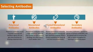
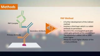
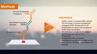
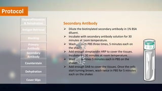
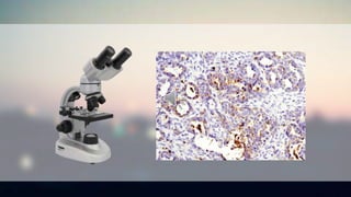
IMMUNOHISTOCHEMISTRY
https://www.creative-bioarray.com/
CONTENTS
Definition
Application
Method
Protocol
Selecting Antibody
Definition
Provide quantitative, qualitative
and temporal information on
processes taking place in
tissues.
Use antibodies conjugated to
enzymes that catalyze reactions
to form detectable compounds
to visualize and localize specific
antigens in a tissue sample.The most common application
of immunostaining.
Definition
Antigen/Antibody
based Tissue
based Reaction
IMMUNOHISTOCHEMISTRY
Applications of IHC
 Prognostic markers in cancer
 Tumors of uncertain histogenesis
 Prediction of response to therapy
 Infections
 In genetics
 Neurodegenerative disorders
 Brain trauma
 IHC in muscle disease
 Research application
Application
Polyclonal antibodies result
in greater staining and
excellent signal, but can
give false positives by
binding unwanted sites.
Polyclonal
Antibodies
Monoclonal antibodies have
a high specificity, reducing
the number of false positive
bindings, but often hive a
much weaker stain.
Monoclonal
Antibodies
Pooled monoclonal antibodies
also give excellent staining and
high specificity however there is
limited availability for those do
not bind noncompetitively.
Pooled Monoclonal
Antibodies
Secondary antibodies, against the
species of primary antibodies, will
be labeled with an enzyme like
HRP or biotin conjugated for
staining or amplifying signal.
Secondary
Antibodies
Selecting Antibodies
IHC Methods
Direct method
PAP method
LSAB method
Methods
ABC method
Indirect method
Methods
• One step staining method
• Labeled antibodies react directly with
the antigen
• This method utilizes only one antibody
and the procedure is short and quick
• It is insensitive due to little signal
amplification and rarely used since the
introduction of indirect method
Direct Method
Primary Antibody
Methods
• An unlabeled primary antibody reacts
with tissue antigen
• A labeled secondary antibody reacts with
primary antibody
• More sensitive due to signal
amplification through several secondary
antibody reactions
• Economy since one labeled secondary
antibody can be used with many primary
antibodies
• The secondary antibody may be labeled
with enzymes or fluorescent dyes
Indirect Method
Primary Antibody
Secondary Antibody
Methods
• A further development of the indirect
method
• Involves a third layer which is a rabbit
antibody to peroxidase.
• Bound to the unconjugated goat anti-
rabbit gaba-globulin of the second layer.
• The sensitivity is about 100 to 1000
times higher
• Allows for much higher dilution of the
primary antibody
PAP Method
Primary Antibody
Secondary Antibody
PAP Complex
Methods
• A standard IHC method
• The technique involves three layers.
• The first layer is unlabeled primary
antibody. The second layer is biotinylated
secondary antibody. The third layer is a
complex of avidin-biotin peroxidase.
• The peroxidase is then developed by the
DAB or other substrate to produce
different colorimetric end products.
ABC Method
Primary Antibody
Biotinylated Secondary
Antibody
Avidin-biotin
Peroxidase Complex
Methods
• LSAB is similar to standard ABC method.
• The third layer is Enzyme-Streptavidin
conjugates (HRP-Streptavidin or AP-
Streptavidin) to replace the complex of
avidin-biotin peroxidase.
• The third layer can also be Fluorescent
dye-Streptavidin such as FITC-
Streptavidin.
• Streptavidin does not contain
carbohydrate groups which might bind to
tissue lectins, avoiding some background
staining.
LSAB Method
Primary Antibody
Biotinylated Secondary
Antibody
Enzyme-Streptavidin
Conjugates
Protocol
 Dip slides in three changes of xylene for 3
minutes each.
 Dip slides in two change of 100% alcohol for 3
minutes each.
 Dip slides in one change of 95% alcohol for 3
minutes.
 Dip slides in one change of 70% alcohol for 3
minutes.
 Rinse slides twice in dH2O for 5 minutes.
Deparaffinization
& Rehydration
Antigen Retrieval
Blocking
Primary
Antibody
Secondary
Antibody
Dehydration
Counterstain
Cover Slips
Deparaffinization & Rehydration
Protocol
 Soak slides in 3% H2O2 for 5 minutes.
 Rinse slides twice in dH2O for 5 minutes.
 Soak the slides in the working citrate buffer and
cover with a lid.
 Microwave until the liquid boils.
 Remove from heat and let it stand at room
temperature for 20 minutes.
 Wash three times for 5 minutes in dH2O
 Remove the liquid and use a PAP pen to circle
around the tissue.
Deparaffinization
& Rehydration
Antigen Retrieval
Blocking
Primary
Antibody
Secondary
Antibody
Dehydration
Counterstain
Cover Slips
Antigen Retrieval
Protocol
 Apply enough 5% BSA with a transfer
pipette to cover the tissues.
 Incubate the slides overnight at 4℃ in
a humid chamber.
Deparaffinization
& Rehydration
Antigen Retrieval
Blocking
Primary
Antibody
Secondary
Antibody
Dehydration
Counterstain
Cover Slips
Bloocking
Protocol
 Dilute the primary antibody to the
recommended concentration in 1%
BSA/PBS diluent.
 Remove the BSA, and incubate with
primary antibody solution for 1 hour at
room temperature.
 Wash slides three times 5 minutes
each on the shaker.
Deparaffinization
& Rehydration
Antigen Retrieval
Blocking
Primary
Antibody
Secondary
Antibody
Dehydration
Counterstain
Cover Slips
Primary Antibody
Protocol
 Dilute the biotinylated secondary antibody in 1% BSA
diluent.
 Incubate with secondary antibody solution for 30
minutes at room temperature.
 Wash slides in PBS three times, 5 minutes each on
the shaker.
 Add enough streptavidin HRP to cover the tissues.
Incubate for 30 minutes at room temperature.
 Wash three times 5 minutes each in PBS on the
shaker.
 Add enough DAB to cover the tissues. Once the cells
start turning brown, wash twice in PBS for 5 minutes
each on the shaker.
Deparaffinization
& Rehydration
Antigen Retrieval
Blocking
Primary
Antibody
Secondary
Antibody
Dehydration
Counterstain
Cover Slips
Secondary Antibody
Protocol
 Dip the slide rack with the slides into a staining dish of
hematoxylin for 30 seconds.
 Dip into an acetic bath (200mL dH2O with one to three
drops of acetic acid). Rinse with dH2O.
Deparaffinization
& Rehydration
Antigen Retrieval
Blocking
Primary
Antibody
Secondary
Antibody
Dehydration
Counterstain
Cover Slips
Counterstain
Protocol
 Dip slides in 70% and 95% alcohol for 3 minutes each.
 Dip slides in 2 changes of 100% alcohol for 3 minutes.
 Dip slides in 3 changes of xylene or xylene substitute
for 3 minutes.
Deparaffinization
& Rehydration
Antigen Retrieval
Blocking
Primary
Antibody
Secondary
Antibody
Dehydration
Counterstain
Cover Slips
Dehydration
Protocol
Deparaffinization
& Rehydration
Antigen Retrieval
Blocking
Primary
Antibody
Secondary
Antibody
Dehydration
Counterstain
 Apply coverslip to slide.
 Let the slides dry overnight.
Cover Slips
Cover Slips
THANK YOU !

Immunohistochemistry

  • 1.
  • 2.
  • 3.
    Definition Provide quantitative, qualitative andtemporal information on processes taking place in tissues. Use antibodies conjugated to enzymes that catalyze reactions to form detectable compounds to visualize and localize specific antigens in a tissue sample.The most common application of immunostaining.
  • 4.
  • 5.
    Applications of IHC Prognostic markers in cancer  Tumors of uncertain histogenesis  Prediction of response to therapy  Infections  In genetics  Neurodegenerative disorders  Brain trauma  IHC in muscle disease  Research application Application
  • 6.
    Polyclonal antibodies result ingreater staining and excellent signal, but can give false positives by binding unwanted sites. Polyclonal Antibodies Monoclonal antibodies have a high specificity, reducing the number of false positive bindings, but often hive a much weaker stain. Monoclonal Antibodies Pooled monoclonal antibodies also give excellent staining and high specificity however there is limited availability for those do not bind noncompetitively. Pooled Monoclonal Antibodies Secondary antibodies, against the species of primary antibodies, will be labeled with an enzyme like HRP or biotin conjugated for staining or amplifying signal. Secondary Antibodies Selecting Antibodies
  • 7.
    IHC Methods Direct method PAPmethod LSAB method Methods ABC method Indirect method
  • 8.
    Methods • One stepstaining method • Labeled antibodies react directly with the antigen • This method utilizes only one antibody and the procedure is short and quick • It is insensitive due to little signal amplification and rarely used since the introduction of indirect method Direct Method Primary Antibody
  • 9.
    Methods • An unlabeledprimary antibody reacts with tissue antigen • A labeled secondary antibody reacts with primary antibody • More sensitive due to signal amplification through several secondary antibody reactions • Economy since one labeled secondary antibody can be used with many primary antibodies • The secondary antibody may be labeled with enzymes or fluorescent dyes Indirect Method Primary Antibody Secondary Antibody
  • 10.
    Methods • A furtherdevelopment of the indirect method • Involves a third layer which is a rabbit antibody to peroxidase. • Bound to the unconjugated goat anti- rabbit gaba-globulin of the second layer. • The sensitivity is about 100 to 1000 times higher • Allows for much higher dilution of the primary antibody PAP Method Primary Antibody Secondary Antibody PAP Complex
  • 11.
    Methods • A standardIHC method • The technique involves three layers. • The first layer is unlabeled primary antibody. The second layer is biotinylated secondary antibody. The third layer is a complex of avidin-biotin peroxidase. • The peroxidase is then developed by the DAB or other substrate to produce different colorimetric end products. ABC Method Primary Antibody Biotinylated Secondary Antibody Avidin-biotin Peroxidase Complex
  • 12.
    Methods • LSAB issimilar to standard ABC method. • The third layer is Enzyme-Streptavidin conjugates (HRP-Streptavidin or AP- Streptavidin) to replace the complex of avidin-biotin peroxidase. • The third layer can also be Fluorescent dye-Streptavidin such as FITC- Streptavidin. • Streptavidin does not contain carbohydrate groups which might bind to tissue lectins, avoiding some background staining. LSAB Method Primary Antibody Biotinylated Secondary Antibody Enzyme-Streptavidin Conjugates
  • 13.
    Protocol  Dip slidesin three changes of xylene for 3 minutes each.  Dip slides in two change of 100% alcohol for 3 minutes each.  Dip slides in one change of 95% alcohol for 3 minutes.  Dip slides in one change of 70% alcohol for 3 minutes.  Rinse slides twice in dH2O for 5 minutes. Deparaffinization & Rehydration Antigen Retrieval Blocking Primary Antibody Secondary Antibody Dehydration Counterstain Cover Slips Deparaffinization & Rehydration
  • 14.
    Protocol  Soak slidesin 3% H2O2 for 5 minutes.  Rinse slides twice in dH2O for 5 minutes.  Soak the slides in the working citrate buffer and cover with a lid.  Microwave until the liquid boils.  Remove from heat and let it stand at room temperature for 20 minutes.  Wash three times for 5 minutes in dH2O  Remove the liquid and use a PAP pen to circle around the tissue. Deparaffinization & Rehydration Antigen Retrieval Blocking Primary Antibody Secondary Antibody Dehydration Counterstain Cover Slips Antigen Retrieval
  • 15.
    Protocol  Apply enough5% BSA with a transfer pipette to cover the tissues.  Incubate the slides overnight at 4℃ in a humid chamber. Deparaffinization & Rehydration Antigen Retrieval Blocking Primary Antibody Secondary Antibody Dehydration Counterstain Cover Slips Bloocking
  • 16.
    Protocol  Dilute theprimary antibody to the recommended concentration in 1% BSA/PBS diluent.  Remove the BSA, and incubate with primary antibody solution for 1 hour at room temperature.  Wash slides three times 5 minutes each on the shaker. Deparaffinization & Rehydration Antigen Retrieval Blocking Primary Antibody Secondary Antibody Dehydration Counterstain Cover Slips Primary Antibody
  • 17.
    Protocol  Dilute thebiotinylated secondary antibody in 1% BSA diluent.  Incubate with secondary antibody solution for 30 minutes at room temperature.  Wash slides in PBS three times, 5 minutes each on the shaker.  Add enough streptavidin HRP to cover the tissues. Incubate for 30 minutes at room temperature.  Wash three times 5 minutes each in PBS on the shaker.  Add enough DAB to cover the tissues. Once the cells start turning brown, wash twice in PBS for 5 minutes each on the shaker. Deparaffinization & Rehydration Antigen Retrieval Blocking Primary Antibody Secondary Antibody Dehydration Counterstain Cover Slips Secondary Antibody
  • 18.
    Protocol  Dip theslide rack with the slides into a staining dish of hematoxylin for 30 seconds.  Dip into an acetic bath (200mL dH2O with one to three drops of acetic acid). Rinse with dH2O. Deparaffinization & Rehydration Antigen Retrieval Blocking Primary Antibody Secondary Antibody Dehydration Counterstain Cover Slips Counterstain
  • 19.
    Protocol  Dip slidesin 70% and 95% alcohol for 3 minutes each.  Dip slides in 2 changes of 100% alcohol for 3 minutes.  Dip slides in 3 changes of xylene or xylene substitute for 3 minutes. Deparaffinization & Rehydration Antigen Retrieval Blocking Primary Antibody Secondary Antibody Dehydration Counterstain Cover Slips Dehydration
  • 20.
  • 22.