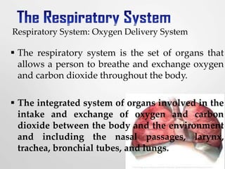
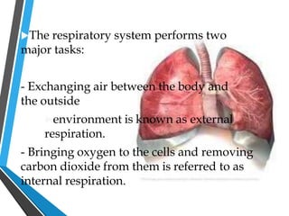
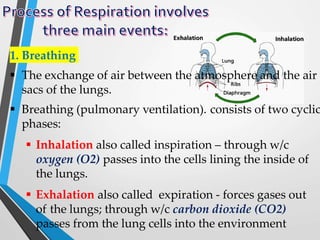
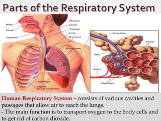
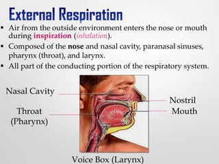
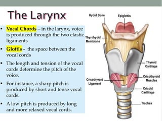
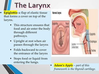
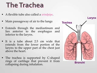
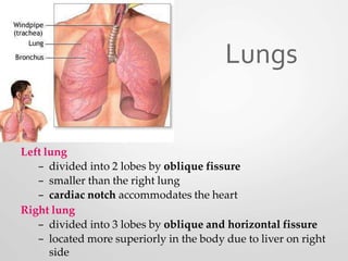
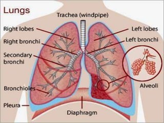
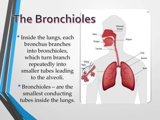
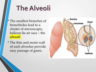
T
h
eRespiratory
system
s
y
s
t
e
m
Respiratory System: Oxygen Delivery System
 The respiratory system is the set of organs that
allows a person to breathe and exchange oxygen
and carbon dioxide throughout the body.
 The integrated system of organs involved in the
intake and exchange of oxygen and carbon
dioxide between the body and the environment
and including the nasal passages, larynx,
trachea, bronchial tubes, and lungs.
The respiratory system performs two
major tasks:
- Exchanging air between the body and
the outside
environment is known as external
respiration.
- Bringing oxygen to the cells and removing
carbon dioxide from them is referred to as
internal respiration.
1. Supplies the body with oxygen and disposes of
carbon dioxide
2. Filters-inspired air
3. Produces sound
4. Contains receptors for smell
5. Rids the body of some excess water and heat
6. Helps regulate blood pH
1. Breathing
 The exchange of air between the atmosphere and the air
sacs of the lungs.
 Breathing (pulmonary ventilation). consists of two cyclic
phases:
 Inhalation also called inspiration – through w/c
oxygen (O2) passes into the cells lining the inside of
the lungs.
 Exhalation also called expiration - forces gases out
of the lungs; through w/c carbon dioxide (CO2)
passes from the lung cells into the environment
 The transfer of O2 and CO2 across
respiratory membranes.
 It involves the diffusion of O2 from the
air sacs into the blood and the release of
CO2 from the blood.
 Oxygen is carried into the cells and
carbon dioxide is delivered from the
cells to the lungs and out of the body.
Human Respiratory System – consists of various cavities and
passages that allow air to reach the lungs.
- The main function is to transport oxygen to the body cells and
to get rid of carbon dioxide.
 Air from the outside environment enters the nose or mouth
during inspiration (inhalation).
 Composed of the nose and nasal cavity, paranasal sinuses,
pharynx (throat), and larynx.
 All part of the conducting portion of the respiratory system.
Nasal Cavity
Throat
(Pharynx)
Nostril
Mouth
Voice Box (Larynx)
 Nostrils - Air enters the body through the openings of the nose.
 Nasal Cavities -pair of passages inside the nostrils
 Nasal Septum – bony partition that separates the nasal cavities
 Cilia –the nasal cavities are lined with mucous membrane and
coarse hairlike projections
Nose and Nasal Cavities
Nasal concha Sphenoid sinus
Internal naris
Nasopharynx
External naris
Frontal sinus
Middle nasal concha
Inferior nasal
concha
 - as you breathe through the nose, air entering the nasal cavity is moistened
and warmed by the blood flowing in the capillaries of the mucous membrane.
 The mucous membrane – protects the lung from microorganisms and
other foreign materials.
 Common space used by both the respiratory and digestive
systems.
 Commonly called the throat.
 Originates posterior to the nasal and oral cavities and extends
inferiorly near the level of the bifurcation of the larynx and
esophagus.
 Common pathway for both air and food.
 Walls are lined by mucosa and contain skeletal muscles that are
primarily used for swallowing.
 Flexible lateral walls are distensible in order to force swallowed
food into the esophagus.
 This is a passageway that is divided into two smaller passages.
Three Sections of the Pharynx
 Nasopharynx
 contains the pharyngeal tonsils (adenoids) which aid in the
body’s immune defense.
 Oropharynx
 back portion of the mouth contains the palatine tonsils
which aid in the body’s immune defense.
 Laryngopharynx
 bottom section of the pharynx where the respiratory tract
divides into the esophagus and the larynx.
Nasopharynx
Oropharynx
Laryngopharynx
L
a
r
y
n
x
d
 Larynx – more commonly known
as the voice box, located bet. the
pharynx and the trachea.
 Prevents swallowed materials from
entering the lower respiratory tract.
 Conducts air into the lower
respiratory tract.
 Produces sounds.
 Supported by a framework of nine
pieces of cartilage (three individual
pieces and three cartilage pairs)
that are held in place by ligaments
and muscles.
L
a
r
y
n
x
d
 Vocal Chords – in the larynx, voice
is produced through the two elastic
ligaments
 Glottis - the space between the
vocal cords
 The length and tension of the vocal
cords determine the pitch of the
voice.
 For instance, a sharp pitch is
produced by short and tense vocal
cords.
 A low pitch is produced by long
and more relaxed vocal cords.
d
 Epiglottis–a flap of elastic tissue
that forms a cover on top of the
larynx.
 This structure ensures that
food and air enter the body
through different
pathways.
 Upright at rest when air
passes through the larynx
 Folds backward to cover
the entrance of the larynx
 Stops food or liquid from
entering the lungs.
 Adam’s Apple – part of this
framework is the thyroid cartilage.
 A flexible tube also called a windpipe.
 Main passageway of air to the lungs
 Extends through the mediastinum and
lies anterior to the esophagus and
inferior to the larynx.
 It is a tube about 2.5 cm wide that
extends from the lower portion of the
larynx to the upper part of the chest just
above the heart.
 The trachea is supported by C-shaped
rings of cartilage that prevent it from
collapsing during inhalation.
Trachea
Bronchi
Larynx
 The trachea divides into two main stems or bronchi, which lead to
the lungs.
 The right bronchus – is larger in diameter than the left.
 The bronchi – are also supported by rings of cartilage.
 Hilus or Hilum – bronchus enters the lung at a depression
 Air that reaches the bronchi is clean, warm, and moist.
 Each lung has a conical shape. Its wide, concave base rests
upon the muscular diaphragm.
 Its superior region called the apex projects superiorly to a
point that is slightly superior and posterior to the clavicle.
 Both lungs are bordered by the thoracic wall anteriorly,
laterally, and posteriorly, and supported by the rib cage.
 Toward the midline, the lungs are separated from each
other by the mediastinum.
 The relatively broad, rounded surface in contact with the
thoracic wall is called the costal surface of the lung.
 Primary organs of respiration.
 Diffusion of gases takes place in the lungs
through extremely thin and delicate tissues.
 The lungs are located side by side in the thoracic
(chest) cavity, which is separated from the
abdominal cavity by the muscular partition called
a diaphragm.
 Pleura – thin tissue layer covering the lungs.
 Keeps each lung moist and enables it to
move, with very little friction, in the
chest cavity during breathing.
Left lung
– divided into 2 lobes by oblique fissure
– smaller than the right lung
– cardiac notch accommodates the heart
Right lung
– divided into 3 lobes by oblique and horizontal fissure
– located more superiorly in the body due to liver on right
side
Lungs
•Inside the lungs, each
bronchus branches
into bronchioles,
which turn branch
repeatedly into
smaller tubes leading
to the alveoli.
•Bronchioles – are the
smallest conducting
tubes inside the lungs.
•The smallest branches of
bronchioles lead to a
cluster of microscopic,
balloon-lie air sacs – the
alveoli
•The thin and moist wall
of each alveolus provide
easy passage of gases.
The Respiratory System.pptx

The Respiratory System.pptx

  • 1.
  • 2.
    Respiratory System: OxygenDelivery System  The respiratory system is the set of organs that allows a person to breathe and exchange oxygen and carbon dioxide throughout the body.  The integrated system of organs involved in the intake and exchange of oxygen and carbon dioxide between the body and the environment and including the nasal passages, larynx, trachea, bronchial tubes, and lungs.
  • 3.
    The respiratory systemperforms two major tasks: - Exchanging air between the body and the outside environment is known as external respiration. - Bringing oxygen to the cells and removing carbon dioxide from them is referred to as internal respiration.
  • 4.
    1. Supplies thebody with oxygen and disposes of carbon dioxide 2. Filters-inspired air 3. Produces sound 4. Contains receptors for smell 5. Rids the body of some excess water and heat 6. Helps regulate blood pH
  • 5.
    1. Breathing  Theexchange of air between the atmosphere and the air sacs of the lungs.  Breathing (pulmonary ventilation). consists of two cyclic phases:  Inhalation also called inspiration – through w/c oxygen (O2) passes into the cells lining the inside of the lungs.  Exhalation also called expiration - forces gases out of the lungs; through w/c carbon dioxide (CO2) passes from the lung cells into the environment
  • 6.
     The transferof O2 and CO2 across respiratory membranes.  It involves the diffusion of O2 from the air sacs into the blood and the release of CO2 from the blood.  Oxygen is carried into the cells and carbon dioxide is delivered from the cells to the lungs and out of the body.
  • 7.
    Human Respiratory System– consists of various cavities and passages that allow air to reach the lungs. - The main function is to transport oxygen to the body cells and to get rid of carbon dioxide.
  • 8.
     Air fromthe outside environment enters the nose or mouth during inspiration (inhalation).  Composed of the nose and nasal cavity, paranasal sinuses, pharynx (throat), and larynx.  All part of the conducting portion of the respiratory system. Nasal Cavity Throat (Pharynx) Nostril Mouth Voice Box (Larynx)
  • 9.
     Nostrils -Air enters the body through the openings of the nose.  Nasal Cavities -pair of passages inside the nostrils  Nasal Septum – bony partition that separates the nasal cavities  Cilia –the nasal cavities are lined with mucous membrane and coarse hairlike projections Nose and Nasal Cavities Nasal concha Sphenoid sinus Internal naris Nasopharynx External naris Frontal sinus Middle nasal concha Inferior nasal concha  - as you breathe through the nose, air entering the nasal cavity is moistened and warmed by the blood flowing in the capillaries of the mucous membrane.  The mucous membrane – protects the lung from microorganisms and other foreign materials.
  • 10.
     Common spaceused by both the respiratory and digestive systems.  Commonly called the throat.  Originates posterior to the nasal and oral cavities and extends inferiorly near the level of the bifurcation of the larynx and esophagus.  Common pathway for both air and food.  Walls are lined by mucosa and contain skeletal muscles that are primarily used for swallowing.  Flexible lateral walls are distensible in order to force swallowed food into the esophagus.  This is a passageway that is divided into two smaller passages.
  • 11.
    Three Sections ofthe Pharynx  Nasopharynx  contains the pharyngeal tonsils (adenoids) which aid in the body’s immune defense.  Oropharynx  back portion of the mouth contains the palatine tonsils which aid in the body’s immune defense.  Laryngopharynx  bottom section of the pharynx where the respiratory tract divides into the esophagus and the larynx. Nasopharynx Oropharynx Laryngopharynx
  • 12.
    L a r y n x d  Larynx –more commonly known as the voice box, located bet. the pharynx and the trachea.  Prevents swallowed materials from entering the lower respiratory tract.  Conducts air into the lower respiratory tract.  Produces sounds.  Supported by a framework of nine pieces of cartilage (three individual pieces and three cartilage pairs) that are held in place by ligaments and muscles.
  • 13.
    L a r y n x d  Vocal Chords– in the larynx, voice is produced through the two elastic ligaments  Glottis - the space between the vocal cords  The length and tension of the vocal cords determine the pitch of the voice.  For instance, a sharp pitch is produced by short and tense vocal cords.  A low pitch is produced by long and more relaxed vocal cords.
  • 14.
    d  Epiglottis–a flapof elastic tissue that forms a cover on top of the larynx.  This structure ensures that food and air enter the body through different pathways.  Upright at rest when air passes through the larynx  Folds backward to cover the entrance of the larynx  Stops food or liquid from entering the lungs.  Adam’s Apple – part of this framework is the thyroid cartilage.
  • 15.
     A flexibletube also called a windpipe.  Main passageway of air to the lungs  Extends through the mediastinum and lies anterior to the esophagus and inferior to the larynx.  It is a tube about 2.5 cm wide that extends from the lower portion of the larynx to the upper part of the chest just above the heart.  The trachea is supported by C-shaped rings of cartilage that prevent it from collapsing during inhalation. Trachea Bronchi Larynx
  • 16.
     The tracheadivides into two main stems or bronchi, which lead to the lungs.  The right bronchus – is larger in diameter than the left.  The bronchi – are also supported by rings of cartilage.  Hilus or Hilum – bronchus enters the lung at a depression  Air that reaches the bronchi is clean, warm, and moist.
  • 17.
     Each lunghas a conical shape. Its wide, concave base rests upon the muscular diaphragm.  Its superior region called the apex projects superiorly to a point that is slightly superior and posterior to the clavicle.  Both lungs are bordered by the thoracic wall anteriorly, laterally, and posteriorly, and supported by the rib cage.  Toward the midline, the lungs are separated from each other by the mediastinum.  The relatively broad, rounded surface in contact with the thoracic wall is called the costal surface of the lung.
  • 18.
     Primary organsof respiration.  Diffusion of gases takes place in the lungs through extremely thin and delicate tissues.  The lungs are located side by side in the thoracic (chest) cavity, which is separated from the abdominal cavity by the muscular partition called a diaphragm.
  • 19.
     Pleura –thin tissue layer covering the lungs.  Keeps each lung moist and enables it to move, with very little friction, in the chest cavity during breathing.
  • 20.
    Left lung – dividedinto 2 lobes by oblique fissure – smaller than the right lung – cardiac notch accommodates the heart Right lung – divided into 3 lobes by oblique and horizontal fissure – located more superiorly in the body due to liver on right side Lungs
  • 22.
    •Inside the lungs,each bronchus branches into bronchioles, which turn branch repeatedly into smaller tubes leading to the alveoli. •Bronchioles – are the smallest conducting tubes inside the lungs.
  • 23.
    •The smallest branchesof bronchioles lead to a cluster of microscopic, balloon-lie air sacs – the alveoli •The thin and moist wall of each alveolus provide easy passage of gases.