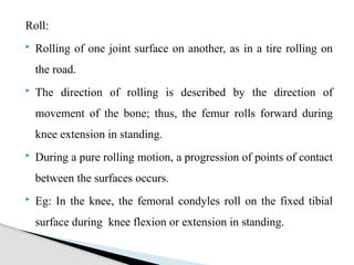
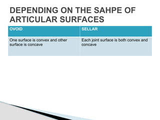
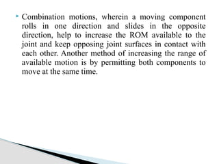
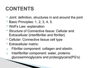
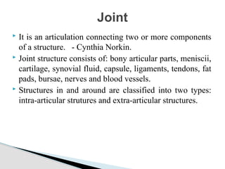
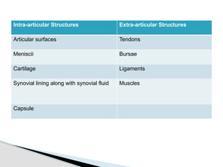
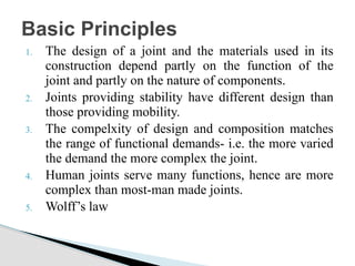
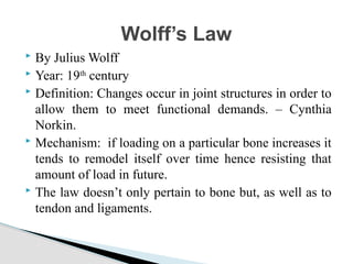
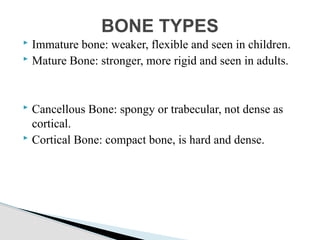
This document provides an overview of joint structure and function. It defines a joint and lists the intra-articular and extra-articular structures. It describes the basic principles of joint design and complexity matching function. It explains Wolff's law relating bone structure to function. It details the cellular and extracellular components of connective tissue, including collagen and elastin fibers. Finally, it discusses joint classification, motions, and the response of connective tissues to loads.
Elbow complex is designed to serve hand.
They provide MOBILITY for Hand in space by apparent shortening and Lengthening of upper extremity.
They provide Stability for skillful and forceful movements
Elbow complex is designed to serve hand.
They provide MOBILITY for Hand in space by apparent shortening and Lengthening of upper extremity.
They provide Stability for skillful and forceful movements
this PPT contain detailed kinetics & kinematics of ankle joint & all joints of foot complex, muscles of ankle & foot complex, plantar arches & weight distribution during standing.
Goniometry is the measuring of angles created by the bones of the body at the joints.1, 2, 3
The term goniometry is derived from two Greek words, gonia meaning angle and metron, meaning measure. 1, 2, 3, 4, 5,
System to measure the joint ranges in each plane of the joint is termed goniometry. 4
These measurements are done with instrument such as goniometer, a tape measure, inclinometers or by visual estimate.
Biomechanics of Tendon, Ligament, Cartilage,, Bone
Wolf's Law
Mechnical Behavior
stress strain curve (Young's Moudulas)
viscoelasticity
time depended and rate depended properties
creep stress relaxation, hyteresis,
Thank You
Femoral Head (Superiorly, Medially, Anteriorly).
Acetabulum (Inferiorly, Laterally, Anteriorly).
Horseshoe-shaped (Acetabular Notch).
The deepest portion (Acetabular Fossa).
Labrum Acetabular:
Is a wedged fibrocartilaginous ring inserted into the acetabular rim to increase the acetabular concavity.
This lecture help the students such as medical ,nursing , and any health care provider to understand the basic information about musculoskeletal system.
what biomechanics is and why it's important in understanding ligaments and tendons. structure and composition of ligaments and tendons. the roles of ligaments and tendons in the body and how they contribute to movement and stability. mechanical properties of ligaments and tendons, including strength, elasticity, and viscoelasticity.
1. Connective tissue is one of the four tissue types found throughout the body. Connective tissue is the most abundant and widely distributed tissue in the body.
2. Connective tissue develop from mesoderm during embryonic development. While some connective tissues are specialized ( bone , blood), all organs have some amount of connective tissue in them which hold their parenchyma together.
A traditional manual therapy technique developed by John Upledger, involving bare hands and stretching the tension membrane so as to ease the tension within
Its a compilation of both traditional and recent advance techniques of not only assessing musculoskeletal but also cardiovascular and respiratory endurance as well as strength
Traction: a basic physiotherapy modality used for inducing space between the joints. this slideshow deals with various types of traction and its application to cervical, thoracic and lumbar spine.
the PPT Describes about various types of dysfunction in mechanical pattern as described by Janda's. it also describes about normal muscle slings prresent within the body and its compensation and decompensation patterns towards the adaptations of the body
this slideshow describes about the hip joint anatomy, biomechanics and its pathomechanics along with angles of hip joint. the slide show also briefs about the pelvic femoral rhythm in daily activities
Thoracic and rib cage anatomy, biomechanics, and pathomechanicsRadhika Chintamani
This slide show describes about thoracic and rib cage in detail with its anatomy, kinetics and kinematics along with force couple. the slideshow also describes about the pathology and pathomechanics related to the topic
Knee joint anatomy, biomechanics, pathomechanics and assessmentRadhika Chintamani
the knee complex complete anatomy, biomechanics, pathomechanics and its physical assessment in one single slideshow.a brief table given for easy understanding of what special test to be performed in which condition along with evidences of each special test.
small correction in slide number: 10
during flexion of tibia over femur in OKC; tibia glides and rolls posteriorly
during extension of tibia over femur in OKC: tibia glides and rolls anteriorly
A very old school of manual therapy which comprises of two main principle centralization and peripheralization thought given by Robin McKenzie. The slideshow explain theoretical and practical part of both entire spine and extremities as well
this is a slide show which gives in brief about anatomy and detailed description about biomechanics as well as pathomechanics of shoulder joint. various rhythms of shoulder complex are discussed as well along with the stability factors
Muscle energy technique, a manual therapy technique with a long term history and 8 variations which can be used in various condition to treat muscle as well as joints. This slide show consists of detailed history, variations/types and summary of MET in detail.
A type of manual therapy in which the muscle or the joint is altered and placed in a position of comfort for certain duration after which the pain disappears completely or gets reduced. this slide show explains about the principles, mechanism and Phases of PRT
Massage of therapeutic form is beneficial in many conditions like stroke, flaccidity, muscle tightness, spasm etc.
it has many physiological effects along with many types for different conditions as well as different body areas.
it is another taping technique which inhibits or control the movement. it is helpful in postural correction and movement pattern correction as well. usually used clinically
Sacroiliac joint biomechanics, dysfunctions, assessment and its manual therapyRadhika Chintamani
Sacroiliac joint: mostly commonly affected joint due to its smaller articular surfaces. this slideshow briefs about its anatomy, biomechanics i.e. movements and axis, muscles, ligaments around it, types of dysfunction of SI joints, its special test and manual therapy management of the dysfunctions.
Lung Cancer: Artificial Intelligence, Synergetics, Complex System Analysis, S...Oleg Kshivets
RESULTS: Overall life span (LS) was 2252.1±1742.5 days and cumulative 5-year survival (5YS) reached 73.2%, 10 years – 64.8%, 20 years – 42.5%. 513 LCP lived more than 5 years (LS=3124.6±1525.6 days), 148 LCP – more than 10 years (LS=5054.4±1504.1 days).199 LCP died because of LC (LS=562.7±374.5 days). 5YS of LCP after bi/lobectomies was significantly superior in comparison with LCP after pneumonectomies (78.1% vs.63.7%, P=0.00001 by log-rank test). AT significantly improved 5YS (66.3% vs. 34.8%) (P=0.00000 by log-rank test) only for LCP with N1-2. Cox modeling displayed that 5YS of LCP significantly depended on: phase transition (PT) early-invasive LC in terms of synergetics, PT N0—N12, cell ratio factors (ratio between cancer cells- CC and blood cells subpopulations), G1-3, histology, glucose, AT, blood cell circuit, prothrombin index, heparin tolerance, recalcification time (P=0.000-0.038). Neural networks, genetic algorithm selection and bootstrap simulation revealed relationships between 5YS and PT early-invasive LC (rank=1), PT N0—N12 (rank=2), thrombocytes/CC (3), erythrocytes/CC (4), eosinophils/CC (5), healthy cells/CC (6), lymphocytes/CC (7), segmented neutrophils/CC (8), stick neutrophils/CC (9), monocytes/CC (10); leucocytes/CC (11). Correct prediction of 5YS was 100% by neural networks computing (area under ROC curve=1.0; error=0.0).
CONCLUSIONS: 5YS of LCP after radical procedures significantly depended on: 1) PT early-invasive cancer; 2) PT N0--N12; 3) cell ratio factors; 4) blood cell circuit; 5) biochemical factors; 6) hemostasis system; 7) AT; 8) LC characteristics; 9) LC cell dynamics; 10) surgery type: lobectomy/pneumonectomy; 11) anthropometric data. Optimal diagnosis and treatment strategies for LC are: 1) screening and early detection of LC; 2) availability of experienced thoracic surgeons because of complexity of radical procedures; 3) aggressive en block surgery and adequate lymph node dissection for completeness; 4) precise prediction; 5) adjuvant chemoimmunoradiotherapy for LCP with unfavorable prognosis.
The prostate is an exocrine gland of the male mammalian reproductive system
It is a walnut-sized gland that forms part of the male reproductive system and is located in front of the rectum and just below the urinary bladder
Function is to store and secrete a clear, slightly alkaline fluid that constitutes 10-30% of the volume of the seminal fluid that along with the spermatozoa, constitutes semen
A healthy human prostate measures (4cm-vertical, by 3cm-horizontal, 2cm ant-post ).
It surrounds the urethra just below the urinary bladder. It has anterior, median, posterior and two lateral lobes
It’s work is regulated by androgens which are responsible for male sex characteristics
Generalised disease of the prostate due to hormonal derangement which leads to non malignant enlargement of the gland (increase in the number of epithelial cells and stromal tissue)to cause compression of the urethra leading to symptoms (LUTS
New Directions in Targeted Therapeutic Approaches for Older Adults With Mantl...i3 Health
i3 Health is pleased to make the speaker slides from this activity available for use as a non-accredited self-study or teaching resource.
This slide deck presented by Dr. Kami Maddocks, Professor-Clinical in the Division of Hematology and
Associate Division Director for Ambulatory Operations
The Ohio State University Comprehensive Cancer Center, will provide insight into new directions in targeted therapeutic approaches for older adults with mantle cell lymphoma.
STATEMENT OF NEED
Mantle cell lymphoma (MCL) is a rare, aggressive B-cell non-Hodgkin lymphoma (NHL) accounting for 5% to 7% of all lymphomas. Its prognosis ranges from indolent disease that does not require treatment for years to very aggressive disease, which is associated with poor survival (Silkenstedt et al, 2021). Typically, MCL is diagnosed at advanced stage and in older patients who cannot tolerate intensive therapy (NCCN, 2022). Although recent advances have slightly increased remission rates, recurrence and relapse remain very common, leading to a median overall survival between 3 and 6 years (LLS, 2021). Though there are several effective options, progress is still needed towards establishing an accepted frontline approach for MCL (Castellino et al, 2022). Treatment selection and management of MCL are complicated by the heterogeneity of prognosis, advanced age and comorbidities of patients, and lack of an established standard approach for treatment, making it vital that clinicians be familiar with the latest research and advances in this area. In this activity chaired by Michael Wang, MD, Professor in the Department of Lymphoma & Myeloma at MD Anderson Cancer Center, expert faculty will discuss prognostic factors informing treatment, the promising results of recent trials in new therapeutic approaches, and the implications of treatment resistance in therapeutic selection for MCL.
Target Audience
Hematology/oncology fellows, attending faculty, and other health care professionals involved in the treatment of patients with mantle cell lymphoma (MCL).
Learning Objectives
1.) Identify clinical and biological prognostic factors that can guide treatment decision making for older adults with MCL
2.) Evaluate emerging data on targeted therapeutic approaches for treatment-naive and relapsed/refractory MCL and their applicability to older adults
3.) Assess mechanisms of resistance to targeted therapies for MCL and their implications for treatment selection
Factory Supply Best Quality Pmk Oil CAS 28578–16–7 PMK Powder in Stockrebeccabio
Factory Supply Best Quality Pmk Oil CAS 28578–16–7 PMK Powder in Stock
Telegram: bmksupplier
signal: +85264872720
threema: TUD4A6YC
You can contact me on Telegram or Threema
Communicate promptly and reply
Free of customs clearance, Double Clearance 100% pass delivery to USA, Canada, Spain, Germany, Netherland, Poland, Italy, Sweden, UK, Czech Republic, Australia, Mexico, Russia, Ukraine, Kazakhstan.Door to door service
Hot Selling Organic intermediates
ARTIFICIAL INTELLIGENCE IN HEALTHCARE.pdfAnujkumaranit
Artificial intelligence (AI) refers to the simulation of human intelligence processes by machines, especially computer systems. It encompasses tasks such as learning, reasoning, problem-solving, perception, and language understanding. AI technologies are revolutionizing various fields, from healthcare to finance, by enabling machines to perform tasks that typically require human intelligence.
MANAGEMENT OF ATRIOVENTRICULAR CONDUCTION BLOCK.pdfJim Jacob Roy
Cardiac conduction defects can occur due to various causes.
Atrioventricular conduction blocks ( AV blocks ) are classified into 3 types.
This document describes the acute management of AV block.
Tom Selleck Health: A Comprehensive Look at the Iconic Actor’s Wellness Journeygreendigital
Tom Selleck, an enduring figure in Hollywood. has captivated audiences for decades with his rugged charm, iconic moustache. and memorable roles in television and film. From his breakout role as Thomas Magnum in Magnum P.I. to his current portrayal of Frank Reagan in Blue Bloods. Selleck's career has spanned over 50 years. But beyond his professional achievements. fans have often been curious about Tom Selleck Health. especially as he has aged in the public eye.
Follow us on: Pinterest
Introduction
Many have been interested in Tom Selleck health. not only because of his enduring presence on screen but also because of the challenges. and lifestyle choices he has faced and made over the years. This article delves into the various aspects of Tom Selleck health. exploring his fitness regimen, diet, mental health. and the challenges he has encountered as he ages. We'll look at how he maintains his well-being. the health issues he has faced, and his approach to ageing .
Early Life and Career
Childhood and Athletic Beginnings
Tom Selleck was born on January 29, 1945, in Detroit, Michigan, and grew up in Sherman Oaks, California. From an early age, he was involved in sports, particularly basketball. which played a significant role in his physical development. His athletic pursuits continued into college. where he attended the University of Southern California (USC) on a basketball scholarship. This early involvement in sports laid a strong foundation for his physical health and disciplined lifestyle.
Transition to Acting
Selleck's transition from an athlete to an actor came with its physical demands. His first significant role in "Magnum P.I." required him to perform various stunts and maintain a fit appearance. This role, which he played from 1980 to 1988. necessitated a rigorous fitness routine to meet the show's demands. setting the stage for his long-term commitment to health and wellness.
Fitness Regimen
Workout Routine
Tom Selleck health and fitness regimen has evolved. adapting to his changing roles and age. During his "Magnum, P.I." days. Selleck's workouts were intense and focused on building and maintaining muscle mass. His routine included weightlifting, cardiovascular exercises. and specific training for the stunts he performed on the show.
Selleck adjusted his fitness routine as he aged to suit his body's needs. Today, his workouts focus on maintaining flexibility, strength, and cardiovascular health. He incorporates low-impact exercises such as swimming, walking, and light weightlifting. This balanced approach helps him stay fit without putting undue strain on his joints and muscles.
Importance of Flexibility and Mobility
In recent years, Selleck has emphasized the importance of flexibility and mobility in his fitness regimen. Understanding the natural decline in muscle mass and joint flexibility with age. he includes stretching and yoga in his routine. These practices help prevent injuries, improve posture, and maintain mobilit
2. Joint: definition, structures in and around the joint
Basic Principles: 1, 2, 3, 4, 5.
Wolf’s Law: explanation
Structure of Connective tissue: Cellular and
Extracellular {interfibrillar and fibrillar}
Cellular: Connective tissue cell type
Extracellular matrix:
a. Fibrillar component: collagen and elastin.
b. Interfibrillar component: water, proteins:
glycosaminoglycans and proteoglycans(PG’s)
CONTENTS
3. It is an articulation connecting two or more components
of a structure. - Cynthia Norkin.
Joint structure consists of: bony articular parts, meniscii,
cartilage, synovial fluid, capsule, ligaments, tendons, fat
pads, bursae, nerves and blood vessels.
Structures in and around are classified into two types:
intra-articular strutures and extra-articular structures.
Joint
4. Intra-articular Structures Extra-articular Structures
Articular surfaces Tendons
Meniscii Bursae
Cartilage Ligaments
Synovial lining along with synovial fluid Muscles
Capsule
5. 1. The design of a joint and the materials used in its
construction depend partly on the function of the
joint and partly on the nature of components.
2. Joints providing stability have different design than
those providing mobility.
3. The compelxity of design and composition matches
the range of functional demands- i.e. the more varied
the demand the more complex the joint.
4. Human joints serve many functions, hence are more
complex than most-man made joints.
5. Wolff’s law
Basic Principles
6. By Julius Wolff
Year: 19th
century
Definition: Changes occur in joint structures in order to
allow them to meet functional demands. – Cynthia
Norkin.
Mechanism: if loading on a particular bone increases it
tends to remodel itself over time hence resisting that
amount of load in future.
The law doesn’t only pertain to bone but, as well as to
tendon and ligaments.
Wolff’s Law
8. It is a type of tissue that supports, binds, connects or
separates other tissue from each other, embedded with
few cells in a matrix.
Connective tissue is further made up of cellular (fixed
and transient) & extracellular (interfibrillar and
fibrillar)
Connective tissue
9.
10. Cellular Matrix: a. Fixed type
NAME LOCATION FUNCTION
Fibroblast Found in tendon, ligament, skin,
bone. etc
Creates mostly type I
collagen
Chondrobalst These are differentiated
fibrobalsts found in cartilage
Type II collagen
Osteoblast bone Type I collagen and
production of
hydroxyapetite
Osteocalst bone Bone resorption
Mast cells Various connective tissue Inflammatory cells
Adipose cells Adipose tissue Produce and store fat
Mesenchyme
cells
Embryos and bone marrow Differentiate into
connective tissue
11. Cellular Matrix: b. Transient type
NAME FUNCTION
Lymphocytes Have surface protein specific for antigen
Neutrophils Involved in fighting infection
Macrophages Involved in immune response
Plasma cells Produce antibodies
12. The interfibrillar component of connective tissue is
composed of hydrated networks of proteins: primarily
glycoproteins and proteoglycans (PGs)
PG: the PGs are found mainly in connective tissues,
where they contribute to the organization and physical
properties of the ECM.
GAG’s: The GAGs are all very similar to glucose in
structure and are distinguished by the number and location
of the amine and sulfate groups that are attached. The
major types of sulfated GAGs include chondroitin 4 and
chondroitin 6 sulfate, keratan sulfate, heparin,
heparan sulfate, and dermatan sulfate.
Extracellular Matrix: a. Interfibrillar
13. Glycoproteins such as fibronectin, laminin,
chondronectin, osteonectin, tenascin, and entactin
play an important role in fastening the various
components of the ECM together and in the adhesion
between collagen and integrin molecules in the cell
membranes of the resident cells of the tissue.
14. The fibrillar, or fibrous, component of the ECM
contains two major classes of structural proteins:
collagen and elastin.2
Collagen: has a tensile strength and is responsible for
the functional integrity of connective tissue structures
and their resistance to tensile forces.
Elastin: Each elastin molecule uncoils into a more
extended conformation when the fiber is stretched and
will recoil spontaneously as soon as the stretching force
is relaxed.
b. Fibrillar
15. Bone contains two principle structural components:
collagen (Type I) and hydroxyapatite (HA).
Organic components of bone make up approximately
40% of the bone’s dry weight, and collagen is
responsible for about 90% of bone’s organic content.
The inorganic, or mineral, components make up
approximately 60% of the bone’s dry weight.
The primary mineral component is HA, which is a
calcium phosphate–based mineral: Ca10(PO4)6(OH)2.
The HA crystals are found primarily between the
collagen fibers
Bone
16. types II, IX, and X are known as cartilage-specific
collagens because they seem to be found only in
cartilage.
Because HA is a ceramic, bone can be expected to have
ceramic-like properties. For example, ceramics are
generally brittle, tolerating little deformation before
fracture.
Ceramics and bone are also relatively strong in
compression but weak in tension.
The structure of human bone changes with age.
REMEMBER
17. Immature bone: weaker, flexible and seen in children.
Mature Bone: stronger, more rigid and seen in adults.
Cancellous Bone: spongy or trabecular, not dense as
cortical.
Cortical Bone: compact bone, is hard and dense.
BONE TYPES
18. Wolff observed that bone, especially cancellous bone,
is oriented to resist the primary forces to which bone is
subjected.
Wolff suggested that “the shape of bone is determined
only by static loading”
Note
20. Provides structural support
Protect the vital organs
Allows body to move through muscles, or perform any
other activity
Depending on kinematic chain it varies: stability and
mobility
Functions of bones
21. Bone is isotropic transversely and anisotropic
longitudinally.
Stress-strain curves for bone demonstrate that cortical
bone is stiffer than cancellous (trabecular) bone,
meaning that cortical bone can withstand greater stress
but less strain than the cancellous bone.
Cancellous bone can sustain strains of 75% before
failing in vivo, but cortical bone will fail if strain
exceeds 2%.
Bone can withstand greater stress, and will undergo
less strain, in compression than in tension.
Principle features of bone
22. The physiologic response of trabecular bone to an
increase in loading is hypertrophy. If loading is
decreased or absent, the trabeculae become smaller and
weaker.
Repeated loadings, either high repetition coupled with
low load or low repetition with high load, can cause
permanent strain and lead to bone failure. Bone loses
stiffness and strength with repetitive loading.
Bone loses stiffness and strength with repetitive
loading as a result of creep strain. Creep strain occurs
when a tissue is loaded repetitively during the time
the material is undergoing creep.
23. Synarthrosis:
a. Fibrous joints: sutures, ghomphoses, syndesmoses.
b. Cartilagenous joints: symphysis, synchondroses.
Diarthrosis:
Classification of joints
24. The material used to connect the bony components in
synarthrodial joints is interosseus connective tissue.
Divided into two types based on the type of connective
tissue used to connect the bones.
a. Fibrous: fibrous tissue directly unites the bone.
b. Cartilagenous: cartilage is used to connect the bones
forming joint; either fibrocartilagenous or hyaline
cartilage.
Synarthrosis
25. 1. Sutures: two bony components are united by
collagenous sutural ligament or membrane.
The ends of the bony components are shaped so that
the edges interlock or overlap one another.
Eg: skull bones
Fibrous
26. 2.Gomphosis joint: surfaces of bony components are
adapted to each other like a peg in hole.
Component parts are connected by fibrous tissue.
Eg: tooth, mandible or maxilla.
3.Syndesmosis: Two bony components are directly
connected by interosseous membrane.
Eg: radio-ulnar joint and tibio-femoral joints.
27. Symphysis: two bony components are covered with a
thin lamina of hyaline cartilage and directly joined by
fibrocartilage in the form of disks or pads
EG: Intervertebral joints, pubic symphysis
Synchondrosis: material used for connecting the two
components is hyaline cartilage. The cartilage forms a
bond between two ossifying centers of bone. The
function of this type of joint is to permit bone growth
while also providing stability and allowing a small
amount of mobility.
Eg: first costosternal joint.
Cartillagenous
28. The system of joints and links is constructed so that motion of
one link at one joint will produce motion at all of the other
joints in the system in a predictable manner. The kinematic
chain can be open or closed.
In an open kinematic chain, one joint can move
independently of others in the chain.
When one end of the chain remains fixed, it creates a closed
system or closed kinematic chain.
Under these conditions, movement at one joint automatically
creates movement in other joints in the chain.
KINEMTICS
29. Range of motion: the range or the arc through which the
movement of one bony lever occurs with respect to another.
There are two types of ROM’s:
a. Anatomical ROM: Movement of the joint within
anatomical limits.
b. Physiological ROM: movement of the joint beyond the
anatomical limit.
Joint motion
30. The extent of the anatomic range is determined by a number
of factors, including the shape of the joint surfaces, the joint
capsule, ligaments, muscle bulk, and surrounding
musculotendinous and bony structures.
Eg:
i. The humeroulnar joint at the elbow is limited in extension
by bony contact of the ulna on the olecranon fossa of the
humerus.
ii. The tibifemoral joint at the knee is limited in flexion by soft
tissue approximation at the popliteal fossa.
31. Given by cyriax
Experience felt by the therapist during the motion carried out
passively at the end of the range of passive physiologic ROM.
End feel
33. Osteokinematics refers to the movement of the bones
in space during physiologic joint motion.
These are the movements in the sagittal, frontal, and
transverse planes that occur at joints.
The movements are typically described by the plane
in which they occur, the axis about which they occur,
and the direction of movement.
Eg: Osteokinematic movements at the ulnohumeral
joint include flexion or extension (direction) of the
ulna on the humerus (or humerus on the ulna) in the
sagittal plane about a frontal axis.
Osteokinematics
34. Physiologic joint motion involves motion of bony
segments (osteokinematics) as well as motion of the
joint surfaces in relation to another.
Accompany voluntary movements, but can’t be
produced voluntarily.
The term arthrokinematics is used to refer to these
movements of joint surfaces on one another.
Arthrokinematics
36. Roll:
Rolling of one joint surface on another, as in a tire rolling on
the road.
The direction of rolling is described by the direction of
movement of the bone; thus, the femur rolls forward during
knee extension in standing.
During a pure rolling motion, a progression of points of contact
between the surfaces occurs.
Eg: In the knee, the femoral condyles roll on the fixed tibial
surface during knee flexion or extension in standing.
37. Slide:
Pure translatory motion.
Gliding of one component over another, as when a
braked wheel skids. The point of contact changes in the
fixed component as the sliding component moves over it.
Eg: In the hand, the proximal phalanx slides over the
fixed end of the metacarpal during flexion and extension.
38. Spin:
Spin is a pure rotatory motion. The same points remain
in contact on both the moving and stationary
components.
Eg: elbow, the head of the radius spins on the
capitulum of the humerus during supination and
pronation of the forearm.
39. Convex-concave rule: Convex joint surfaces roll
and glide in opposite directions, whereas concave
joint surfaces roll and slide in the same direction.
CONVEX: OPPOSITE
CONCAVE: SAME
CONCAVE-CONVEX RULE
40. OVOID SELLAR
One surface is convex and other
surface is concave
Each joint surface is both convex and
concave
DEPENDING ON THE SAHPE OF
ARTICULAR SURFACES
41. Joint motions commonly include a combination of
sliding, spinning, and rolling.
Although we typically describe the axis of rotation for
various joints in the body and use anatomical
landmarks to represent these axes, the combination of
sliding and spinning or rolling produces curvilinear
motion and a moving axis of motion.
The axis of rotation at any particular point in the
motion is called the instantaneous axis of rotation
(IAR).
NOTE
42. Combination motions, wherein a moving component
rolls in one direction and slides in the opposite
direction, help to increase the ROM available to the
joint and keep opposing joint surfaces in contact with
each other. Another method of increasing the range of
available motion is by permitting both components to
move at the same time.
43. All connective tissues will adapt to increased load
through changes in structural and/or material properties
(form follows function).
The load must be gradual and progressive; as the tissue
adapts to the new loading conditions, the load must
change to induce further adaptation.
The type of connective tissue formed will match the
type and volume of the load:
i. compression: cartilage or bone
ii. tension: ligament or tendon
Connective Tissue Response to
loads