Download as PDF, PPTX


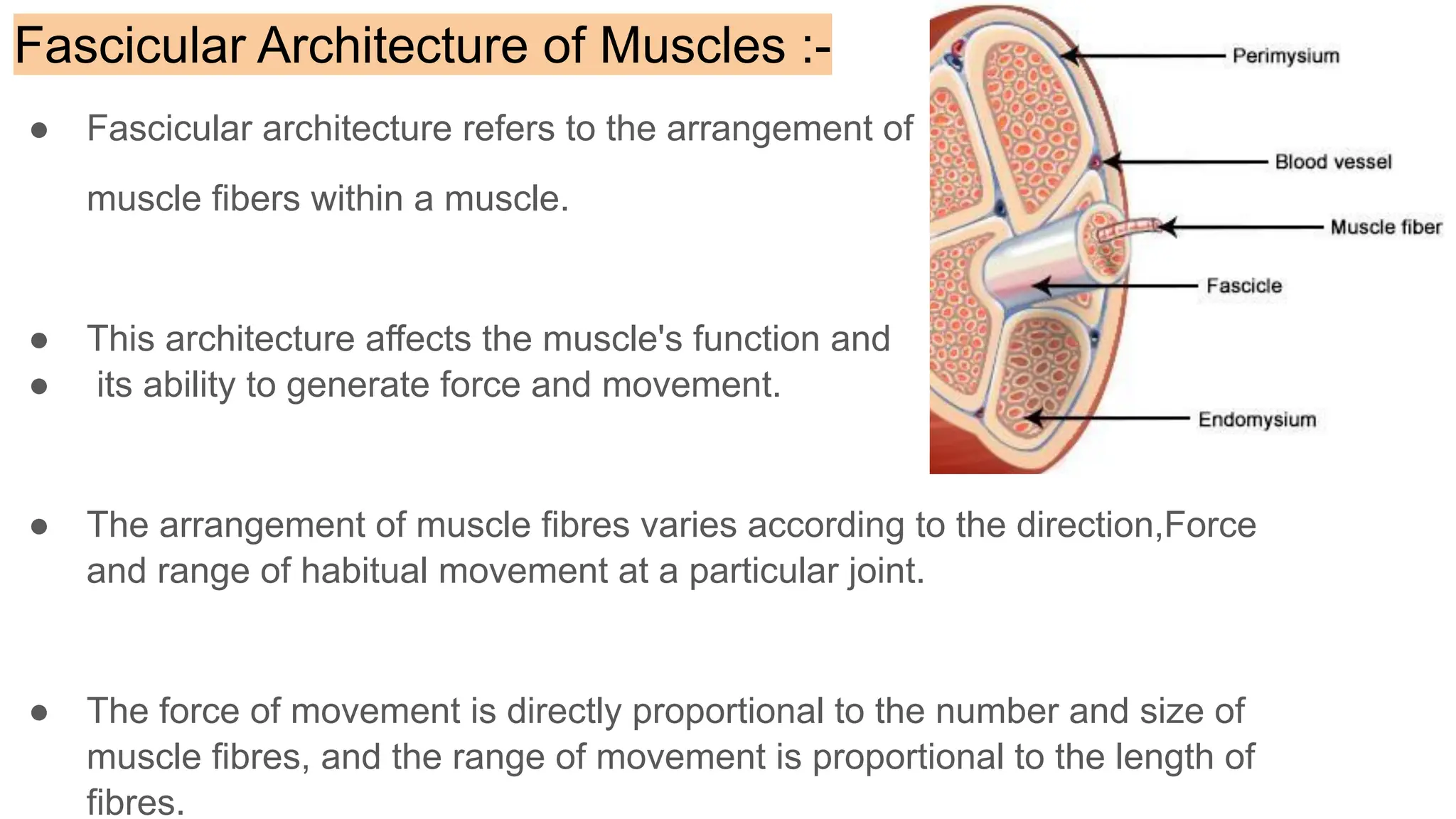


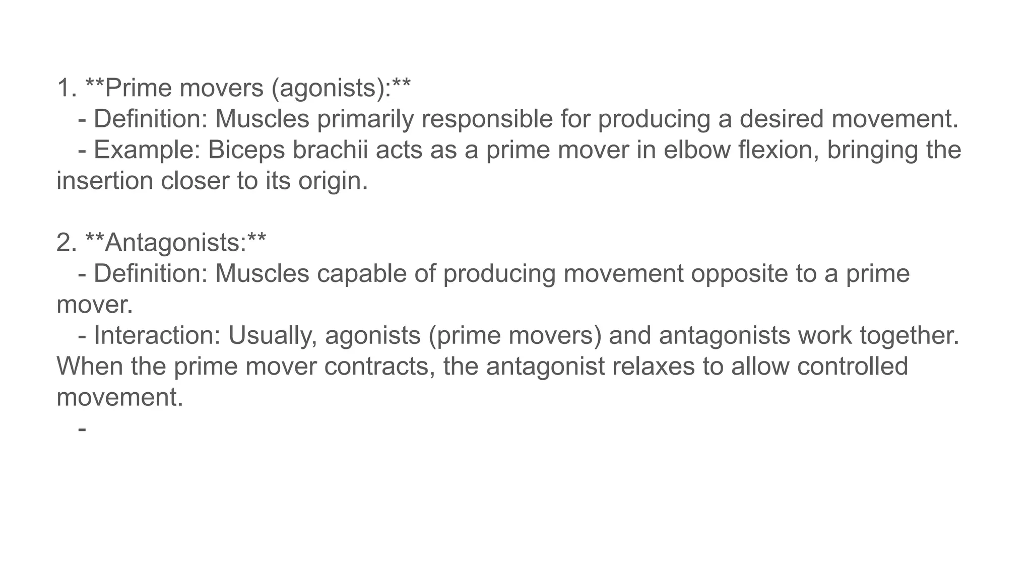


This document provides an overview of muscle classification, fascicular architecture, and their blood and nerve supply. It details types of muscle fasciculi, their arrangement, actions, and how muscle contraction works, including the roles of motor units and the concepts of muscle tone, active insufficiency, and passive insufficiency. Additionally, it explains the interaction between different muscle groups during movement, defining prime movers, antagonists, fixators, and synergists.


