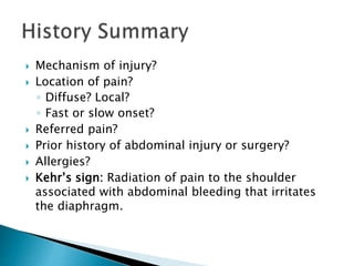
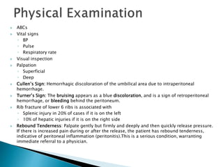
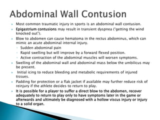
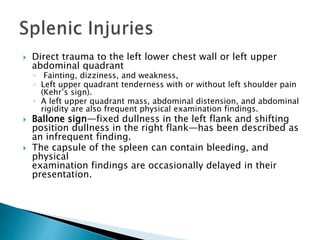
Emergency Care in Athletic Training
Chapter 10
Keith Gorse, Robert Blanc, Francis Feld and Mathew Radelet
Presentation Prepared by:
Dr Asma Lashari
University of Health Sciences
 In a 30-year period, Bergovist et al concluded that most common diagnosis was an
abdominal wall contusion.
◦ Soccer most commonly involved sport.
◦ Kidney was the most commonly injured organ
◦ Spleen, the next most commonly injured organ.
◦ Peritonitis and delayed splenic rupture are less common.
 Wan et al. found that sports were responsible for only 6.64%
of the cases reported to the trauma centers.
◦ Of the injuries, 84% occurred in children between the ages of 12 and 18 years.
◦ American football was the most frequently involved sport, with baseball and
basketball the next most common.
◦ The spleen was the most frequently injured organ across all age groups, with 96
cases,
◦ Kidney was second.
 Ryan noted that the relative infrequency of abdominal injuries put them at risk to go
undiagnosed because they are internal and concealed.
 Repeated monitoring of vital signs as well as the abdominal examination is imperative
regardless of the severity of the trauma involved, and should be repeated at intervals
until the athlete has either clearly improved, or clearly requires transport.
 In the general population unrecognized abdominal injury is a frequent cause of
preventable death, and can be masked by head injury, orthopedic injury, medications, or
ergogenic aids.
 Mechanism of injury?
 Location of pain?
◦ Diffuse? Local?
◦ Fast or slow onset?
 Referred pain?
 Prior history of abdominal injury or surgery?
 Allergies?
 Kehr’s sign: Radiation of pain to the shoulder
associated with abdominal bleeding that irritates
the diaphragm.
 ABCs
 Vital signs
◦ BP
◦ Pulse
◦ Respiratory rate
 Visual inspection
 Palpation
◦ Superficial
◦ Deep
 Cullen’s Sign: Hemorrhagic discoloration of the umbilical area due to intraperitoneal
hemorrhage.
 Turner’s Sign: The bruising appears as a blue discoloration, and is a sign of retroperitoneal
hemorrhage, or bleeding behind the peritoneum.
 Rib fracture of lower 6 ribs is associated with
◦ Splenic injury in 20% of cases if it is on the left
◦ 10% of hepatic injuries if it is on the right side
 Rebound Tenderness: Palpate gently but firmly and deeply and then quickly release pressure.
If there is increased pain during or after the release, the patient has rebound tenderness,
indicative of peritoneal inflammation (peritonitis).This is a serious condition, warranting
immediate referral to a physician.
 Most common traumatic injury in sports is an abdominal wall contusion.
 Epigastrium contusions may result in transient dyspnea (“getting the wind
knocked out”).
 Blow to abdomen can cause hematoma in the rectus abdominus, which can
mimic an acute abdominal internal injury.
◦ Sudden abdominal pain
◦ Rapid swelling but will improve by a forward flexed position.
◦ Active contraction of the abdominal muscles will worsen symptoms.
 Swelling of the abdominal wall and abdominal mass below the umbilicus may
be present.
 Initial icing to reduce bleeding and metabolic requirements of injured
tissues.
 Padding for protection or a flak jacket if available may further reduce risk of
reinjury if the athlete decides to return to play.
 It is possible for a player to suffer a direct blow to the abdomen, recover
adequately to return to play only to have symptoms later in the game or
afterwards and ultimately be diagnosed with a hollow viscus injury or injury
to a solid organ.
 Direct trauma to the left lower chest wall or left upper
abdominal quadrant
◦ Fainting, dizziness, and weakness,
◦ Left upper quadrant tenderness with or without left shoulder pain
(Kehr’s sign).
◦ A left upper quadrant mass, abdominal distension, and abdominal
rigidity are also frequent physical examination findings.
 Ballone sign—fixed dullness in the left flank and shifting
position dullness in the right flank—has been described as
an infrequent finding.
 The capsule of the spleen can contain bleeding, and
physical
examination findings are occasionally delayed in their
presentation.
 Plain x-rays may demonstrate
◦ Enlarged spleen if a subcapsular hematoma with an intact capsule is
present.
◦ Enlarged spleen may also displace the stomach anteromedially and the left
kidney, left transverse colon, and splenic flexure inferiorly.
◦ Haziness of the abdomen, bulging flanks, and displacement of small
bowel loops are associated with signs of free peritoneal fluid, such as
blood.
 Sensitivity of ultrasound in diagnosing splenic injuries is greater
than for other abdominal organs.
 The “gold standard” test to evaluate an injury to the spleen is
computed tomography (CT) scanning.
◦ Capsular disruptions
◦ Subcapsular and intrasplenic hematomas
◦ Single and multiple fractures
◦ Shattered and fragmented spleens
◦ Sensitivity and specificity of CT scan in the diagnosis of splenic injury are
generally in the range of 96%.
 Majority of injuries to the spleen more than half of the
higher-grade injuries can manage nonoperatively.
 Some patients predict the failure of nonoperative
management
◦ Who do not stabilize with minimal resuscitation,
◦ Those with recurrent hemodynamic instability,
◦ Those with the presence of pooling or “blush” in the spleen on the
initial CT scan with intravenous contrast all.
 On the field, the athletic trainer should obtain baseline
vital signs, perform an abdominal examination, reassess
vital signs for evidence of vascular instability, and decide if
triage to the emergency room is required.
 If vital signs appear stable and abdominal pain does not
increase with cough, sneeze, or rapid movements.
 If there is any evidence of vascular instability, transport to
hospital should be considered.
 Liver injuries are uncommon in sports.
 Direct blow to the right upper quadrant of the abdomen or a deceleration
type of injury that lacerates the relatively thin capsule of the liver.
 Right upper quadrant tenderness
 Physical examination:
◦ ABCs followed by blood pressure, pulse rate, and respirations.
 In cases of blunt abdominal trauma, associated injuries to other abdominal
structures are seen 4% to 15% of the time.
 Transport are based on hemodynamic stability.
 CT scan is regarded as the best test to evaluate liver injuries and for the
presence of blood in the abdomen.
 Use of alanine aminotransferase (ALT) and aspartate aminotransferase (AST)
has been shown to correlate with the presence of abdominal injury in both
adults and children and can be used as screening laboratory tests in patients
who are
hemodynamically stable. These enzymes are elevated in cases without a
radiologically definable liver injury.
 Most common abdominal emergencies in sports.
 Symptom is flank pain, and hematuria
 Nephrectomy can be as high as 10% to 12%,
 10% will have injuries severe enough to require surgery.
 16% of football players had gross hematuria and that hematuria
peaked during games.
 Sports hematuria resolves with rest.
 Evaluations of vital signs and abdominal examinations.
 CT scan remains the test of choice for renal injuries.
 In confusing cases with a history of trauma and sports
hematuria, repeated urinalysis with and without activity can be
used to distinguish sports hematuria from abdominal trauma
with acute renal injury.
 Renal vein thrombosis following martial arts trauma is a rare
entity and presents with flank pain and microscopic hematuria.
 Rarely is reported with abdominal trauma in sports.
 Duodenal injury is associated with a direct blow to the epigastric
area and is in conjunction with an injury to the pancreas.
 Signs and symptoms are notoriously subtle
 CT scan with contrast is best test.
 A high degree of suspicion is important in these cases because a
delay in diagnosis and surgical intervention can increase
mortality.
 Jejunal injuries are also extremely rare and have variable
presentations.
 In some cases athletes have returned to play, only to have
symptoms after play and ultimately require surgery.
 Although CT scan with contrast is the test of choice, it may
initially be negative.
 Pancreas injuries are extremely rare and often
difficult to
diagnose
 Onset is often gradual with slow progression of
symptoms.
 Serum amylase levels can be diagnostic, but the
levels are often slow to rise.
 CT scan is the initial imaging study of choice and
can show lacerations of the pancreas
 but injuries to the pancreatic duct is visible by
endoscopic retrograde cholangeography (ERCP).
 Appendicitis often presents with atypical symptoms, and diagnosis is often in
doubt.
 Use of laboratory and imaging studies used to support the clinical
impression.
◦ Right lower quadrant tenderness,
◦ Abdominal rigidity,
◦ Guarding,
◦ Rebound tenderness,
◦ Pain aggravated by coughing or movement
◦ Duration of pain are the classic findings on examination
 Symptoms of loss of appetite, nausea, and a low-grade fever are often
present at the onset.
 Pain that begins in the periumbilical area and then shifts to the right lower
quadrant of the abdomen is also highly predictive.
 Although CT scans are generally considered an excellent diagnostic test for
appendicitis.
 Implantation of a fertilized ovum outside of the endometrial cavity
 Undiagnosed can result in rupture of the fallopian tube, massive hemorrhage, and
death.
 Major risk factors include
◦ Prior damage to the fallopian tube from pelvic inflammatory disease,
◦ Prior ectopic pregnancy,
◦ Prior tubal surgery.
◦ Cigarette smoking,
◦ Increasing age
 Hypotension, elevated pulse rate, and abdominal rigidity and guarding
 Diagnosis by the use of “discriminatory cutoff.”
◦ beta-human chorionic gonadatropin (-hCG) is often used as a marker of gestational
age.
◦ When -hCG reaches a specific level, usually 1500 to 2500, an intrauterine pregnancy
should be visualized.
◦ The absence of an intrauterine pregnancy implies an abnormal location and increases
concern for an ectopic pregnancy.
◦ When levels are below the discriminatory cutoff, serial measurements can be made
and the athlete can be observed.
◦ but when levels are above the cutoff and no intrauterine pregnancy is seen on
ultrasound, surgery is recommended.
 Some types of abdominal injuries can be fatal. Fortunately,
injuries to the abdomen are relatively uncommon.
 The kidney is the most frequently injured organ, followed by the spleen.
 Injuries to the liver, pancreas, and intestines are rare.
 A good history and physical examination, although usually not conclusive in
abdominal injuries, are management tools for athletic trainers.
Chapter 10 Abdominal Emergencies

Chapter 10 Abdominal Emergencies

  • 1.
    Emergency Care inAthletic Training Chapter 10 Keith Gorse, Robert Blanc, Francis Feld and Mathew Radelet Presentation Prepared by: Dr Asma Lashari University of Health Sciences
  • 2.
     In a30-year period, Bergovist et al concluded that most common diagnosis was an abdominal wall contusion. ◦ Soccer most commonly involved sport. ◦ Kidney was the most commonly injured organ ◦ Spleen, the next most commonly injured organ. ◦ Peritonitis and delayed splenic rupture are less common.  Wan et al. found that sports were responsible for only 6.64% of the cases reported to the trauma centers. ◦ Of the injuries, 84% occurred in children between the ages of 12 and 18 years. ◦ American football was the most frequently involved sport, with baseball and basketball the next most common. ◦ The spleen was the most frequently injured organ across all age groups, with 96 cases, ◦ Kidney was second.  Ryan noted that the relative infrequency of abdominal injuries put them at risk to go undiagnosed because they are internal and concealed.  Repeated monitoring of vital signs as well as the abdominal examination is imperative regardless of the severity of the trauma involved, and should be repeated at intervals until the athlete has either clearly improved, or clearly requires transport.  In the general population unrecognized abdominal injury is a frequent cause of preventable death, and can be masked by head injury, orthopedic injury, medications, or ergogenic aids.
  • 3.
     Mechanism ofinjury?  Location of pain? ◦ Diffuse? Local? ◦ Fast or slow onset?  Referred pain?  Prior history of abdominal injury or surgery?  Allergies?  Kehr’s sign: Radiation of pain to the shoulder associated with abdominal bleeding that irritates the diaphragm.
  • 4.
     ABCs  Vitalsigns ◦ BP ◦ Pulse ◦ Respiratory rate  Visual inspection  Palpation ◦ Superficial ◦ Deep  Cullen’s Sign: Hemorrhagic discoloration of the umbilical area due to intraperitoneal hemorrhage.  Turner’s Sign: The bruising appears as a blue discoloration, and is a sign of retroperitoneal hemorrhage, or bleeding behind the peritoneum.  Rib fracture of lower 6 ribs is associated with ◦ Splenic injury in 20% of cases if it is on the left ◦ 10% of hepatic injuries if it is on the right side  Rebound Tenderness: Palpate gently but firmly and deeply and then quickly release pressure. If there is increased pain during or after the release, the patient has rebound tenderness, indicative of peritoneal inflammation (peritonitis).This is a serious condition, warranting immediate referral to a physician.
  • 6.
     Most commontraumatic injury in sports is an abdominal wall contusion.  Epigastrium contusions may result in transient dyspnea (“getting the wind knocked out”).  Blow to abdomen can cause hematoma in the rectus abdominus, which can mimic an acute abdominal internal injury. ◦ Sudden abdominal pain ◦ Rapid swelling but will improve by a forward flexed position. ◦ Active contraction of the abdominal muscles will worsen symptoms.  Swelling of the abdominal wall and abdominal mass below the umbilicus may be present.  Initial icing to reduce bleeding and metabolic requirements of injured tissues.  Padding for protection or a flak jacket if available may further reduce risk of reinjury if the athlete decides to return to play.  It is possible for a player to suffer a direct blow to the abdomen, recover adequately to return to play only to have symptoms later in the game or afterwards and ultimately be diagnosed with a hollow viscus injury or injury to a solid organ.
  • 7.
     Direct traumato the left lower chest wall or left upper abdominal quadrant ◦ Fainting, dizziness, and weakness, ◦ Left upper quadrant tenderness with or without left shoulder pain (Kehr’s sign). ◦ A left upper quadrant mass, abdominal distension, and abdominal rigidity are also frequent physical examination findings.  Ballone sign—fixed dullness in the left flank and shifting position dullness in the right flank—has been described as an infrequent finding.  The capsule of the spleen can contain bleeding, and physical examination findings are occasionally delayed in their presentation.
  • 8.
     Plain x-raysmay demonstrate ◦ Enlarged spleen if a subcapsular hematoma with an intact capsule is present. ◦ Enlarged spleen may also displace the stomach anteromedially and the left kidney, left transverse colon, and splenic flexure inferiorly. ◦ Haziness of the abdomen, bulging flanks, and displacement of small bowel loops are associated with signs of free peritoneal fluid, such as blood.  Sensitivity of ultrasound in diagnosing splenic injuries is greater than for other abdominal organs.  The “gold standard” test to evaluate an injury to the spleen is computed tomography (CT) scanning. ◦ Capsular disruptions ◦ Subcapsular and intrasplenic hematomas ◦ Single and multiple fractures ◦ Shattered and fragmented spleens ◦ Sensitivity and specificity of CT scan in the diagnosis of splenic injury are generally in the range of 96%.
  • 10.
     Majority ofinjuries to the spleen more than half of the higher-grade injuries can manage nonoperatively.  Some patients predict the failure of nonoperative management ◦ Who do not stabilize with minimal resuscitation, ◦ Those with recurrent hemodynamic instability, ◦ Those with the presence of pooling or “blush” in the spleen on the initial CT scan with intravenous contrast all.  On the field, the athletic trainer should obtain baseline vital signs, perform an abdominal examination, reassess vital signs for evidence of vascular instability, and decide if triage to the emergency room is required.  If vital signs appear stable and abdominal pain does not increase with cough, sneeze, or rapid movements.  If there is any evidence of vascular instability, transport to hospital should be considered.
  • 11.
     Liver injuriesare uncommon in sports.  Direct blow to the right upper quadrant of the abdomen or a deceleration type of injury that lacerates the relatively thin capsule of the liver.  Right upper quadrant tenderness  Physical examination: ◦ ABCs followed by blood pressure, pulse rate, and respirations.  In cases of blunt abdominal trauma, associated injuries to other abdominal structures are seen 4% to 15% of the time.  Transport are based on hemodynamic stability.  CT scan is regarded as the best test to evaluate liver injuries and for the presence of blood in the abdomen.  Use of alanine aminotransferase (ALT) and aspartate aminotransferase (AST) has been shown to correlate with the presence of abdominal injury in both adults and children and can be used as screening laboratory tests in patients who are hemodynamically stable. These enzymes are elevated in cases without a radiologically definable liver injury.
  • 13.
     Most commonabdominal emergencies in sports.  Symptom is flank pain, and hematuria  Nephrectomy can be as high as 10% to 12%,  10% will have injuries severe enough to require surgery.  16% of football players had gross hematuria and that hematuria peaked during games.  Sports hematuria resolves with rest.  Evaluations of vital signs and abdominal examinations.  CT scan remains the test of choice for renal injuries.  In confusing cases with a history of trauma and sports hematuria, repeated urinalysis with and without activity can be used to distinguish sports hematuria from abdominal trauma with acute renal injury.  Renal vein thrombosis following martial arts trauma is a rare entity and presents with flank pain and microscopic hematuria.
  • 14.
     Rarely isreported with abdominal trauma in sports.  Duodenal injury is associated with a direct blow to the epigastric area and is in conjunction with an injury to the pancreas.  Signs and symptoms are notoriously subtle  CT scan with contrast is best test.  A high degree of suspicion is important in these cases because a delay in diagnosis and surgical intervention can increase mortality.  Jejunal injuries are also extremely rare and have variable presentations.  In some cases athletes have returned to play, only to have symptoms after play and ultimately require surgery.  Although CT scan with contrast is the test of choice, it may initially be negative.
  • 15.
     Pancreas injuriesare extremely rare and often difficult to diagnose  Onset is often gradual with slow progression of symptoms.  Serum amylase levels can be diagnostic, but the levels are often slow to rise.  CT scan is the initial imaging study of choice and can show lacerations of the pancreas  but injuries to the pancreatic duct is visible by endoscopic retrograde cholangeography (ERCP).
  • 17.
     Appendicitis oftenpresents with atypical symptoms, and diagnosis is often in doubt.  Use of laboratory and imaging studies used to support the clinical impression. ◦ Right lower quadrant tenderness, ◦ Abdominal rigidity, ◦ Guarding, ◦ Rebound tenderness, ◦ Pain aggravated by coughing or movement ◦ Duration of pain are the classic findings on examination  Symptoms of loss of appetite, nausea, and a low-grade fever are often present at the onset.  Pain that begins in the periumbilical area and then shifts to the right lower quadrant of the abdomen is also highly predictive.  Although CT scans are generally considered an excellent diagnostic test for appendicitis.
  • 18.
     Implantation ofa fertilized ovum outside of the endometrial cavity  Undiagnosed can result in rupture of the fallopian tube, massive hemorrhage, and death.  Major risk factors include ◦ Prior damage to the fallopian tube from pelvic inflammatory disease, ◦ Prior ectopic pregnancy, ◦ Prior tubal surgery. ◦ Cigarette smoking, ◦ Increasing age  Hypotension, elevated pulse rate, and abdominal rigidity and guarding  Diagnosis by the use of “discriminatory cutoff.” ◦ beta-human chorionic gonadatropin (-hCG) is often used as a marker of gestational age. ◦ When -hCG reaches a specific level, usually 1500 to 2500, an intrauterine pregnancy should be visualized. ◦ The absence of an intrauterine pregnancy implies an abnormal location and increases concern for an ectopic pregnancy. ◦ When levels are below the discriminatory cutoff, serial measurements can be made and the athlete can be observed. ◦ but when levels are above the cutoff and no intrauterine pregnancy is seen on ultrasound, surgery is recommended.
  • 19.
     Some typesof abdominal injuries can be fatal. Fortunately, injuries to the abdomen are relatively uncommon.  The kidney is the most frequently injured organ, followed by the spleen.  Injuries to the liver, pancreas, and intestines are rare.  A good history and physical examination, although usually not conclusive in abdominal injuries, are management tools for athletic trainers.