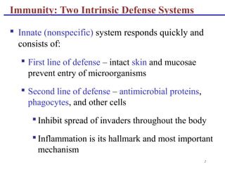
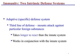
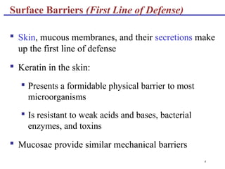
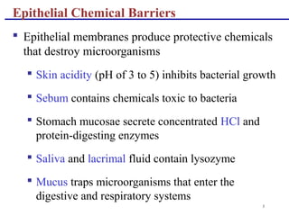
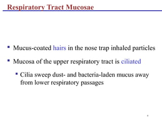
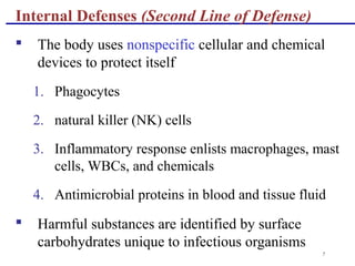
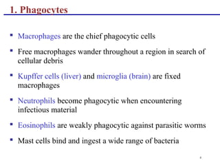
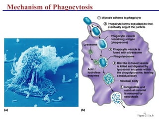
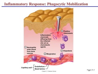
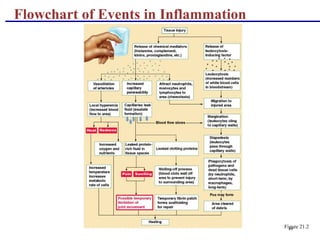
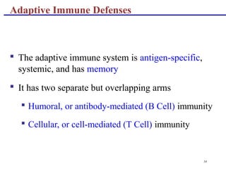
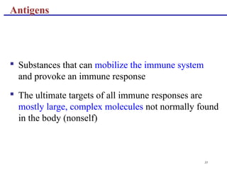
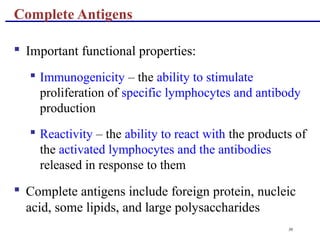
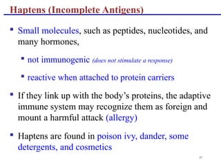
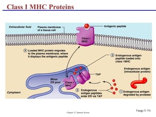
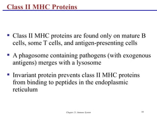
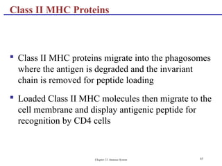
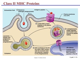
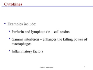
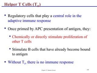
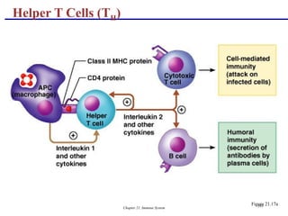
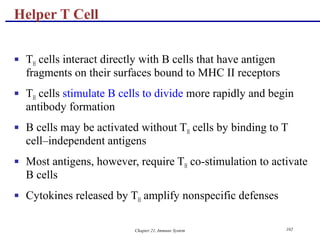
1
The Immune System:
Innate and Adaptive
Body Defenses
2
Immunity: Two Intrinsic Defense Systems
 Innate (nonspecific) system responds quickly and
consists of:
 First line of defense – intact skin and mucosae
prevent entry of microorganisms
 Second line of defense – antimicrobial proteins,
phagocytes, and other cells
 Inhibit spread of invaders throughout the body
 Inflammation is its hallmark and most important
mechanism
3
Immunity: Two Intrinsic Defense Systems
 Adaptive (specific) defense system
 Third line of defense – mounts attack against
particular foreign substances
 Takes longer to react than the innate system
 Works in conjunction with the innate system
4
Surface Barriers (First Line of Defense)
 Skin, mucous membranes, and their secretions make
up the first line of defense
 Keratin in the skin:
 Presents a formidable physical barrier to most
microorganisms
 Is resistant to weak acids and bases, bacterial
enzymes, and toxins
 Mucosae provide similar mechanical barriers
5
Epithelial Chemical Barriers
 Epithelial membranes produce protective chemicals
that destroy microorganisms
 Skin acidity (pH of 3 to 5) inhibits bacterial growth
 Sebum contains chemicals toxic to bacteria
 Stomach mucosae secrete concentrated HCl and
protein-digesting enzymes
 Saliva and lacrimal fluid contain lysozyme
 Mucus traps microorganisms that enter the
digestive and respiratory systems
6
Respiratory Tract Mucosae
 Mucus-coated hairs in the nose trap inhaled particles
 Mucosa of the upper respiratory tract is ciliated
 Cilia sweep dust- and bacteria-laden mucus away
from lower respiratory passages
7
Internal Defenses (Second Line of Defense)
 The body uses nonspecific cellular and chemical
devices to protect itself
1. Phagocytes
2. natural killer (NK) cells
3. Inflammatory response enlists macrophages, mast
cells, WBCs, and chemicals
4. Antimicrobial proteins in blood and tissue fluid
 Harmful substances are identified by surface
carbohydrates unique to infectious organisms
8
1. Phagocytes
 Macrophages are the chief phagocytic cells
 Free macrophages wander throughout a region in search of
cellular debris
 Kupffer cells (liver) and microglia (brain) are fixed
macrophages
 Neutrophils become phagocytic when encountering
infectious material
 Eosinophils are weakly phagocytic against parasitic worms
 Mast cells bind and ingest a wide range of bacteria
9
Mechanism of Phagocytosis
 Microbes adhere to the phagocyte
 Pseudopods engulf the particle (antigen) into a
phagosome
 Phagosomes fuse with a lysosome to form a
phagolysosome
 Invaders in the phagolysosome are digested by
proteolytic enzymes
 Indigestible and residual material is removed by
exocytosis
10
Mechanism of Phagocytosis
Figure 21.1a, b
11
2. Natural Killer (NK) Cells
 Cells that can lyse and kill cancer cells and virus-infected
cells
 Natural killer cells:
 Are a small, distinct group of large granular lymphocytes
 React nonspecifically and eliminate cancerous and virus-
infected cells
 Kill their target cells by releasing perforins and other
cytolytic chemicals
 Secrete potent chemicals that enhance the inflammatory
response
12
3. Inflammation: Tissue Response to Injury
 The inflammatory response is triggered whenever
body tissues are injured
 Prevents the spread of damaging agents to nearby
tissues
 Disposes of cell debris and pathogens
 Sets the stage for repair processes
 The four cardinal signs of acute inflammation are
redness, heat, swelling, and pain
13
Inflammation Response
 Begins with a flood of inflammatory chemicals
released into the extracellular fluid
 Inflammatory mediators (chemicals) :
 Include kinins, prostaglandins (PGs), complement,
and cytokines
 Are released by injured tissue, phagocytes,
lymphocytes, and mast cells
 Cause local small blood vessels to dilate, resulting
in hyperemia
14
Toll-like Receptors (TLRs)
 Macrophages and cells lining the gastrointestinal
and respiratory tracts bear TLRs
 TLRs recognize specific classes of infecting
microbes
 Activated TLRs trigger the release of cytokines that
promote inflammation
15
Inflammatory Response: Vascular
Permeability
 Chemicals liberated by the inflammatory response
increase the permeability of local capillaries
 Exudate (fluid containing proteins, clotting factors,
and antibodies):
 Seeps into tissue spaces causing local edema
(swelling), which contributes to the sensation of
pain
16
Inflammatory Response: Edema
 The surge of protein-rich fluids into tissue spaces
(edema):
 Helps to dilute harmful substances
 Brings in large quantities of oxygen and nutrients
needed for repair
 Allows entry of clotting proteins, which prevents
the spread of bacteria
17
 Occurs in four main phases:
 Leukocytosis – neutrophils are released from the
bone marrow in response to leukocytosis-inducing
factors released by injured cells
 Margination – neutrophils cling to the walls of
capillaries in the injured area
 Diapedesis – neutrophils squeeze through capillary
walls and begin phagocytosis
 Chemotaxis – inflammatory chemicals attract
neutrophils to the injury site
Inflammatory Response: Phagocytic Mobilization
Chapter 21, Immune System 18
Neutrophils
enter blood
from bone
marrow
1
2
3
4
Margination
Diapedesis
Positive
chemotaxis
Capillary wall
Endothelium
Basal lamina
Inflammatory
chemicals
diffusing from
the inflamed
site act as
chemotactic
agents
Inflammatory Response: Phagocytic Mobilization
Figure 21.3
19
Flowchart of Events in Inflammation
Figure 21.2
20
4. Antimicrobial Proteins
 Enhance the innate defenses by:
 Attacking microorganisms directly
 Hindering microorganisms’ ability to reproduce
 The most important antimicrobial proteins are:
 Interferon
 Complement proteins
21
 Genes that synthesize IFN are activated when a host
cell is invaded by a virus
 Interferon molecules leave the infected cell and
enter neighboring cells
 Interferon stimulates the neighboring cells to
activate genes for PKR (an antiviral protein)
 PKR nonspecifically blocks viral reproduction in
the neighboring cell
4 a. Interferon (IFN)
22
Interferon (IFN)
Figure 21.4
23
Interferon Family
 Interferons are a family of related proteins each with
slightly different physiological effects
 Lymphocytes secrete gamma (γ) interferon, but most
other WBCs secrete alpha (α) interferon
 Fibroblasts secrete beta (β) interferon
 Interferons also activate macrophages and mobilize
NKs
 FDA-approved alpha IFN is used:
 As an antiviral drug against hepatitis C virus
 To treat genital warts caused by the herpes virus
24
 20 or so proteins that circulate in the blood in an
inactive form
 Proteins include C1 through C9, factors B, D, and P,
and regulatory proteins
 Provides a major mechanism for destroying foreign
substances in the body
4 b. Complement
25
 Amplifies all aspects of the inflammatory response
 Kills bacteria and certain other cell types (our cells
are immune to complement)
 Enhances the effectiveness of both nonspecific and
specific defenses
Complement
26
 Complement can be activated by two pathways:
classical and alternative
 Classical pathway is linked to the immune system
 Depends on the binding of antibodies to invading
organisms
 Subsequent binding of C1 to the antigen-antibody
complexes (complement fixation)
 Alternative pathway is triggered by interaction
among factors B, D, and P, and polysaccharide
molecules present on microorganisms
Complement Pathways
27
 Each pathway involves a cascade in which complement
proteins are activated in an orderly sequence and where each
step catalyzes the next
 Both pathways converge on C3, which cleaves into C3a and
C3b
 C3b initiates formation of a membrane attack complex
(MAC)
 MAC causes cell lysis by interfering with a cell’s ability to
eject Ca2+
 C3b also causes opsonization, and C3a causes inflammation
Complement Pathways
28
Complement Pathways
Figure 21.5
29
 CRP is produced by the liver in response to
inflammatory molecules
 CRP is a clinical marker used to assess for:
 The presence of an acute infection
 An inflammatory condition and its response to
treatment
C-reactive Protein (CRP)
30
 Binds to PC receptor of pathogens and exposed self-
antigens
 Plays a surveillance role in targeting damaged cells
for disposal
 Activates complement
Functions of C-reactive Protein
31
 Abnormally high body temperature in response to
invading microorganisms
 The body’s thermostat is reset upwards in response
to pyrogens, chemicals secreted by leukocytes and
macrophages exposed to bacteria and other foreign
substances
Fever
32
 High fevers are dangerous as they can denature
enzymes
 Moderate fever can be beneficial, as it causes:
 The liver and spleen to sequester iron and zinc
(needed by microorganisms)
 An increase in the metabolic rate, which speeds up
tissue repair
Fever
33
 The adaptive immune system is a functional system
that:
 Recognizes specific foreign substances
 Acts to immobilize, neutralize, or destroy foreign
substances
 Amplifies inflammatory response and activates
complement
Adaptive (Specific) Defenses (Third Line of Defense)
34
 The adaptive immune system is antigen-specific,
systemic, and has memory
 It has two separate but overlapping arms
 Humoral, or antibody-mediated (B Cell) immunity
 Cellular, or cell-mediated (T Cell) immunity
Adaptive Immune Defenses
35
 Substances that can mobilize the immune system
and provoke an immune response
 The ultimate targets of all immune responses are
mostly large, complex molecules not normally found
in the body (nonself)
Antigens
36
 Important functional properties:
 Immunogenicity – the ability to stimulate
proliferation of specific lymphocytes and antibody
production
 Reactivity – the ability to react with the products of
the activated lymphocytes and the antibodies
released in response to them
 Complete antigens include foreign protein, nucleic
acid, some lipids, and large polysaccharides
Complete Antigens
37
 Small molecules, such as peptides, nucleotides, and
many hormones,
 not immunogenic (does not stimulate a response)
 reactive when attached to protein carriers
 If they link up with the body’s proteins, the adaptive
immune system may recognize them as foreign and
mount a harmful attack (allergy)
 Haptens are found in poison ivy, dander, some
detergents, and cosmetics
Haptens (Incomplete Antigens)
38
 Only certain parts of an entire antigen are
immunogenic
 Antibodies and activated lymphocytes bind to these
antigenic determinants
 Most naturally occurring antigens have numerous
antigenic determinants that:
 Mobilize several different lymphocyte populations
 Form different kinds of antibodies against it
 Large, chemically simple molecules (e.g., plastics)
have little or no immunogenicity
Antigenic Determinants
39
Antigenic Determinants
Figure 21.6
40
 Our cells are dotted with protein molecules (self-
antigens) that are not antigenic to us but are strongly
antigenic to others (reason for transplant rejection)
 One type of these, MHC proteins, mark a cell as self
 The two classes of MHC proteins are:
 Class I MHC proteins – found on virtually all body
cells
 Class II MHC proteins – found on certain cells in
the immune response
Self-Antigens: MHC Proteins
41
 Are coded for by genes of the major
histocompatibility complex (MHC) and are unique
to an individual
 Each MHC molecule has a deep groove that displays
a peptide, which is a normal cellular product of
protein recycling
 In infected cells, MHC proteins bind to fragments of
foreign antigens, which play a crucial role in
mobilizing the immune system
MHC Proteins
42
 Two types of lymphocytes
 B lymphocytes – oversee humoral immunity
 T lymphocytes – non-antibody-producing cells that
constitute the cell-mediated arm of immunity
 Antigen-presenting cells (APCs):
 Do not respond to specific antigens
 Play essential auxiliary roles in immunity
Cells of the Adaptive Immune System
43
 Immature lymphocytes released from bone marrow
are essentially identical
 Whether a lymphocyte matures into a B cell or a T
cell depends on where in the body it becomes
immunocompetent
 B cells mature in the bone marrow
 T cells mature in the thymus
Lymphocytes
44
T Cell Selection in the Thymus
Figure 21.7
45
 T cells mature in the thymus under negative and
positive selection pressures
 Negative selection – eliminates T cells that are
strongly anti-self
 Positive selection – selects T cells with a weak
response to self-antigens, which thus become both
immunocompetent and self-tolerant
T Cells
46
 B cells become immunocompetent and self-tolerant
in bone marrow
 Some self-reactive B cells are inactivated (anergy)
while others are killed
 Other B cells undergo receptor editing in which
there is a rearrangement of their receptors
B Cells
47
 Display a unique type of receptor that responds to a
distinct antigen
 Become immunocompetent before they encounter
antigens they may later attack
 Are exported to secondary lymphoid tissue where
encounters with antigens occur
 Mature into fully functional antigen-activated cells
upon binding with their recognized antigen
 It is genes, not antigens, that determine which
foreign substances our immune system will
recognize and resist
Immunocompetent B or T cells
48
Red
bone marrow
1
2
3
Immunocompetent,
but still naive,
lymphocyte
migrates via blood
Mature (antigen-activated)
immunocompetent lymphocytes
circulate continuously in the
bloodstream and lymph and
throughout the lymphoid organs of
the body.
Key: = Site of lymphocyte origin
= Site of development of immunocompetence
as B or T cells; primary lymphoid organs
= Site of antigen challenge and final
differentiation to activated B and T cellsImmature
lymphocytesCirculation in
blood
1
1 Lymphocytes destined to become T
cells migrate to the thymus and develop
immunocompetence there. B cells
develop immunocompetence in red
bone marrow.
Thymus
Bone
marrow
Lymph nodes,
spleen, and other
lymphoid tissues
2 2 After leaving the thymus or bone
marrow as naive immunocompetent
cells, lymphocytes “seed” the lymph
nodes, spleen, and other lymphoid
tissues where the antigen challenge
occurs.
3 3
Activated
immunocompetent
B and T cells
recirculate in blood
and lymph
Immunocompetent B or T cells
Figure 21.8
49
 Major roles in immunity are:
 To engulf foreign particles
 To present fragments of antigens on their own
surfaces, to be recognized by T cells
 Major APCs are dendritic cells (DCs), macrophages,
and activated B cells
 The major initiators of adaptive immunity are DCs,
which actively migrate to the lymph nodes and
secondary lymphoid organs and present antigens to
T and B cells
Antigen-Presenting Cells (APCs)
50
 Secrete soluble proteins that activate T cells
 Activated T cells in turn release chemicals that:
 Rev up the maturation and mobilization of DCs
 Prod macrophages to become activated
macrophages, which are insatiable phagocytes that
secrete bactericidal chemicals
Macrophages and Dendritic Cells
51
 Antigen challenge – first encounter between an
antigen and a naive immunocompetent cell
 Takes place in the spleen or other lymphoid organ
 If the lymphocyte is a B cell:
 The challenging antigen provokes a humoral
immune response
 Antibodies are produced against the challenger
Humoral Immunity Response
52
 Stimulated B cell growth forms clones bearing the
same antigen-specific receptors
 A naive, immunocompetent B cell is activated when
antigens bind to its surface receptors and cross-link
adjacent receptors
 Antigen binding is followed by receptor-mediated
endocytosis of the cross-linked antigen-receptor
complexes
 These activating events, plus T cell interactions,
trigger clonal selection
Clonal Selection
53
Clonal Selection
Figure 21.9
54
 Most clone cells become antibody-secreting plasma
cells
 Plasma cells secrete specific antibody at the rate of
2000 molecules per second
Fate of the Clones
55
 Secreted antibodies:
 Bind to free antigens
 Mark the antigens for destruction by specific or
nonspecific mechanisms
 Clones that do not become plasma cells become
memory cells that can mount an immediate response
to subsequent exposures of the same antigen
Fate of the Clones
Chapter 21, Immune System
56
 Primary immune response – cellular differentiation
and proliferation, which occurs on the first exposure
to a specific antigen
 Lag period: 3 to 6 days after antigen challenge
 Peak levels of plasma antibody are achieved in 10
days
 Antibody levels then decline
Immunological Memory
57
 Secondary immune response – re-exposure to the
same antigen
 Sensitized memory cells respond within hours
 Antibody levels peak in 2 to 3 days at much higher
levels than in the primary response
 Antibodies bind with greater affinity, and their
levels in the blood can remain high for weeks to
months
Immunological Memory
Chapter 21, Immune System 58
Primary and Secondary Humoral Responses
Figure 21.10
Chapter 21, Immune System 59
 B cells encounter antigens and produce antibodies
against them
 Naturally acquired – response to a bacterial or viral
infection
 Artificially acquired – response to a vaccine of
dead or attenuated pathogens
 Vaccines – spare us the symptoms of disease,
and their weakened antigens provide antigenic
determinants that are immunogenic and reactive
Active Humoral Immunity
Chapter 21, Immune System 60
 Differs from active immunity in the antibody source
and the degree of protection
 B cells are not challenged by antigens
 Immunological memory does not occur
 Protection ends when antigens naturally degrade in
the body
 Naturally acquired – from the mother to her fetus via
the placenta
 Artificially acquired – from the injection of serum,
such as gamma globulin
Passive Humoral Immunity
Chapter 21, Immune System 61
Types of Acquired Immunity
Figure 21.11
Chapter 21, Immune System 62
 Also called immunoglobulins
 Constitute the gamma globulin portion of blood
proteins
 Are soluble proteins secreted by activated B cells
and plasma cells in response to an antigen
 Are capable of binding specifically with that
antigen
 There are five classes of antibodies: IgD, IgM, IgG,
IgA, and IgE
Antibodies
Chapter 21, Immune System 63
 IgD – monomer attached to the surface of B cells, important
in B cell activation
 IgM – pentamer released by plasma cells during the primary
immune response
 IgG – monomer that is the most abundant and diverse
antibody in primary and secondary response; crosses the
placenta and confers passive immunity
 IgA – dimer that helps prevent attachment of pathogens to
epithelial cell surfaces
 IgE – monomer that binds to mast cells and basophils,
causing histamine release when activated
Classes of Antibodies
Chapter 21, Immune System 64
 Consists of four looping polypeptide chains linked
together with disulfide bonds
 Two identical heavy (H) chains and two identical
light (L) chains
 The four chains bound together form an antibody
monomer
 Each chain has a variable (V) region at one end and
a constant (C) region at the other
 Variable regions of the heavy and light chains
combine to form the antigen-binding site
Basic Antibody Structure
Chapter 21, Immune System 65
Basic Antibody Structure
Figure 21.12a, b
Chapter 21, Immune System 66
 Antibodies responding to different antigens have different V
regions but the C region is the same for all antibodies in a
given class
 C regions form the stem of the Y-shaped antibody and:
 Determine the class of the antibody
 Serve common functions in all antibodies
 Dictate the cells and chemicals that the antibody can bind to
 Determine how the antibody class will function in
elimination of antigens
Antibody Structure
Chapter 21, Immune System 67
 Plasma cells make over a billion different types of antibodies
 Each cell, however, only contains 100,000 genes that code
for these polypeptides
 To code for this many antibodies, somatic recombination
takes place
 Gene segments are shuffled and combined in different ways
by each B cell as it becomes immunocompetent
 Information of the newly assembled genes is expressed as B
cell receptors and as antibodies
Mechanisms of Antibody Diversity
Chapter 21, Immune System 68
 Random mixing of gene segments makes unique
antibody genes that:
 Code for H and L chains
 Account for part of the variability in antibodies
 V gene segments, called hypervariable regions,
mutate and increase antibody variation
 Plasma cells can switch H chains, making two or
more classes with the same V region
Antibody Diversity
Chapter 21, Immune System 69
 Antibodies themselves do not destroy antigen; they
inactivate and tag it for destruction
 All antibodies form an antigen-antibody (immune)
complex
 Defensive mechanisms used by antibodies are
neutralization, agglutination, precipitation, and
complement fixation
Antibody Targets
Chapter 21, Immune System 70
 Complement fixation is the main mechanism used against
cellular antigens
 Antibodies bound to cells change shape and expose
complement binding sites
 This triggers complement fixation and cell lysis
 Complement activation:
 Enhances the inflammatory response
 Uses a positive feedback cycle to promote phagocytosis
 Enlists more and more defensive elements
Complement Fixation and Activation
Chapter 21, Immune System 71
 Neutralization – antibodies bind to and block
specific sites on viruses or exotoxins, thus
preventing these antigens from binding to receptors
on tissue cells
Other Mechanisms of Antibody Action
Chapter 21, Immune System 72
 Agglutination – antibodies bind the same
determinant on more than one antigen
 Makes antigen-antibody complexes that are cross-
linked into large lattices
 Cell-bound antigens are cross-linked, causing
clumping (agglutination)
 Precipitation – soluble molecules are cross-linked
into large insoluble complexes
Other Mechanisms of Antibody Action
Chapter 21, Immune System 73
Mechanisms of Antibody Action
Figure 21.13
Chapter 21, Immune System 74
 Commercially prepared antibodies are used:
 To provide passive immunity
 In research, clinical testing, and treatment of certain
cancers
 Monoclonal antibodies are pure antibody
preparations
 Specific for a single antigenic determinant
 Produced from descendents of a single cell
Monoclonal Antibodies
Chapter 21, Immune System 75
 Hybridomas – cell hybrids made from a fusion of a
tumor cell and a B cell
 Have desirable properties of both parent cells –
indefinite proliferation as well as the ability to
produce a single type of antibody
Monoclonal Antibodies
Chapter 21, Immune System 76
 Since antibodies are useless against intracellular antigens,
cell-mediated immunity is needed
 Two major populations of T cells mediate cellular immunity
 CD4 cells (T4 cells) are primarily helper T cells (TH)
 CD8 cells (T8 cells) are cytotoxic T cells (TC) that destroy
cells harboring foreign antigens
 Other types of T cells are:
 Suppressor T cells (TS)
 Memory T cells
Cell-Mediated Immune Response
Chapter 21, Immune System 77
Major Types of T Cells
Figure 21.14
Chapter 21, Immune System 78
 Soluble antibodies
 The simplest ammunition of the immune response
 Interact in extracellular environments such as body
secretions, tissue fluid, blood, and lymph
Importance of Humoral Response
Chapter 21, Immune System 79
 T cells recognize and respond only to processed
fragments of antigen displayed on the surface of
body cells
 T cells are best suited for cell-to-cell interactions,
and target:
 Cells infected with viruses, bacteria, or intracellular
parasites
 Abnormal or cancerous cells
 Cells of infused or transplanted foreign tissue
Importance of Cellular Response
Chapter 21, Immune System 80
 Immunocompetent T cells are activated when the V
regions of their surface receptors bind to a
recognized antigen
 T cells must simultaneously recognize:
 Nonself (the antigen)
 Self (a MHC protein of a body cell)
Antigen Recognition and MHC Restriction
Chapter 21, Immune System 81
 Both types of MHC proteins are important to T cell
activation
 Class I MHC proteins
 Always recognized by CD8 T cells
 Display peptides from endogenous antigens
MHC Proteins
Chapter 21, Immune System 82
 Endogenous antigens are:
 Degraded by proteases and enter the endoplasmic
reticulum
 Transported via TAP (transporter associated with
antigen processing)
 Loaded onto class I MHC molecules
 Displayed on the cell surface in association with a
class I MHC molecule
Class I MHC Proteins
Chapter 21, Immune System 83
Class I MHC Proteins
Figure 21.15a
Chapter 21, Immune System 84
 Class II MHC proteins are found only on mature B
cells, some T cells, and antigen-presenting cells
 A phagosome containing pathogens (with exogenous
antigens) merges with a lysosome
 Invariant protein prevents class II MHC proteins
from binding to peptides in the endoplasmic
reticulum
Class II MHC Proteins
Chapter 21, Immune System 85
 Class II MHC proteins migrate into the phagosomes
where the antigen is degraded and the invariant
chain is removed for peptide loading
 Loaded Class II MHC molecules then migrate to the
cell membrane and display antigenic peptide for
recognition by CD4 cells
Class II MHC Proteins
Chapter 21, Immune System 86
Class II MHC Proteins
Figure 21.15b
Chapter 21, Immune System 87
 Provides the key for the immune system to
recognize the presence of intracellular
microorganisms
 MHC proteins are ignored by T cells if they are
complexed with self protein fragments
Antigen Recognition
Chapter 21, Immune System 88
 If MHC proteins are complexed with endogenous or
exogenous antigenic peptides, they:
 Indicate the presence of intracellular infectious
microorganisms
 Act as antigen holders
 Form the self part of the self-antiself complexes
recognized by T cells
Antigen Recognition
Chapter 21, Immune System 89
 T cell antigen receptors (TCRs):
 Bind to an antigen-MHC protein complex
 Have variable and constant regions consisting of
two chains (alpha and beta)
T Cell Activation: Step One – Antigen Binding
Chapter 21, Immune System 90
 MHC restriction – TH and TC bind to different classes
of MHC proteins
 TH cells bind to antigen linked to class II MHC
proteins
 Mobile APCs (Langerhans’ cells) quickly alert the
body to the presence of antigen by migrating to the
lymph nodes and presenting antigen
T Cell Activation: Step One – Antigen Binding
Chapter 21, Immune System 91
 TC cells are activated by antigen fragments
complexed with class I MHC proteins
 APCs produce co-stimulatory molecules that are
required for TC activation
 TCR that acts to recognize the self-antiself complex
is linked to multiple intracellular signaling pathways
 Other T cell surface proteins are involved in antigen
binding (e.g., CD4 and CD8 help maintain coupling
during antigen recognition)
T Cell Activation: Step One – Antigen Binding
Chapter 21, Immune System 92
T Cell Activation: Step One – Antigen Binding
Figure 21.16
Chapter 21, Immune System 93
 Before a T cell can undergo clonal expansion, it
must recognize one or more co-stimulatory signals
 This recognition may require binding to other
surface receptors on an APC
 Macrophages produce surface B7 proteins when
nonspecific defenses are mobilized
 B7 binding with the CD28 receptor on the surface of
T cells is a crucial co-stimulatory signal
 Other co-stimulatory signals include cytokines and
interleukin 1 and 2
T Cell Activation: Step Two – Co-stimulation
Chapter 21, Immune System 94
 Depending on receptor type, co-stimulators can
cause T cells to complete their activation or abort
activation
 Without co-stimulation, T cells:
 Become tolerant to that antigen
 Are unable to divide
 Do not secrete cytokines
T Cell Activation: Step Two – Co-stimulation
Chapter 21, Immune System 95
 T cells that are activated:
 Enlarge, proliferate, and form clones
 Differentiate and perform functions according to
their T cell class
T Cell Activation: Step Two – Co-stimulation
Chapter 21, Immune System 96
 Primary T cell response peaks within a week after
signal exposure
 T cells then undergo apoptosis between days 7 and
30
 Effector activity wanes as the amount of antigen
declines
 The disposal of activated effector cells is a
protective mechanism for the body
 Memory T cells remain and mediate secondary
responses to the same antigen
T Cell Activation: Step Two – Co-stimulation
Chapter 21, Immune System 97
 Mediators involved in cellular immunity, including
hormonelike glycoproteins released by activated T
cells and macrophages
 Some are co-stimulators of T cells and T cell
proliferation
 Interleukin 1 (IL-1) released by macrophages co-
stimulates bound T cells to:
 Release interleukin 2 (IL-2)
 Synthesize more IL-2 receptors
Cytokines
Chapter 21, Immune System 98
 IL-2 is a key growth factor, which sets up a positive
feedback cycle that encourages activated T cells to
divide
 It is used therapeutically to enhance the body’s
defenses against cancer
 Other cytokines amplify and regulate immune and
nonspecific responses
Cytokines
Chapter 21, Immune System 99
 Examples include:
 Perforin and lymphotoxin – cell toxins
 Gamma interferon – enhances the killing power of
macrophages
 Inflammatory factors
Cytokines
Chapter 21, Immune System 100
 Regulatory cells that play a central role in the
adaptive immune response
 Once primed by APC presentation of antigen, they:
 Chemically or directly stimulate proliferation of
other T cells
 Stimulate B cells that have already become bound
to antigen
 Without TH, there is no immune response
Helper T Cells (TH)
Chapter 21, Immune System 101
Helper T Cells (TH)
Figure 21.17a
Chapter 21, Immune System 102
 TH cells interact directly with B cells that have antigen
fragments on their surfaces bound to MHC II receptors
 TH cells stimulate B cells to divide more rapidly and begin
antibody formation
 B cells may be activated without TH cells by binding to T
cell–independent antigens
 Most antigens, however, require TH co-stimulation to activate
B cells
 Cytokines released by TH amplify nonspecific defenses
Helper T Cell
Chapter 21, Immune System 103
Helper T Cells
Figure 21.17b
Chapter 21, Immune System 104
 TC cells, or killer T cells, are the only T cells that can directly
attack and kill other cells
 They circulate throughout the body in search of body cells
that display the antigen to which they have been sensitized
 Their targets include:
 Virus-infected cells
 Cells with intracellular bacteria or parasites
 Cancer cells
 Foreign cells from blood transfusions or transplants
Cytotoxic T Cell (Tc)
Chapter 21, Immune System 105
 Bind to self-antiself complexes on all body cells
 Infected or abnormal cells can be destroyed as long
as appropriate antigen and co-stimulatory stimuli
(e.g., IL-2) are present
 Natural killer cells activate their killing machinery
when they bind to MICA receptor
 MICA receptor – MHC-related cell surface protein
in cancer cells, virus-infected cells, and cells of
transplanted organs
Cytotoxic T Cells
Chapter 21, Immune System 106
 In some cases, TC cells:
 Bind to the target cell and release perforin into its
membrane
 In the presence of Ca2+
perforin causes cell lysis
by creating transmembrane pores
 Other TC cells induce cell death by:
 Secreting lymphotoxin, which fragments the target
cell’s DNA
 Secreting gamma interferon, which stimulates
phagocytosis by macrophages
Mechanisms of Tc Action
Chapter 21, Immune System 107
Mechanisms of Tc Action
Figure 21.18a, b
Chapter 21, Immune System 108
 Suppressor T cells (TS) – regulatory cells that release
cytokines, which suppress the activity of both T cells
and B cells
 Gamma delta T cells (Tgd) – 10% of all T cells found
in the intestines that are triggered by binding to
MICA receptors
Other T Cells
Chapter 21, Immune System 109
Summary of the Primary Immune Response
Figure 21.19
Chapter 21, Immune System 110
 The four major types of grafts are:
 Autografts – graft transplanted from one site on the
body to another in the same person
 Isografts – grafts between identical twins
 Allografts – transplants between individuals that
are not identical twins, but belong to same species
 Xenografts – grafts taken from another animal
species
Organ Transplants
Chapter 21, Immune System 111
 Prevention of tissue rejection is accomplished by
using immunosuppressive drugs
 However, these drugs depress patient’s immune
system so it cannot fight off foreign agents
Prevention of Rejection
Chapter 21, Immune System 112
 Congenital or acquired conditions in which the function or
production of immune cells, phagocytes, or complement is
abnormal
Immunodeficiencies
Chapter 21, Immune System 113
Severe Combined Immunodeficiency (SCID)
 SCID – severe combined immunodeficiency (SCID)
syndromes; genetic defects that produce:
 A marked deficit in B and T cells
 Abnormalities in interleukin receptors
 Defective adenosine deaminase (ADA) enzyme
 Metabolites lethal to T cells accumulate
 SCID is fatal if untreated; treatment is with bone
marrow transplants
Chapter 21, Immune System 114
Severe Combined Immunodeficiency (SCID)
Chapter 21, Immune System 115
 Hodgkin’s disease – cancer of the lymph nodes leads
to immunodeficiency by depressing lymph node
cells
 Acquired immune deficiency syndrome (AIDS) –
cripples the immune system by interfering with the
activity of helper T (CD4) cells
 Characterized by severe weight loss, night sweats,
and swollen lymph nodes
 Opportunistic infections occur, including
pneumocystis pneumonia and Kaposi’s
sarcoma
Acquired Immunodeficiencies
Chapter 21, Immune System 116
 Caused by human immunodeficiency virus (HIV)
transmitted via body fluids – blood, semen, and vaginal
secretions
 HIV enters the body via:
 Blood transfusions
 Contaminated needles
 Intimate sexual contact, including oral sex
 HIV:
 Destroys TH cells
 Depresses cell-mediated immunity
AIDS
Chapter 21, Immune System 117
 HIV multiplies in lymph nodes throughout the
asymptomatic period
 Symptoms appear in a few months to 10 years
 Attachment
 HIV’s coat protein (gp120) attaches to the CD4
receptor
 A nearby protein (gp41) fuses the virus to the target
cell
AIDS
Chapter 21, Immune System 118
 HIV enters the cell and uses reverse transcriptase to
produce DNA from viral RNA
 This DNA (provirus) directs the host cell to make
viral RNA (and proteins), enabling the virus to
reproduce and infect other cells
AIDS
Chapter 21, Immune System 119
 HIV reverse transcriptase is not accurate and
produces frequent transcription errors
 This high mutation rate causes resistance to drugs
 Treatments include:
 Reverse transcriptase inhibitors (AZT)
 Protease inhibitors (saquinavir and ritonavir)
 New drugs currently being developed that block
HIV’s entry to helper T cells
AIDS
Chapter 21, Immune System 120
 Loss of the immune system’s ability to distinguish self from
nonself
 The body produces autoantibodies and sensitized TC cells
that destroy its own tissues
 Examples include:
 multiple sclerosis
 myasthenia gravis
 Graves’ disease
 Type I (juvenile) diabetes mellitus
 systemic lupus erythematosus (SLE)
 Glomerulonephritis
 rheumatoid arthritis
Autoimmune Diseases
Chapter 21, Immune System 121
 Ineffective lymphocyte programming – self-reactive
T and B cells that should have been eliminated in the
thymus and bone marrow escape into the circulation
 New self-antigens appear, generated by:
 Gene mutations that cause new proteins to appear
 Changes in self-antigens by hapten attachment or as
a result of infectious damage
Mechanisms of Autoimmune Diseases
Chapter 21, Immune System 122
 If the determinants on foreign antigens resemble
self-antigens:
 Antibodies made against foreign antigens cross-
react with self-antigens
Mechanisms of Autoimmune Diseases
Chapter 21, Immune System 123
 Immune responses that cause tissue damage
 Different types of hypersensitivity reactions are
distinguished by:
 Their time course
 Whether antibodies or T cells are the principle
immune elements involved
 Antibody-mediated allergies are immediate and
subacute hypersensitivities
 The most important cell-mediated allergic condition
is delayed hypersensitivity
Hypersensitivity
Chapter 21, Immune System 124
 Acute (type I) hypersensitivities begin in seconds
after contact with allergen
 Anaphylaxis – initial allergen contact is
asymptomatic but sensitizes the person
 Subsequent exposures to allergen cause:
 Release of histamine and inflammatory
chemicals
 Systemic or local responses
Immediate Hypersensitivity
Chapter 21, Immune System 125
 The mechanism involves IL-4 secreted by T cells
 IL-4 stimulates B cells to produce IgE
 IgE binds to mast cells and basophils causing them
to degranulate, resulting in a flood of histamine
release and inducing the inflammatory response
Immediate Hypersensitivity
Chapter 21, Immune System 126
Acute Allergic Response
Figure 21.20
Chapter 21, Immune System 127
 Reactions include runny nose, itching reddened skin,
and watery eyes
 If allergen is inhaled, asthmatic symptoms appear –
constriction of bronchioles and restricted airflow
 If allergen is ingested, cramping, vomiting, or
diarrhea occur
 Antihistamines counteract these effects
Anaphylaxis
Chapter 21, Immune System 128
 Response to allergen that directly enters the blood (e.g.,
insect bite, injection)
 Basophils and mast cells are enlisted throughout the body
 Systemic histamine releases may result in:
 Constriction of bronchioles
 Sudden vasodilation and fluid loss from the bloodstream
 Hypotensive shock and death
 Treatment – epinephrine is the drug of choice
Anaphylactic Shock
Chapter 21, Immune System 129
 Caused by IgM and IgG, and transferred via blood
plasma or serum
 Onset is slow (1–3 hours) after antigen exposure
 Duration is long lasting (10–15 hours)
 Cytotoxic (type II) reactions
 Antibodies bind to antigens on specific body cells,
stimulating phagocytosis and complement-mediated
lysis of the cellular antigens
 Example: mismatched blood transfusion reaction
Subacute Hypersensitivities
Chapter 21, Immune System 130
 Immune complex (type III) hypersensitivity
 Antigens are widely distributed through the body or
blood
 Insoluble antigen-antibody complexes form
 Complexes cannot be cleared from a particular area
of the body
 Intense inflammation, local cell lysis, and death
may result
 Example: systemic lupus erythematosus (SLE)
Subacute Hypersensitivities
Chapter 21, Immune System 131
 Onset is slow (1–3 days)
 Mediated by mechanisms involving delayed
hypersensitivity T cells and cytotoxic T cells
 Cytokines from activated TCare the mediators of the
inflammatory response
 Antihistamines are ineffective and corticosteroid
drugs are used to provide relief
Delayed Hypersensitivities (Type IV)
Chapter 21, Immune System 132
 Example: allergic contact dermatitis (e.g., poison
ivy)
 Involved in protective reactions against viruses,
bacteria, fungi, protozoa, cancer, and rejection of
foreign grafts or transplants
Delayed Hypersensitivities (Type IV)

The Immune system

  • 1.
    1 The Immune System: Innateand Adaptive Body Defenses
  • 2.
    2 Immunity: Two IntrinsicDefense Systems  Innate (nonspecific) system responds quickly and consists of:  First line of defense – intact skin and mucosae prevent entry of microorganisms  Second line of defense – antimicrobial proteins, phagocytes, and other cells  Inhibit spread of invaders throughout the body  Inflammation is its hallmark and most important mechanism
  • 3.
    3 Immunity: Two IntrinsicDefense Systems  Adaptive (specific) defense system  Third line of defense – mounts attack against particular foreign substances  Takes longer to react than the innate system  Works in conjunction with the innate system
  • 4.
    4 Surface Barriers (FirstLine of Defense)  Skin, mucous membranes, and their secretions make up the first line of defense  Keratin in the skin:  Presents a formidable physical barrier to most microorganisms  Is resistant to weak acids and bases, bacterial enzymes, and toxins  Mucosae provide similar mechanical barriers
  • 5.
    5 Epithelial Chemical Barriers Epithelial membranes produce protective chemicals that destroy microorganisms  Skin acidity (pH of 3 to 5) inhibits bacterial growth  Sebum contains chemicals toxic to bacteria  Stomach mucosae secrete concentrated HCl and protein-digesting enzymes  Saliva and lacrimal fluid contain lysozyme  Mucus traps microorganisms that enter the digestive and respiratory systems
  • 6.
    6 Respiratory Tract Mucosae Mucus-coated hairs in the nose trap inhaled particles  Mucosa of the upper respiratory tract is ciliated  Cilia sweep dust- and bacteria-laden mucus away from lower respiratory passages
  • 7.
    7 Internal Defenses (SecondLine of Defense)  The body uses nonspecific cellular and chemical devices to protect itself 1. Phagocytes 2. natural killer (NK) cells 3. Inflammatory response enlists macrophages, mast cells, WBCs, and chemicals 4. Antimicrobial proteins in blood and tissue fluid  Harmful substances are identified by surface carbohydrates unique to infectious organisms
  • 8.
    8 1. Phagocytes  Macrophagesare the chief phagocytic cells  Free macrophages wander throughout a region in search of cellular debris  Kupffer cells (liver) and microglia (brain) are fixed macrophages  Neutrophils become phagocytic when encountering infectious material  Eosinophils are weakly phagocytic against parasitic worms  Mast cells bind and ingest a wide range of bacteria
  • 9.
    9 Mechanism of Phagocytosis Microbes adhere to the phagocyte  Pseudopods engulf the particle (antigen) into a phagosome  Phagosomes fuse with a lysosome to form a phagolysosome  Invaders in the phagolysosome are digested by proteolytic enzymes  Indigestible and residual material is removed by exocytosis
  • 10.
  • 11.
    11 2. Natural Killer(NK) Cells  Cells that can lyse and kill cancer cells and virus-infected cells  Natural killer cells:  Are a small, distinct group of large granular lymphocytes  React nonspecifically and eliminate cancerous and virus- infected cells  Kill their target cells by releasing perforins and other cytolytic chemicals  Secrete potent chemicals that enhance the inflammatory response
  • 12.
    12 3. Inflammation: TissueResponse to Injury  The inflammatory response is triggered whenever body tissues are injured  Prevents the spread of damaging agents to nearby tissues  Disposes of cell debris and pathogens  Sets the stage for repair processes  The four cardinal signs of acute inflammation are redness, heat, swelling, and pain
  • 13.
    13 Inflammation Response  Beginswith a flood of inflammatory chemicals released into the extracellular fluid  Inflammatory mediators (chemicals) :  Include kinins, prostaglandins (PGs), complement, and cytokines  Are released by injured tissue, phagocytes, lymphocytes, and mast cells  Cause local small blood vessels to dilate, resulting in hyperemia
  • 14.
    14 Toll-like Receptors (TLRs) Macrophages and cells lining the gastrointestinal and respiratory tracts bear TLRs  TLRs recognize specific classes of infecting microbes  Activated TLRs trigger the release of cytokines that promote inflammation
  • 15.
    15 Inflammatory Response: Vascular Permeability Chemicals liberated by the inflammatory response increase the permeability of local capillaries  Exudate (fluid containing proteins, clotting factors, and antibodies):  Seeps into tissue spaces causing local edema (swelling), which contributes to the sensation of pain
  • 16.
    16 Inflammatory Response: Edema The surge of protein-rich fluids into tissue spaces (edema):  Helps to dilute harmful substances  Brings in large quantities of oxygen and nutrients needed for repair  Allows entry of clotting proteins, which prevents the spread of bacteria
  • 17.
    17  Occurs infour main phases:  Leukocytosis – neutrophils are released from the bone marrow in response to leukocytosis-inducing factors released by injured cells  Margination – neutrophils cling to the walls of capillaries in the injured area  Diapedesis – neutrophils squeeze through capillary walls and begin phagocytosis  Chemotaxis – inflammatory chemicals attract neutrophils to the injury site Inflammatory Response: Phagocytic Mobilization
  • 18.
    Chapter 21, ImmuneSystem 18 Neutrophils enter blood from bone marrow 1 2 3 4 Margination Diapedesis Positive chemotaxis Capillary wall Endothelium Basal lamina Inflammatory chemicals diffusing from the inflamed site act as chemotactic agents Inflammatory Response: Phagocytic Mobilization Figure 21.3
  • 19.
    19 Flowchart of Eventsin Inflammation Figure 21.2
  • 20.
    20 4. Antimicrobial Proteins Enhance the innate defenses by:  Attacking microorganisms directly  Hindering microorganisms’ ability to reproduce  The most important antimicrobial proteins are:  Interferon  Complement proteins
  • 21.
    21  Genes thatsynthesize IFN are activated when a host cell is invaded by a virus  Interferon molecules leave the infected cell and enter neighboring cells  Interferon stimulates the neighboring cells to activate genes for PKR (an antiviral protein)  PKR nonspecifically blocks viral reproduction in the neighboring cell 4 a. Interferon (IFN)
  • 22.
  • 23.
    23 Interferon Family  Interferonsare a family of related proteins each with slightly different physiological effects  Lymphocytes secrete gamma (γ) interferon, but most other WBCs secrete alpha (α) interferon  Fibroblasts secrete beta (β) interferon  Interferons also activate macrophages and mobilize NKs  FDA-approved alpha IFN is used:  As an antiviral drug against hepatitis C virus  To treat genital warts caused by the herpes virus
  • 24.
    24  20 orso proteins that circulate in the blood in an inactive form  Proteins include C1 through C9, factors B, D, and P, and regulatory proteins  Provides a major mechanism for destroying foreign substances in the body 4 b. Complement
  • 25.
    25  Amplifies allaspects of the inflammatory response  Kills bacteria and certain other cell types (our cells are immune to complement)  Enhances the effectiveness of both nonspecific and specific defenses Complement
  • 26.
    26  Complement canbe activated by two pathways: classical and alternative  Classical pathway is linked to the immune system  Depends on the binding of antibodies to invading organisms  Subsequent binding of C1 to the antigen-antibody complexes (complement fixation)  Alternative pathway is triggered by interaction among factors B, D, and P, and polysaccharide molecules present on microorganisms Complement Pathways
  • 27.
    27  Each pathwayinvolves a cascade in which complement proteins are activated in an orderly sequence and where each step catalyzes the next  Both pathways converge on C3, which cleaves into C3a and C3b  C3b initiates formation of a membrane attack complex (MAC)  MAC causes cell lysis by interfering with a cell’s ability to eject Ca2+  C3b also causes opsonization, and C3a causes inflammation Complement Pathways
  • 28.
  • 29.
    29  CRP isproduced by the liver in response to inflammatory molecules  CRP is a clinical marker used to assess for:  The presence of an acute infection  An inflammatory condition and its response to treatment C-reactive Protein (CRP)
  • 30.
    30  Binds toPC receptor of pathogens and exposed self- antigens  Plays a surveillance role in targeting damaged cells for disposal  Activates complement Functions of C-reactive Protein
  • 31.
    31  Abnormally highbody temperature in response to invading microorganisms  The body’s thermostat is reset upwards in response to pyrogens, chemicals secreted by leukocytes and macrophages exposed to bacteria and other foreign substances Fever
  • 32.
    32  High feversare dangerous as they can denature enzymes  Moderate fever can be beneficial, as it causes:  The liver and spleen to sequester iron and zinc (needed by microorganisms)  An increase in the metabolic rate, which speeds up tissue repair Fever
  • 33.
    33  The adaptiveimmune system is a functional system that:  Recognizes specific foreign substances  Acts to immobilize, neutralize, or destroy foreign substances  Amplifies inflammatory response and activates complement Adaptive (Specific) Defenses (Third Line of Defense)
  • 34.
    34  The adaptiveimmune system is antigen-specific, systemic, and has memory  It has two separate but overlapping arms  Humoral, or antibody-mediated (B Cell) immunity  Cellular, or cell-mediated (T Cell) immunity Adaptive Immune Defenses
  • 35.
    35  Substances thatcan mobilize the immune system and provoke an immune response  The ultimate targets of all immune responses are mostly large, complex molecules not normally found in the body (nonself) Antigens
  • 36.
    36  Important functionalproperties:  Immunogenicity – the ability to stimulate proliferation of specific lymphocytes and antibody production  Reactivity – the ability to react with the products of the activated lymphocytes and the antibodies released in response to them  Complete antigens include foreign protein, nucleic acid, some lipids, and large polysaccharides Complete Antigens
  • 37.
    37  Small molecules,such as peptides, nucleotides, and many hormones,  not immunogenic (does not stimulate a response)  reactive when attached to protein carriers  If they link up with the body’s proteins, the adaptive immune system may recognize them as foreign and mount a harmful attack (allergy)  Haptens are found in poison ivy, dander, some detergents, and cosmetics Haptens (Incomplete Antigens)
  • 38.
    38  Only certainparts of an entire antigen are immunogenic  Antibodies and activated lymphocytes bind to these antigenic determinants  Most naturally occurring antigens have numerous antigenic determinants that:  Mobilize several different lymphocyte populations  Form different kinds of antibodies against it  Large, chemically simple molecules (e.g., plastics) have little or no immunogenicity Antigenic Determinants
  • 39.
  • 40.
    40  Our cellsare dotted with protein molecules (self- antigens) that are not antigenic to us but are strongly antigenic to others (reason for transplant rejection)  One type of these, MHC proteins, mark a cell as self  The two classes of MHC proteins are:  Class I MHC proteins – found on virtually all body cells  Class II MHC proteins – found on certain cells in the immune response Self-Antigens: MHC Proteins
  • 41.
    41  Are codedfor by genes of the major histocompatibility complex (MHC) and are unique to an individual  Each MHC molecule has a deep groove that displays a peptide, which is a normal cellular product of protein recycling  In infected cells, MHC proteins bind to fragments of foreign antigens, which play a crucial role in mobilizing the immune system MHC Proteins
  • 42.
    42  Two typesof lymphocytes  B lymphocytes – oversee humoral immunity  T lymphocytes – non-antibody-producing cells that constitute the cell-mediated arm of immunity  Antigen-presenting cells (APCs):  Do not respond to specific antigens  Play essential auxiliary roles in immunity Cells of the Adaptive Immune System
  • 43.
    43  Immature lymphocytesreleased from bone marrow are essentially identical  Whether a lymphocyte matures into a B cell or a T cell depends on where in the body it becomes immunocompetent  B cells mature in the bone marrow  T cells mature in the thymus Lymphocytes
  • 44.
    44 T Cell Selectionin the Thymus Figure 21.7
  • 45.
    45  T cellsmature in the thymus under negative and positive selection pressures  Negative selection – eliminates T cells that are strongly anti-self  Positive selection – selects T cells with a weak response to self-antigens, which thus become both immunocompetent and self-tolerant T Cells
  • 46.
    46  B cellsbecome immunocompetent and self-tolerant in bone marrow  Some self-reactive B cells are inactivated (anergy) while others are killed  Other B cells undergo receptor editing in which there is a rearrangement of their receptors B Cells
  • 47.
    47  Display aunique type of receptor that responds to a distinct antigen  Become immunocompetent before they encounter antigens they may later attack  Are exported to secondary lymphoid tissue where encounters with antigens occur  Mature into fully functional antigen-activated cells upon binding with their recognized antigen  It is genes, not antigens, that determine which foreign substances our immune system will recognize and resist Immunocompetent B or T cells
  • 48.
    48 Red bone marrow 1 2 3 Immunocompetent, but stillnaive, lymphocyte migrates via blood Mature (antigen-activated) immunocompetent lymphocytes circulate continuously in the bloodstream and lymph and throughout the lymphoid organs of the body. Key: = Site of lymphocyte origin = Site of development of immunocompetence as B or T cells; primary lymphoid organs = Site of antigen challenge and final differentiation to activated B and T cellsImmature lymphocytesCirculation in blood 1 1 Lymphocytes destined to become T cells migrate to the thymus and develop immunocompetence there. B cells develop immunocompetence in red bone marrow. Thymus Bone marrow Lymph nodes, spleen, and other lymphoid tissues 2 2 After leaving the thymus or bone marrow as naive immunocompetent cells, lymphocytes “seed” the lymph nodes, spleen, and other lymphoid tissues where the antigen challenge occurs. 3 3 Activated immunocompetent B and T cells recirculate in blood and lymph Immunocompetent B or T cells Figure 21.8
  • 49.
    49  Major rolesin immunity are:  To engulf foreign particles  To present fragments of antigens on their own surfaces, to be recognized by T cells  Major APCs are dendritic cells (DCs), macrophages, and activated B cells  The major initiators of adaptive immunity are DCs, which actively migrate to the lymph nodes and secondary lymphoid organs and present antigens to T and B cells Antigen-Presenting Cells (APCs)
  • 50.
    50  Secrete solubleproteins that activate T cells  Activated T cells in turn release chemicals that:  Rev up the maturation and mobilization of DCs  Prod macrophages to become activated macrophages, which are insatiable phagocytes that secrete bactericidal chemicals Macrophages and Dendritic Cells
  • 51.
    51  Antigen challenge– first encounter between an antigen and a naive immunocompetent cell  Takes place in the spleen or other lymphoid organ  If the lymphocyte is a B cell:  The challenging antigen provokes a humoral immune response  Antibodies are produced against the challenger Humoral Immunity Response
  • 52.
    52  Stimulated Bcell growth forms clones bearing the same antigen-specific receptors  A naive, immunocompetent B cell is activated when antigens bind to its surface receptors and cross-link adjacent receptors  Antigen binding is followed by receptor-mediated endocytosis of the cross-linked antigen-receptor complexes  These activating events, plus T cell interactions, trigger clonal selection Clonal Selection
  • 53.
  • 54.
    54  Most clonecells become antibody-secreting plasma cells  Plasma cells secrete specific antibody at the rate of 2000 molecules per second Fate of the Clones
  • 55.
    55  Secreted antibodies: Bind to free antigens  Mark the antigens for destruction by specific or nonspecific mechanisms  Clones that do not become plasma cells become memory cells that can mount an immediate response to subsequent exposures of the same antigen Fate of the Clones
  • 56.
    Chapter 21, ImmuneSystem 56  Primary immune response – cellular differentiation and proliferation, which occurs on the first exposure to a specific antigen  Lag period: 3 to 6 days after antigen challenge  Peak levels of plasma antibody are achieved in 10 days  Antibody levels then decline Immunological Memory
  • 57.
    57  Secondary immuneresponse – re-exposure to the same antigen  Sensitized memory cells respond within hours  Antibody levels peak in 2 to 3 days at much higher levels than in the primary response  Antibodies bind with greater affinity, and their levels in the blood can remain high for weeks to months Immunological Memory
  • 58.
    Chapter 21, ImmuneSystem 58 Primary and Secondary Humoral Responses Figure 21.10
  • 59.
    Chapter 21, ImmuneSystem 59  B cells encounter antigens and produce antibodies against them  Naturally acquired – response to a bacterial or viral infection  Artificially acquired – response to a vaccine of dead or attenuated pathogens  Vaccines – spare us the symptoms of disease, and their weakened antigens provide antigenic determinants that are immunogenic and reactive Active Humoral Immunity
  • 60.
    Chapter 21, ImmuneSystem 60  Differs from active immunity in the antibody source and the degree of protection  B cells are not challenged by antigens  Immunological memory does not occur  Protection ends when antigens naturally degrade in the body  Naturally acquired – from the mother to her fetus via the placenta  Artificially acquired – from the injection of serum, such as gamma globulin Passive Humoral Immunity
  • 61.
    Chapter 21, ImmuneSystem 61 Types of Acquired Immunity Figure 21.11
  • 62.
    Chapter 21, ImmuneSystem 62  Also called immunoglobulins  Constitute the gamma globulin portion of blood proteins  Are soluble proteins secreted by activated B cells and plasma cells in response to an antigen  Are capable of binding specifically with that antigen  There are five classes of antibodies: IgD, IgM, IgG, IgA, and IgE Antibodies
  • 63.
    Chapter 21, ImmuneSystem 63  IgD – monomer attached to the surface of B cells, important in B cell activation  IgM – pentamer released by plasma cells during the primary immune response  IgG – monomer that is the most abundant and diverse antibody in primary and secondary response; crosses the placenta and confers passive immunity  IgA – dimer that helps prevent attachment of pathogens to epithelial cell surfaces  IgE – monomer that binds to mast cells and basophils, causing histamine release when activated Classes of Antibodies
  • 64.
    Chapter 21, ImmuneSystem 64  Consists of four looping polypeptide chains linked together with disulfide bonds  Two identical heavy (H) chains and two identical light (L) chains  The four chains bound together form an antibody monomer  Each chain has a variable (V) region at one end and a constant (C) region at the other  Variable regions of the heavy and light chains combine to form the antigen-binding site Basic Antibody Structure
  • 65.
    Chapter 21, ImmuneSystem 65 Basic Antibody Structure Figure 21.12a, b
  • 66.
    Chapter 21, ImmuneSystem 66  Antibodies responding to different antigens have different V regions but the C region is the same for all antibodies in a given class  C regions form the stem of the Y-shaped antibody and:  Determine the class of the antibody  Serve common functions in all antibodies  Dictate the cells and chemicals that the antibody can bind to  Determine how the antibody class will function in elimination of antigens Antibody Structure
  • 67.
    Chapter 21, ImmuneSystem 67  Plasma cells make over a billion different types of antibodies  Each cell, however, only contains 100,000 genes that code for these polypeptides  To code for this many antibodies, somatic recombination takes place  Gene segments are shuffled and combined in different ways by each B cell as it becomes immunocompetent  Information of the newly assembled genes is expressed as B cell receptors and as antibodies Mechanisms of Antibody Diversity
  • 68.
    Chapter 21, ImmuneSystem 68  Random mixing of gene segments makes unique antibody genes that:  Code for H and L chains  Account for part of the variability in antibodies  V gene segments, called hypervariable regions, mutate and increase antibody variation  Plasma cells can switch H chains, making two or more classes with the same V region Antibody Diversity
  • 69.
    Chapter 21, ImmuneSystem 69  Antibodies themselves do not destroy antigen; they inactivate and tag it for destruction  All antibodies form an antigen-antibody (immune) complex  Defensive mechanisms used by antibodies are neutralization, agglutination, precipitation, and complement fixation Antibody Targets
  • 70.
    Chapter 21, ImmuneSystem 70  Complement fixation is the main mechanism used against cellular antigens  Antibodies bound to cells change shape and expose complement binding sites  This triggers complement fixation and cell lysis  Complement activation:  Enhances the inflammatory response  Uses a positive feedback cycle to promote phagocytosis  Enlists more and more defensive elements Complement Fixation and Activation
  • 71.
    Chapter 21, ImmuneSystem 71  Neutralization – antibodies bind to and block specific sites on viruses or exotoxins, thus preventing these antigens from binding to receptors on tissue cells Other Mechanisms of Antibody Action
  • 72.
    Chapter 21, ImmuneSystem 72  Agglutination – antibodies bind the same determinant on more than one antigen  Makes antigen-antibody complexes that are cross- linked into large lattices  Cell-bound antigens are cross-linked, causing clumping (agglutination)  Precipitation – soluble molecules are cross-linked into large insoluble complexes Other Mechanisms of Antibody Action
  • 73.
    Chapter 21, ImmuneSystem 73 Mechanisms of Antibody Action Figure 21.13
  • 74.
    Chapter 21, ImmuneSystem 74  Commercially prepared antibodies are used:  To provide passive immunity  In research, clinical testing, and treatment of certain cancers  Monoclonal antibodies are pure antibody preparations  Specific for a single antigenic determinant  Produced from descendents of a single cell Monoclonal Antibodies
  • 75.
    Chapter 21, ImmuneSystem 75  Hybridomas – cell hybrids made from a fusion of a tumor cell and a B cell  Have desirable properties of both parent cells – indefinite proliferation as well as the ability to produce a single type of antibody Monoclonal Antibodies
  • 76.
    Chapter 21, ImmuneSystem 76  Since antibodies are useless against intracellular antigens, cell-mediated immunity is needed  Two major populations of T cells mediate cellular immunity  CD4 cells (T4 cells) are primarily helper T cells (TH)  CD8 cells (T8 cells) are cytotoxic T cells (TC) that destroy cells harboring foreign antigens  Other types of T cells are:  Suppressor T cells (TS)  Memory T cells Cell-Mediated Immune Response
  • 77.
    Chapter 21, ImmuneSystem 77 Major Types of T Cells Figure 21.14
  • 78.
    Chapter 21, ImmuneSystem 78  Soluble antibodies  The simplest ammunition of the immune response  Interact in extracellular environments such as body secretions, tissue fluid, blood, and lymph Importance of Humoral Response
  • 79.
    Chapter 21, ImmuneSystem 79  T cells recognize and respond only to processed fragments of antigen displayed on the surface of body cells  T cells are best suited for cell-to-cell interactions, and target:  Cells infected with viruses, bacteria, or intracellular parasites  Abnormal or cancerous cells  Cells of infused or transplanted foreign tissue Importance of Cellular Response
  • 80.
    Chapter 21, ImmuneSystem 80  Immunocompetent T cells are activated when the V regions of their surface receptors bind to a recognized antigen  T cells must simultaneously recognize:  Nonself (the antigen)  Self (a MHC protein of a body cell) Antigen Recognition and MHC Restriction
  • 81.
    Chapter 21, ImmuneSystem 81  Both types of MHC proteins are important to T cell activation  Class I MHC proteins  Always recognized by CD8 T cells  Display peptides from endogenous antigens MHC Proteins
  • 82.
    Chapter 21, ImmuneSystem 82  Endogenous antigens are:  Degraded by proteases and enter the endoplasmic reticulum  Transported via TAP (transporter associated with antigen processing)  Loaded onto class I MHC molecules  Displayed on the cell surface in association with a class I MHC molecule Class I MHC Proteins
  • 83.
    Chapter 21, ImmuneSystem 83 Class I MHC Proteins Figure 21.15a
  • 84.
    Chapter 21, ImmuneSystem 84  Class II MHC proteins are found only on mature B cells, some T cells, and antigen-presenting cells  A phagosome containing pathogens (with exogenous antigens) merges with a lysosome  Invariant protein prevents class II MHC proteins from binding to peptides in the endoplasmic reticulum Class II MHC Proteins
  • 85.
    Chapter 21, ImmuneSystem 85  Class II MHC proteins migrate into the phagosomes where the antigen is degraded and the invariant chain is removed for peptide loading  Loaded Class II MHC molecules then migrate to the cell membrane and display antigenic peptide for recognition by CD4 cells Class II MHC Proteins
  • 86.
    Chapter 21, ImmuneSystem 86 Class II MHC Proteins Figure 21.15b
  • 87.
    Chapter 21, ImmuneSystem 87  Provides the key for the immune system to recognize the presence of intracellular microorganisms  MHC proteins are ignored by T cells if they are complexed with self protein fragments Antigen Recognition
  • 88.
    Chapter 21, ImmuneSystem 88  If MHC proteins are complexed with endogenous or exogenous antigenic peptides, they:  Indicate the presence of intracellular infectious microorganisms  Act as antigen holders  Form the self part of the self-antiself complexes recognized by T cells Antigen Recognition
  • 89.
    Chapter 21, ImmuneSystem 89  T cell antigen receptors (TCRs):  Bind to an antigen-MHC protein complex  Have variable and constant regions consisting of two chains (alpha and beta) T Cell Activation: Step One – Antigen Binding
  • 90.
    Chapter 21, ImmuneSystem 90  MHC restriction – TH and TC bind to different classes of MHC proteins  TH cells bind to antigen linked to class II MHC proteins  Mobile APCs (Langerhans’ cells) quickly alert the body to the presence of antigen by migrating to the lymph nodes and presenting antigen T Cell Activation: Step One – Antigen Binding
  • 91.
    Chapter 21, ImmuneSystem 91  TC cells are activated by antigen fragments complexed with class I MHC proteins  APCs produce co-stimulatory molecules that are required for TC activation  TCR that acts to recognize the self-antiself complex is linked to multiple intracellular signaling pathways  Other T cell surface proteins are involved in antigen binding (e.g., CD4 and CD8 help maintain coupling during antigen recognition) T Cell Activation: Step One – Antigen Binding
  • 92.
    Chapter 21, ImmuneSystem 92 T Cell Activation: Step One – Antigen Binding Figure 21.16
  • 93.
    Chapter 21, ImmuneSystem 93  Before a T cell can undergo clonal expansion, it must recognize one or more co-stimulatory signals  This recognition may require binding to other surface receptors on an APC  Macrophages produce surface B7 proteins when nonspecific defenses are mobilized  B7 binding with the CD28 receptor on the surface of T cells is a crucial co-stimulatory signal  Other co-stimulatory signals include cytokines and interleukin 1 and 2 T Cell Activation: Step Two – Co-stimulation
  • 94.
    Chapter 21, ImmuneSystem 94  Depending on receptor type, co-stimulators can cause T cells to complete their activation or abort activation  Without co-stimulation, T cells:  Become tolerant to that antigen  Are unable to divide  Do not secrete cytokines T Cell Activation: Step Two – Co-stimulation
  • 95.
    Chapter 21, ImmuneSystem 95  T cells that are activated:  Enlarge, proliferate, and form clones  Differentiate and perform functions according to their T cell class T Cell Activation: Step Two – Co-stimulation
  • 96.
    Chapter 21, ImmuneSystem 96  Primary T cell response peaks within a week after signal exposure  T cells then undergo apoptosis between days 7 and 30  Effector activity wanes as the amount of antigen declines  The disposal of activated effector cells is a protective mechanism for the body  Memory T cells remain and mediate secondary responses to the same antigen T Cell Activation: Step Two – Co-stimulation
  • 97.
    Chapter 21, ImmuneSystem 97  Mediators involved in cellular immunity, including hormonelike glycoproteins released by activated T cells and macrophages  Some are co-stimulators of T cells and T cell proliferation  Interleukin 1 (IL-1) released by macrophages co- stimulates bound T cells to:  Release interleukin 2 (IL-2)  Synthesize more IL-2 receptors Cytokines
  • 98.
    Chapter 21, ImmuneSystem 98  IL-2 is a key growth factor, which sets up a positive feedback cycle that encourages activated T cells to divide  It is used therapeutically to enhance the body’s defenses against cancer  Other cytokines amplify and regulate immune and nonspecific responses Cytokines
  • 99.
    Chapter 21, ImmuneSystem 99  Examples include:  Perforin and lymphotoxin – cell toxins  Gamma interferon – enhances the killing power of macrophages  Inflammatory factors Cytokines
  • 100.
    Chapter 21, ImmuneSystem 100  Regulatory cells that play a central role in the adaptive immune response  Once primed by APC presentation of antigen, they:  Chemically or directly stimulate proliferation of other T cells  Stimulate B cells that have already become bound to antigen  Without TH, there is no immune response Helper T Cells (TH)
  • 101.
    Chapter 21, ImmuneSystem 101 Helper T Cells (TH) Figure 21.17a
  • 102.
    Chapter 21, ImmuneSystem 102  TH cells interact directly with B cells that have antigen fragments on their surfaces bound to MHC II receptors  TH cells stimulate B cells to divide more rapidly and begin antibody formation  B cells may be activated without TH cells by binding to T cell–independent antigens  Most antigens, however, require TH co-stimulation to activate B cells  Cytokines released by TH amplify nonspecific defenses Helper T Cell
  • 103.
    Chapter 21, ImmuneSystem 103 Helper T Cells Figure 21.17b
  • 104.
    Chapter 21, ImmuneSystem 104  TC cells, or killer T cells, are the only T cells that can directly attack and kill other cells  They circulate throughout the body in search of body cells that display the antigen to which they have been sensitized  Their targets include:  Virus-infected cells  Cells with intracellular bacteria or parasites  Cancer cells  Foreign cells from blood transfusions or transplants Cytotoxic T Cell (Tc)
  • 105.
    Chapter 21, ImmuneSystem 105  Bind to self-antiself complexes on all body cells  Infected or abnormal cells can be destroyed as long as appropriate antigen and co-stimulatory stimuli (e.g., IL-2) are present  Natural killer cells activate their killing machinery when they bind to MICA receptor  MICA receptor – MHC-related cell surface protein in cancer cells, virus-infected cells, and cells of transplanted organs Cytotoxic T Cells
  • 106.
    Chapter 21, ImmuneSystem 106  In some cases, TC cells:  Bind to the target cell and release perforin into its membrane  In the presence of Ca2+ perforin causes cell lysis by creating transmembrane pores  Other TC cells induce cell death by:  Secreting lymphotoxin, which fragments the target cell’s DNA  Secreting gamma interferon, which stimulates phagocytosis by macrophages Mechanisms of Tc Action
  • 107.
    Chapter 21, ImmuneSystem 107 Mechanisms of Tc Action Figure 21.18a, b
  • 108.
    Chapter 21, ImmuneSystem 108  Suppressor T cells (TS) – regulatory cells that release cytokines, which suppress the activity of both T cells and B cells  Gamma delta T cells (Tgd) – 10% of all T cells found in the intestines that are triggered by binding to MICA receptors Other T Cells
  • 109.
    Chapter 21, ImmuneSystem 109 Summary of the Primary Immune Response Figure 21.19
  • 110.
    Chapter 21, ImmuneSystem 110  The four major types of grafts are:  Autografts – graft transplanted from one site on the body to another in the same person  Isografts – grafts between identical twins  Allografts – transplants between individuals that are not identical twins, but belong to same species  Xenografts – grafts taken from another animal species Organ Transplants
  • 111.
    Chapter 21, ImmuneSystem 111  Prevention of tissue rejection is accomplished by using immunosuppressive drugs  However, these drugs depress patient’s immune system so it cannot fight off foreign agents Prevention of Rejection
  • 112.
    Chapter 21, ImmuneSystem 112  Congenital or acquired conditions in which the function or production of immune cells, phagocytes, or complement is abnormal Immunodeficiencies
  • 113.
    Chapter 21, ImmuneSystem 113 Severe Combined Immunodeficiency (SCID)  SCID – severe combined immunodeficiency (SCID) syndromes; genetic defects that produce:  A marked deficit in B and T cells  Abnormalities in interleukin receptors  Defective adenosine deaminase (ADA) enzyme  Metabolites lethal to T cells accumulate  SCID is fatal if untreated; treatment is with bone marrow transplants
  • 114.
    Chapter 21, ImmuneSystem 114 Severe Combined Immunodeficiency (SCID)
  • 115.
    Chapter 21, ImmuneSystem 115  Hodgkin’s disease – cancer of the lymph nodes leads to immunodeficiency by depressing lymph node cells  Acquired immune deficiency syndrome (AIDS) – cripples the immune system by interfering with the activity of helper T (CD4) cells  Characterized by severe weight loss, night sweats, and swollen lymph nodes  Opportunistic infections occur, including pneumocystis pneumonia and Kaposi’s sarcoma Acquired Immunodeficiencies
  • 116.
    Chapter 21, ImmuneSystem 116  Caused by human immunodeficiency virus (HIV) transmitted via body fluids – blood, semen, and vaginal secretions  HIV enters the body via:  Blood transfusions  Contaminated needles  Intimate sexual contact, including oral sex  HIV:  Destroys TH cells  Depresses cell-mediated immunity AIDS
  • 117.
    Chapter 21, ImmuneSystem 117  HIV multiplies in lymph nodes throughout the asymptomatic period  Symptoms appear in a few months to 10 years  Attachment  HIV’s coat protein (gp120) attaches to the CD4 receptor  A nearby protein (gp41) fuses the virus to the target cell AIDS
  • 118.
    Chapter 21, ImmuneSystem 118  HIV enters the cell and uses reverse transcriptase to produce DNA from viral RNA  This DNA (provirus) directs the host cell to make viral RNA (and proteins), enabling the virus to reproduce and infect other cells AIDS
  • 119.
    Chapter 21, ImmuneSystem 119  HIV reverse transcriptase is not accurate and produces frequent transcription errors  This high mutation rate causes resistance to drugs  Treatments include:  Reverse transcriptase inhibitors (AZT)  Protease inhibitors (saquinavir and ritonavir)  New drugs currently being developed that block HIV’s entry to helper T cells AIDS
  • 120.
    Chapter 21, ImmuneSystem 120  Loss of the immune system’s ability to distinguish self from nonself  The body produces autoantibodies and sensitized TC cells that destroy its own tissues  Examples include:  multiple sclerosis  myasthenia gravis  Graves’ disease  Type I (juvenile) diabetes mellitus  systemic lupus erythematosus (SLE)  Glomerulonephritis  rheumatoid arthritis Autoimmune Diseases
  • 121.
    Chapter 21, ImmuneSystem 121  Ineffective lymphocyte programming – self-reactive T and B cells that should have been eliminated in the thymus and bone marrow escape into the circulation  New self-antigens appear, generated by:  Gene mutations that cause new proteins to appear  Changes in self-antigens by hapten attachment or as a result of infectious damage Mechanisms of Autoimmune Diseases
  • 122.
    Chapter 21, ImmuneSystem 122  If the determinants on foreign antigens resemble self-antigens:  Antibodies made against foreign antigens cross- react with self-antigens Mechanisms of Autoimmune Diseases
  • 123.
    Chapter 21, ImmuneSystem 123  Immune responses that cause tissue damage  Different types of hypersensitivity reactions are distinguished by:  Their time course  Whether antibodies or T cells are the principle immune elements involved  Antibody-mediated allergies are immediate and subacute hypersensitivities  The most important cell-mediated allergic condition is delayed hypersensitivity Hypersensitivity
  • 124.
    Chapter 21, ImmuneSystem 124  Acute (type I) hypersensitivities begin in seconds after contact with allergen  Anaphylaxis – initial allergen contact is asymptomatic but sensitizes the person  Subsequent exposures to allergen cause:  Release of histamine and inflammatory chemicals  Systemic or local responses Immediate Hypersensitivity
  • 125.
    Chapter 21, ImmuneSystem 125  The mechanism involves IL-4 secreted by T cells  IL-4 stimulates B cells to produce IgE  IgE binds to mast cells and basophils causing them to degranulate, resulting in a flood of histamine release and inducing the inflammatory response Immediate Hypersensitivity
  • 126.
    Chapter 21, ImmuneSystem 126 Acute Allergic Response Figure 21.20
  • 127.
    Chapter 21, ImmuneSystem 127  Reactions include runny nose, itching reddened skin, and watery eyes  If allergen is inhaled, asthmatic symptoms appear – constriction of bronchioles and restricted airflow  If allergen is ingested, cramping, vomiting, or diarrhea occur  Antihistamines counteract these effects Anaphylaxis
  • 128.
    Chapter 21, ImmuneSystem 128  Response to allergen that directly enters the blood (e.g., insect bite, injection)  Basophils and mast cells are enlisted throughout the body  Systemic histamine releases may result in:  Constriction of bronchioles  Sudden vasodilation and fluid loss from the bloodstream  Hypotensive shock and death  Treatment – epinephrine is the drug of choice Anaphylactic Shock
  • 129.
    Chapter 21, ImmuneSystem 129  Caused by IgM and IgG, and transferred via blood plasma or serum  Onset is slow (1–3 hours) after antigen exposure  Duration is long lasting (10–15 hours)  Cytotoxic (type II) reactions  Antibodies bind to antigens on specific body cells, stimulating phagocytosis and complement-mediated lysis of the cellular antigens  Example: mismatched blood transfusion reaction Subacute Hypersensitivities
  • 130.
    Chapter 21, ImmuneSystem 130  Immune complex (type III) hypersensitivity  Antigens are widely distributed through the body or blood  Insoluble antigen-antibody complexes form  Complexes cannot be cleared from a particular area of the body  Intense inflammation, local cell lysis, and death may result  Example: systemic lupus erythematosus (SLE) Subacute Hypersensitivities
  • 131.
    Chapter 21, ImmuneSystem 131  Onset is slow (1–3 days)  Mediated by mechanisms involving delayed hypersensitivity T cells and cytotoxic T cells  Cytokines from activated TCare the mediators of the inflammatory response  Antihistamines are ineffective and corticosteroid drugs are used to provide relief Delayed Hypersensitivities (Type IV)
  • 132.
    Chapter 21, ImmuneSystem 132  Example: allergic contact dermatitis (e.g., poison ivy)  Involved in protective reactions against viruses, bacteria, fungi, protozoa, cancer, and rejection of foreign grafts or transplants Delayed Hypersensitivities (Type IV)