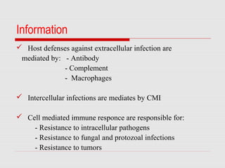
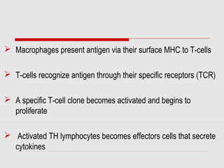
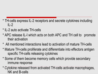
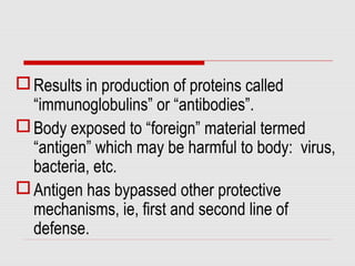
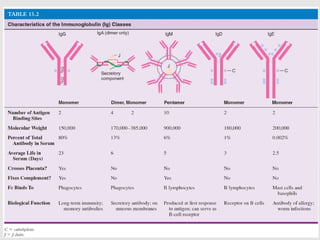
The immune response is how our body recognizes and defends itself against pathogens like bacteria, viruses, and substances that appear foreign and harmful.
4. Information
Host defenses against extracellular infection are
mediated by: - Antibody
- Complement
- Macrophages
Intercellular infections are mediates by CMI
Cell mediated immune responce are responsible for:
- Resistance to intracellular pathogens
- Resistance to fungal and protozoal infections
- Resistance to tumors
5. CMI may play a role in some harmful conditions:
Hypersensitivity reactions type IV (contact dermatitis)
Graft rejection
Autoimmune diseases
Cell mediated Cytotoxicity mediated by:
T-cytotoxic cells
Natural killer cells
Activated macrophages
6. Characters Of CMI
Cellular immune response is mediated by:
Subpopulation of T-lymphocytes
Macrophages and their products
7. Macrophages present antigen via their surface MHC to T-cells
T-cells recognize antigen through their specific receptors (TCR)
A specific T-cell clone becomes activated and begins to
proliferate
Activated TH lymphocytes becomes effectors cells that secrete
cytokines
8. Characters Of Cellular immune response
Cytokines stimulate other effectors cells of CMI and humoral immune
response and mediate the following:
- Attract monocytes, macrophages and lymphocytes to the site
- Activate macrophages to kill intracellular microbes
- Promote activity of CD8 CTLs which directly kill virus infected cells, tumour
cells, and graft rejection
- They activate NK cells increasing their cytotoxic functions
- Stimulate B-cells to differentiate into plasma cells that secret antibodies
9. Phases Of Cell Mediated Immunity
1) Antigen processing and presentation
Protein antigens processed and converted to
peptides then bind to MHC molecules on Antigen
Presenting Cell (APCs ) to be presented to T-
cells
10. 1) Antigen Processing and Presentation
a- Extracellular proteins are internalized into vesicular
compartment of APCs (Dentritic, macrophages,B-cells)
- They are degraded to generate peptides
- These peptides bind into class II MHC molecules
- Peptide-MHC II complex is transported to surface
of APCs to be presented to CD4 TH cells (T Helper cell)
Outcome:
Secretion of cytokines by TH cells
11. b- Endogenously synthesized proteins are degraded to peptides (all
nucleated cells e.g virus infected cells)
- They bind to class I MHC in endoplasmic reticulum
- Peptide-MHC I complex is expressed on surface
of nucleotide cells to be represented to CD8
cytotoxic cells
Outcome:
Killing of presenting cells by CTLs
13. * Mature CD4 and CD8 cells are activated by two signals:
- First signal is recognition of antigenic peptide-MHC complex
on surface of APC by TCR-CD3 complex
- CD4 and CD8 molecules are co-receptors that stabilize the
interaction of TH cells and TC-cells respectively with APCs
- CD3,CD4, and CD8 act as signal transduction molecules
- Second co-stimulatory signal is:
interaction of CD28 on T-cells with CD7 on APCs
14. * TH-cells express IL-2 receptors and secrete cytokines including
IL-2
* IL-2 auto activate TH-cells
* APC release IL-I which acts on both APC and TH cell to promote
their activation
* All mentioned interactions lead to activation of mature TH-cells
* Mature TH-cells proliferate and differentiate into effectors antigen
specific TH-cells releasing cytokines
* Some of them become memory cells which provide secondary
immune response
* Cytokine released from activated TH-cells activate macrophages,
NK and B-cells
16. * Activated CD8 TC-cells proliferate and differentiate into a
clone of effectors cells CTLs
* Effectors CTLs kill target cells
i.e. nucleated cells (expressing MHC-I) infected with
viruses, tumor cells or graft cells
17. Activation of Macrophages and Delayed Type
Hypersensitivity (DTH)
Activated TH cells (TH1) secrete IFN-γ which
activates macrophages and increase their ability to kill
ingested intracellular pathogens
18. The process of activation of macrophages, NK cell
and cytotoxic T-cells, infiltration and proliferation of
inflammatory cells, stimulated by cytokines released
from TH-cells (TH1) is important protective
mechanism against intracellular pathogen
19. Activated macrophages can also kill abnormal host cells
(abnormal or tumor cells)
Its Cytotoxicity is non specific and stimulated by TNF, nitric
oxide, enzymes and oxygen metabolites
If infection is not fully resolved, activated macrophages
cause tissue injury and fibrosis i.e. DTH reaction
21. Results in production of proteins called
“immunoglobulins” or “antibodies”.
Body exposed to “foreign” material termed
“antigen” which may be harmful to body: virus,
bacteria, etc.
Antigen has bypassed other protective
mechanisms, ie, first and second line of
defense.
22. Dynamics of Antibody Production
Primary immune response
Latent period
Gradual rise in antibody production taking days to
weeks
Plateau reached
Antibody level declines
23. Antibody production
Initial antibody produced in IgM
Lasts 10-12 days
Followed by production of IgG
Lasts 4-5 days
Without continued antigenic challenge
antibody levels drop off, although IgG
may continue to be produced.
24. Secondary Response
Second exposure to SAME antigen.
Memory cells Have memory of the antigen.
Recognition of antigen is immediate.
Results in immediate production of
protective antibody, mainly IgG but may see
some IgM
25.
26.
27. Cellular Events
Antigen is “processed” by T lymphocytes and
macrophages.
Possess special receptors on surface.
Termed “antigen presenter cell” APC.
Antigen presented to B cell
28. Basic Antibody Structure
Two identical heavy chains
Gamma
Delta
Alpha
Mu
Epsilon
Two identical light chains
Kappa OR
Lambda
29.
30. Papain Cleavage
Breaks disulfide bonds at hinge region
Results in 2 “fragment antigen binding” (Fab)
fragments.
Contains variable region of antibody molecule
Variable region is part of antibody molecule which
binds to antigen.
31. Pepsin
Breaks antibody above disulfide bond.
Two F(ab’)2 molecules
The rest fragments
Has the ability to bind with antigen and cause
agglutination or precipitation
32.
33. IgG
This monomer is the most abundant and diverse accounts
for about 80-85% of allantibodies.
These monomers can crosses the placenta and confers
passive immunity, as well as, blood vessels.
Monomers activate complement and increase
phagocytosis.
These monomers convey long term immunity
IgG protect against circulating bacteria and viruses, and
bacterial toxins.
34. IgM
This monomer comprises about 5-10% of the antibodies
in serum.
IgM are the first ones to appear in response to the initial
exposure to an antigen.
IgM have a pentamer structure.
They large size prevent IgM from moving about as
freely as IgG, therefore, they remain in the blood
vessels.
They fix complement
35. IgA
This monomer accounts for about 15% of the
antibodies in serum.
IgA can be monomers or dimers.
IgA monomers are found in salvia, sweat, breast
milk, and secretions of the G.I. tract.
They do not fix complement
The main function of IgA is to prevent the
attachment of pathogens to mucosal surfaces.
36. IgD
IgD atibodies comprise only about 0.2% of the total
serum antibodies.
IgD antibodies are found in blood and lymph and on
the surfaces of B cells.
They do not fix complement.
They help to initiate the immune response.
37. IgE
IgE antibodies comprise about 0.002% of the total
number of antibodies.
IgE molecules bind tightly by their Fc ends to
receptors on mast cells and basophils. This causes
the release of histamine.
They do not fix complement.
40. 1. Primary response – after first exposure to an
Ag immune system produces IgM and a
gradual increase in Ab titer.
2. Secondary response –after second contact
with the same Ag, immune system produces
a more rapid, stronger response due to
memory cells.