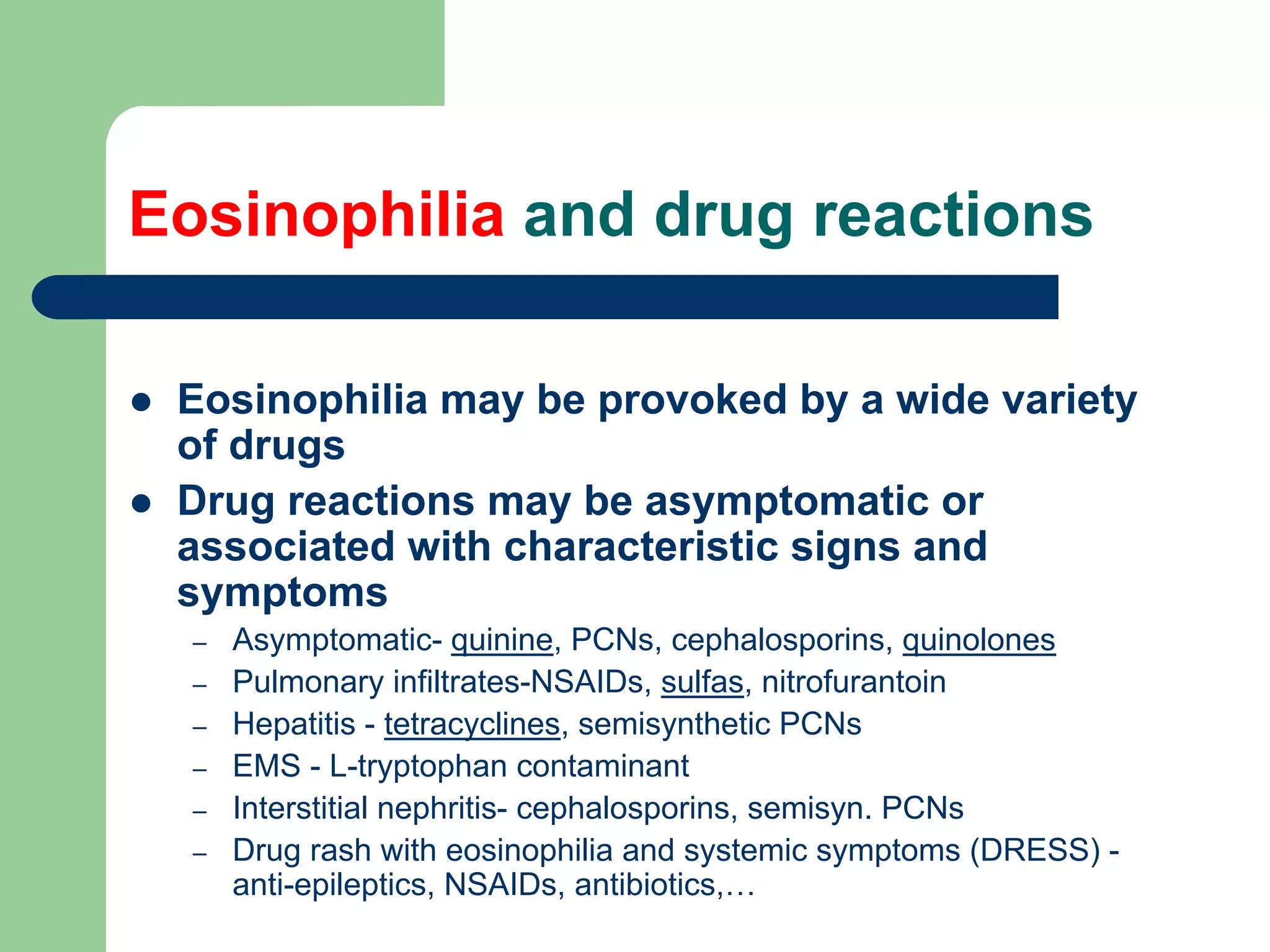
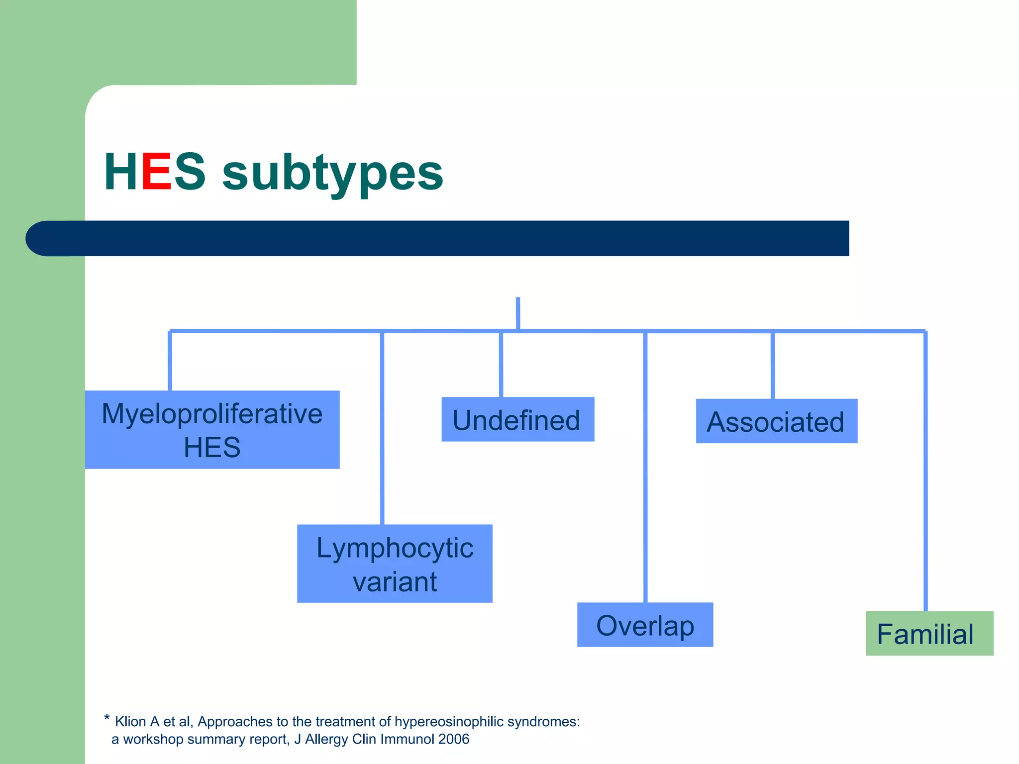
This document discusses eosinophils and eosinophilia. It begins with two case studies, one involving a 57-year-old man with a history of asthma and eosinophilia, and another involving a 28-year-old man diagnosed with hypereosinophilic syndrome after presenting with heart failure and marked eosinophilia. It then provides information on what eosinophils are, their role in the immune system, common causes of eosinophilia including allergies, parasites, and certain drugs. It discusses different subtypes of hypereosinophilic syndrome and their treatment options. It concludes by explaining how eosinophil activation can lead to tissue damage in some individuals with eosinophilia
We studied the review article about How I investigate eosinophilia, which was published in the International Journal of Laboratory Hematology in August 2018. This paper has clearly and simply introduced how clinicians investigate eosinophilia. Hopefully, it can be helpful to everyone who interested in this field.
This is a lecture on Lymphoma, exploring the different types and subtypes of Lymphomas. It also discusses the epidemiology, stages, clinical features, diagnosis, treatment and prognosis.
This was presented to undergraduate medical students at University Teaching Hospital (UTH), department of Cancer Disease Hospital by Nghitukuhamba Tangi Elikana Kalipi (6th year medical student) at Cavendish University Zambia, School of Medicine.
This Presentation of Hemolytic Anemia try to cover important Hemato-pathological aspects of Red cell membrane disorders ( Hereditary Spherocytosis, others ) , Enzymopathies ( G6PD deficieny, others ) and Hemoglobinopathies ( Thallasemia, SCA) and their differentiation. References includes Robbins pathology, Wintrobes atlas and text, and others
Chronic myelogenous leukemia (CML) - pluripotential stem cell disease
A malignancy the treatment of which has been revolutionised over the last decade.
Here is a comprehensive discussion on the disease
MULTIPLE MYELOMA -HEMATOLOGIC MALIGNANCY-DISEASE OF BONE MARROW - PLASMA CELL DISORDER
PATHOLOGY, .CLINICAL FEATURES , AND ITS MANAGMENT REFERENCE HARRISON
Hypersensitivity pneumonitis and pulmonary eosinophilia syndromesdocaneesh
an inflammatory disorder of the lung involving alveolar walls and terminal airways that is induced by repeated inhalation of a variety of organic agents in a susceptible host
INCONTINENTIA PIGMENTI: A CASE REPORT- serena gianfaldoniVR Foundation
INCONTINENTIA PIGMENTI: A CASE REPORT
Incontinentia Pigmenti is a rare X-linked genodermatosis, caused by mutations in the NEMO/IKKγ/IKBKG gene.
It is a systemic disease that involves tissue of ectodermic and mesodermic origin, including cutaneous tissue, teeth, eyes and the central nervous system, amongst other organs.
The name “incontinentia pigmenti” is related to the histological characteristics of the lesions during the third, pigmentary stage, of the disease. It is melanin incontinence by melanocytes in the basal epidermal layer and its presence in the superficial dermis.
Incontinentia pigmenti is really an uncommon disease.
IP occurs in approximately 1 in 10.000 of female newborns.
The disease is predominant in women: less than 3% of cases are male and derives by other genetical disorder, not completely understood.
Most cases have been described in white persons. Also other races (e.g. Korean, brasilian, cinese) are affected.
About 50% of the IP cases have a positive family history.
The clinical presentation of IP vary considerably even among family members. They range from subtle cutaneous and dental involvement to a complex syndrome, sometimes deadly.
Although IP affects many organ systems, the skin manifestations are the most common. Characteristic skin lesions evolve through four stages: 1)bullous stage; 2)verrucous stage; 3) pigmentary stage; 4) atretic stage. Tipically, the skin abnormalities occur along the Blaschko’s lines.
The hair is usually normal but sometimes we describe hair alteration. Alopecia may occur on the scalp and also on the trunk and extremities. Hair may be also thin or lusteriess, wiry and coarse (“woolly hair”). About 40% of patients have nail abnormalities. The most common nail alterations are onychodystrophy, onychogryphosis, pitting, yellow discoloration. Subungeal and periungueal keratotic tumors may appear at the later stage.
Extracutaneous manifestation can also been documented in patients with IP (dental abnormalities, ocular defects, neurological disorders, abnormalities of the musculoskeletal or cardiovascular system).
The diagnosis is mainly clinical. Family history consistent with X-linked inheritance or a history of multiple miscarriages also supports the diagnosis. Peripheral eosinophilia is a suggestive sign in early diagnosis. The histological examination of a skin biopsy could confirm the diagnosis. Also the molecular genetic testing (NEMO mutation) and the X-chromosome inactivation studies are important to confirm the diagnosis.
The prognosis is generally good and depends on extracutaneous manifestations.
Women with IP have a higher than usual risk of pregnancy loss, presumably related to low viability of male fetuses.
Because IP is a systemic disorder, a multidisciplinary approach to management is crucial.
Spontaneous improvement and resolution of skin lesions is general the rule. - Disclaimer-
This PPT is loaded as student material "as is", from the VRF Vitiligo Master Class Barcelona
EOSINOPHILS are granulocyte type of WBCs.
WHITE BLOOD CELLS –
There are five types of white blood cell (leucocyte). These are divided into two main classes
Granulocytes (includes Neutrophils, Eosinophils and Basophils)
Agranulocytes (includes Lymphocytes and Monocytes).
This classification depends on whether granules can be distinguished in their cytoplasm using a light microscope and conventional staining methods).
Hemophagocytic lymphohistiocytosis (hlh), Langerhans cell histiocytosis dr vi...Vijitha A S
Hemophagocytic lymphohistiocytosis (hlh)
Langerhans cell histiocytosis,Benign proliferation of mature histiocytes and uncontrolled phagocytosis of the platelet, erythrocytes, lymphocytes, and their hematopoietic precursors in the bonemarrow & other tissues
Basavarajeeyam is an important text for ayurvedic physician belonging to andhra pradehs. It is a popular compendium in various parts of our country as well as in andhra pradesh. The content of the text was presented in sanskrit and telugu language (Bilingual). One of the most famous book in ayurvedic pharmaceutics and therapeutics. This book contains 25 chapters called as prakaranas. Many rasaoushadis were explained, pioneer of dhatu druti, nadi pareeksha, mutra pareeksha etc. Belongs to the period of 15-16 century. New diseases like upadamsha, phiranga rogas are explained.
263778731218 Abortion Clinic /Pills In Harare ,sisternakatoto
263778731218 Abortion Clinic /Pills In Harare ,ABORTION WOMEN’S CLINIC +27730423979 IN women clinic we believe that every woman should be able to make choices in her pregnancy. Our job is to provide compassionate care, safety,affordable and confidential services. That’s why we have won the trust from all generations of women all over the world. we use non surgical method(Abortion pills) to terminate…Dr.LISA +27730423979women Clinic is committed to providing the highest quality of obstetrical and gynecological care to women of all ages. Our dedicated staff aim to treat each patient and her health concerns with compassion and respect.Our dedicated group ABORTION WOMEN’S CLINIC +27730423979 IN women clinic we believe that every woman should be able to make choices in her pregnancy. Our job is to provide compassionate care, safety,affordable and confidential services. That’s why we have won the trust from all generations of women all over the world. we use non surgical method(Abortion pills) to terminate…Dr.LISA +27730423979women Clinic is committed to providing the highest quality of obstetrical and gynecological care to women of all ages. Our dedicated staff aim to treat each patient and her health concerns with compassion and respect.Our dedicated group of receptionists, nurses, and physicians have worked together as a teamof receptionists, nurses, and physicians have worked together as a team wwww.lisywomensclinic.co.za/
micro teaching on communication m.sc nursing.pdfAnurag Sharma
Microteaching is a unique model of practice teaching. It is a viable instrument for the. desired change in the teaching behavior or the behavior potential which, in specified types of real. classroom situations, tends to facilitate the achievement of specified types of objectives.
Title: Sense of Smell
Presenter: Dr. Faiza, Assistant Professor of Physiology
Qualifications:
MBBS (Best Graduate, AIMC Lahore)
FCPS Physiology
ICMT, CHPE, DHPE (STMU)
MPH (GC University, Faisalabad)
MBA (Virtual University of Pakistan)
Learning Objectives:
Describe the primary categories of smells and the concept of odor blindness.
Explain the structure and location of the olfactory membrane and mucosa, including the types and roles of cells involved in olfaction.
Describe the pathway and mechanisms of olfactory signal transmission from the olfactory receptors to the brain.
Illustrate the biochemical cascade triggered by odorant binding to olfactory receptors, including the role of G-proteins and second messengers in generating an action potential.
Identify different types of olfactory disorders such as anosmia, hyposmia, hyperosmia, and dysosmia, including their potential causes.
Key Topics:
Olfactory Genes:
3% of the human genome accounts for olfactory genes.
400 genes for odorant receptors.
Olfactory Membrane:
Located in the superior part of the nasal cavity.
Medially: Folds downward along the superior septum.
Laterally: Folds over the superior turbinate and upper surface of the middle turbinate.
Total surface area: 5-10 square centimeters.
Olfactory Mucosa:
Olfactory Cells: Bipolar nerve cells derived from the CNS (100 million), with 4-25 olfactory cilia per cell.
Sustentacular Cells: Produce mucus and maintain ionic and molecular environment.
Basal Cells: Replace worn-out olfactory cells with an average lifespan of 1-2 months.
Bowman’s Gland: Secretes mucus.
Stimulation of Olfactory Cells:
Odorant dissolves in mucus and attaches to receptors on olfactory cilia.
Involves a cascade effect through G-proteins and second messengers, leading to depolarization and action potential generation in the olfactory nerve.
Quality of a Good Odorant:
Small (3-20 Carbon atoms), volatile, water-soluble, and lipid-soluble.
Facilitated by odorant-binding proteins in mucus.
Membrane Potential and Action Potential:
Resting membrane potential: -55mV.
Action potential frequency in the olfactory nerve increases with odorant strength.
Adaptation Towards the Sense of Smell:
Rapid adaptation within the first second, with further slow adaptation.
Psychological adaptation greater than receptor adaptation, involving feedback inhibition from the central nervous system.
Primary Sensations of Smell:
Camphoraceous, Musky, Floral, Pepperminty, Ethereal, Pungent, Putrid.
Odor Detection Threshold:
Examples: Hydrogen sulfide (0.0005 ppm), Methyl-mercaptan (0.002 ppm).
Some toxic substances are odorless at lethal concentrations.
Characteristics of Smell:
Odor blindness for single substances due to lack of appropriate receptor protein.
Behavioral and emotional influences of smell.
Transmission of Olfactory Signals:
From olfactory cells to glomeruli in the olfactory bulb, involving lateral inhibition.
Primitive, less old, and new olfactory systems with different path
CDSCO and Phamacovigilance {Regulatory body in India}NEHA GUPTA
The Central Drugs Standard Control Organization (CDSCO) is India's national regulatory body for pharmaceuticals and medical devices. Operating under the Directorate General of Health Services, Ministry of Health & Family Welfare, Government of India, the CDSCO is responsible for approving new drugs, conducting clinical trials, setting standards for drugs, controlling the quality of imported drugs, and coordinating the activities of State Drug Control Organizations by providing expert advice.
Pharmacovigilance, on the other hand, is the science and activities related to the detection, assessment, understanding, and prevention of adverse effects or any other drug-related problems. The primary aim of pharmacovigilance is to ensure the safety and efficacy of medicines, thereby protecting public health.
In India, pharmacovigilance activities are monitored by the Pharmacovigilance Programme of India (PvPI), which works closely with CDSCO to collect, analyze, and act upon data regarding adverse drug reactions (ADRs). Together, they play a critical role in ensuring that the benefits of drugs outweigh their risks, maintaining high standards of patient safety, and promoting the rational use of medicines.
These lecture slides, by Dr Sidra Arshad, offer a quick overview of the physiological basis of a normal electrocardiogram.
Learning objectives:
1. Define an electrocardiogram (ECG) and electrocardiography
2. Describe how dipoles generated by the heart produce the waveforms of the ECG
3. Describe the components of a normal electrocardiogram of a typical bipolar lead (limb II)
4. Differentiate between intervals and segments
5. Enlist some common indications for obtaining an ECG
6. Describe the flow of current around the heart during the cardiac cycle
7. Discuss the placement and polarity of the leads of electrocardiograph
8. Describe the normal electrocardiograms recorded from the limb leads and explain the physiological basis of the different records that are obtained
9. Define mean electrical vector (axis) of the heart and give the normal range
10. Define the mean QRS vector
11. Describe the axes of leads (hexagonal reference system)
12. Comprehend the vectorial analysis of the normal ECG
13. Determine the mean electrical axis of the ventricular QRS and appreciate the mean axis deviation
14. Explain the concepts of current of injury, J point, and their significance
Study Resources:
1. Chapter 11, Guyton and Hall Textbook of Medical Physiology, 14th edition
2. Chapter 9, Human Physiology - From Cells to Systems, Lauralee Sherwood, 9th edition
3. Chapter 29, Ganong’s Review of Medical Physiology, 26th edition
4. Electrocardiogram, StatPearls - https://www.ncbi.nlm.nih.gov/books/NBK549803/
5. ECG in Medical Practice by ABM Abdullah, 4th edition
6. Chapter 3, Cardiology Explained, https://www.ncbi.nlm.nih.gov/books/NBK2214/
7. ECG Basics, http://www.nataliescasebook.com/tag/e-c-g-basics
Local Advanced Lung Cancer: Artificial Intelligence, Synergetics, Complex Sys...Oleg Kshivets
Overall life span (LS) was 1671.7±1721.6 days and cumulative 5YS reached 62.4%, 10 years – 50.4%, 20 years – 44.6%. 94 LCP lived more than 5 years without cancer (LS=2958.6±1723.6 days), 22 – more than 10 years (LS=5571±1841.8 days). 67 LCP died because of LC (LS=471.9±344 days). AT significantly improved 5YS (68% vs. 53.7%) (P=0.028 by log-rank test). Cox modeling displayed that 5YS of LCP significantly depended on: N0-N12, T3-4, blood cell circuit, cell ratio factors (ratio between cancer cells-CC and blood cells subpopulations), LC cell dynamics, recalcification time, heparin tolerance, prothrombin index, protein, AT, procedure type (P=0.000-0.031). Neural networks, genetic algorithm selection and bootstrap simulation revealed relationships between 5YS and N0-12 (rank=1), thrombocytes/CC (rank=2), segmented neutrophils/CC (3), eosinophils/CC (4), erythrocytes/CC (5), healthy cells/CC (6), lymphocytes/CC (7), stick neutrophils/CC (8), leucocytes/CC (9), monocytes/CC (10). Correct prediction of 5YS was 100% by neural networks computing (error=0.000; area under ROC curve=1.0).
Recomendações da OMS sobre cuidados maternos e neonatais para uma experiência pós-natal positiva.
Em consonância com os ODS – Objetivos do Desenvolvimento Sustentável e a Estratégia Global para a Saúde das Mulheres, Crianças e Adolescentes, e aplicando uma abordagem baseada nos direitos humanos, os esforços de cuidados pós-natais devem expandir-se para além da cobertura e da simples sobrevivência, de modo a incluir cuidados de qualidade.
Estas diretrizes visam melhorar a qualidade dos cuidados pós-natais essenciais e de rotina prestados às mulheres e aos recém-nascidos, com o objetivo final de melhorar a saúde e o bem-estar materno e neonatal.
Uma “experiência pós-natal positiva” é um resultado importante para todas as mulheres que dão à luz e para os seus recém-nascidos, estabelecendo as bases para a melhoria da saúde e do bem-estar a curto e longo prazo. Uma experiência pós-natal positiva é definida como aquela em que as mulheres, pessoas que gestam, os recém-nascidos, os casais, os pais, os cuidadores e as famílias recebem informação consistente, garantia e apoio de profissionais de saúde motivados; e onde um sistema de saúde flexível e com recursos reconheça as necessidades das mulheres e dos bebês e respeite o seu contexto cultural.
Estas diretrizes consolidadas apresentam algumas recomendações novas e já bem fundamentadas sobre cuidados pós-natais de rotina para mulheres e neonatos que recebem cuidados no pós-parto em unidades de saúde ou na comunidade, independentemente dos recursos disponíveis.
É fornecido um conjunto abrangente de recomendações para cuidados durante o período puerperal, com ênfase nos cuidados essenciais que todas as mulheres e recém-nascidos devem receber, e com a devida atenção à qualidade dos cuidados; isto é, a entrega e a experiência do cuidado recebido. Estas diretrizes atualizam e ampliam as recomendações da OMS de 2014 sobre cuidados pós-natais da mãe e do recém-nascido e complementam as atuais diretrizes da OMS sobre a gestão de complicações pós-natais.
O estabelecimento da amamentação e o manejo das principais intercorrências é contemplada.
Recomendamos muito.
Vamos discutir essas recomendações no nosso curso de pós-graduação em Aleitamento no Instituto Ciclos.
Esta publicação só está disponível em inglês até o momento.
Prof. Marcus Renato de Carvalho
www.agostodourado.com
2. Disclosure
Employment
– National Institutes of Health
Financial Interests
– None
Research Interests
– GlaxoSmithKline, MedImmune
Organizational Interests
– Member of ASTMH, IES, IDSA
Gifts
– Nothing to Disclose
Other Interests
– Nothing to Disclose
3. Case 1
57 year old man with history of mild asthma,
eczema, allergic rhinitis and eosinophilia of >20
year’s duration
– wbc 10,000 - 40,000 with 30-40% eosinophils (4,000-
8,000/mm3)
– numerous extensive evaluations since 1982, including bone
marrow examinations, parasite serologies, rheumatologic
evaluation, CT scans, echocardiograms, PFTs,… reveal
only mild reversible obstructive pulmonary disease and mild
eczema
4. Case 2
28 year old man with no past medical history who
presented with signs and symptoms of severe
right-sided congestive heart failure in the setting
of marked eosinophilia
– wbc 14,400 with 63% eosinophils (9,072/mm3), aneia,
platelets 90k
– echocardiogram: dilated cardiomyopathy, fibrotic
material filling the right ventricle, moderate to severe
MR and TR, small pericardial effusion
– endomyocardial biopsy: focal fibrosis
– bone marrow: hypercellular with marked eosinophilia,
no blasts, mild fibrosis
5. Case 2 (continued)
A diagnosis of hypereosinophilic
syndrome was made and he was
treated with high dose steroids and
hydrea without response
Approximately 10 months after he
presented, imatinib mesylate therapy
was started with complete
hematologic remission
One month later he developed
hemoptysis, fever and chills and died
of presumed sepsis
6. Questions
What are eosinophils?
What do eosinophils do?
What are the causes of eosinophilia?
Why do some people develop problems
from eosinophilia and others not?
7. What are eosinophils?
Eosinophils are terminally differentiated cells of the
myeloid lineage that stain red with negatively
charged eosin
PMN
cytokines
IL3, GM-CSF, IL5
IL5
CD34+
HSC CMP GMP
No or low GATA-1
GATA-1, FOG,
GATA-1, PU.1,
PU.1, C/EBPα
C/EBPα
transcription
factors MΦ
8. Eosinophil life cycle
1-2% of peripheral
blood leukocytes; t1/2
in blood = 18 hours
>90% of eosinophils are
found in the tissues,
particularly those tissues
which interface with the
environment (BM, lymphoid
tissue, lower GI tract, and
uterus)
(from Rothenberg 1998 NEJM)
10. Questions
What are eosinophils?
What do eosinophils do?
What causes eosinophilia?
Why do some people develop problems
from eosinophilia and others not?
11. What we can learn from ?
there are no reported cases of a congenital
lack of eosinophils in humans, although
eosinophil-free mice have recently been
engineered (EPO-DTN mice, Lee et al. Science 2004; Δdbl
GATA Humbles et al. Science 2004)
eosinophil-free mice appear normal, but have
altered responses to allergen challenge
effect in helminth infection depends on model
schistosomiasis – no effect (Swartz et al. Blood 2006)
strongyloidiasis – impaired resistance to seocndary infection
(Knott et al. Int J Parasitol 2007)
12. What about humans?
– Host defense against helminth infection
– Anti-tumor effector cells
– Reproduction/mammary gland
development
– Wound repair and tissue remodeling
– MHC class I-restricted thymocyte
depletion
– Modulation of the immune response
Antigen presentation
Cytokine secretion
…..
13. Questions
What are eosinophils?
What do eosinophils do?
What causes eosinophilia?
Why do some people develop problems
from eosinophilia and others not?
14. Eosinophilia
Peripheral blood eosinophil count > 450/μl
Occurs in 5-10% of travelers
May be suppressed by:
– bacterial and viral infections
– fever
– corticosteroids
Diurnal variation
16. Eosinophilia and drug reactions
Eosinophilia may be provoked by a wide variety
of drugs
Drug reactions may be asymptomatic or
associated with characteristic signs and
symptoms
– Asymptomatic- quinine, PCNs, cephalosporins, quinolones
– Pulmonary infiltrates-NSAIDs, sulfas, nitrofurantoin
– Hepatitis - tetracyclines, semisynthetic PCNs
– EMS - L-tryptophan contaminant
– Interstitial nephritis- cephalosporins, semisyn. PCNs
– Drug rash with eosinophilia and systemic symptoms (DRESS) -
anti-epileptics, NSAIDs, antibiotics,…
17. Eosinophilia and nonparasitic
infectious diseases
Most acute bacterial and viral diseases
are associated with eosinopenia
Exceptions include:
– Bacteria: resolving scarlet fever, chronic
tuberculosis
– Fungi: coccidiomycosis, allergic
bronchopulmonary aspergillosis (APBA)
– Viruses: HIV
19. Eosinophilia and ectoparasites
Scabies
– sensitization to the mites and
their eggs causes itching,
erythema, rash, and may cause
eosinophilia
Myiasis
– Infestation by fly larvae has rarely
been associated with
hypereosinophilic syndrome
(images courtesy of CDC DPDx)
21. Eosinophilia of several years’
duration
Cysticercosis Flukes
Echinococcosis – schisto,
Filariasis fascioliasis,
– loiasis, clonochiasis,
onchocerciasis, paragonimiasis
mansonellosis, Hookworm
TPE
Strongyloidiasis
Gnathostomiasis
(image courtesy of CDC DPDx)
22. Unexplained eosinophilia
≥ 50% of cases of eosinophilia remain
unexplained despite exhaustive
diagnostic evaluation
Empiric anthelmintic therapy vs.
observation?
23. Hypereosinophilic syndromes
A group of heterogeneous disorders characterized
by:
– the presence of marked peripheral eosinophilia
>1500 eosinophils/mm3 on at least two occasions 6 months apart
– the lack of a secondary cause
ex. drug reaction, parasite infection, neoplasm
– evidence of end organ dysfunction
attributable to the eosinophilia or
otherwise unexplained in the clinical setting
25. Treatment of HES
Prednisone
Hydroxyurea
Interferon-α
Imatinib mesylate
(monoclonal anti-IL5 antibody)
Other agents for which there is data from case
reports and small series
–cyclosporin A, 2-CDA, vincristine,
chlorambucil, bone marrow transplant,
allogeneic stem cell transplant, alemtuzumab
26. HES subtypes
Myeloproliferative Undefined Associated
variant
Lymphocytic
variant
Overlap Familial
* Klion A et al, Approaches to the treatment of hypereosinophilic syndromes:
a workshop summary report, J Allergy Clin Immunol 2006
27. Myeloproliferative variant HES/CEL
FIP1L1/PDGFRA-associated CEL
Etiology unknown
– FIP1L1/PDGFRA-negative
– Clonal eosinophils OR > 4 of the following:
dysplastic eosinophils on peripheral smear
serum B12 >1000 pg/ml
serum tryptase > 12
anemia and/or thrombocytopenia
hepatosplenomegaly
bone marrow cellularity > 80%
spindle-shaped mast cells
myelofibrosis
Chronic eosinophilic leukemia
– Demonstrable cytogenetic abnormalities and/or increased
blasts
28. FIP1L1/PDGFRA-associated CEL
Caused by an interstitial deletion in chromosome 4
that leads to a constitutively activated fusion tyrosine
kinase
Almost exclusively in men, 20-40 years of age
50% 5 year fatality rate prior to the discovery of the
tyrosine kinase inhibitor, imatinib mesylate
29. Tissue fibrosis is a common
sequela of F/P-HES
F/P-HES HES*
(n=14) (n=29)
Endomyocardial 6/14 3/29
fibrosis (43%) (10%)
Restrictive 5/14 2/29
pulmonary (35%) (7%)
disease
Myelofibrosis 6/11 rare
(54%)
* one patient presented with liver
abnormalities; eosinophilia and bridging
fibrosis was seen on biopsy biopsy
30. HES subtypes
Myeloproliferative Undefined Associated
HES
Lymphocytic
variant
Overlap Familial
* Klion A et al, Approaches to the treatment of hypereosinophilic syndromes:
a workshop summary report, J Allergy Clin Immunol 2006
31. Lymphocytic variant HES
Associated with the presence of clonal
lymphocyte populations with aberrant
phenotypes (ex. CD3+CD4-CD8- and CD3-
CD4+) that produce eosinophilopoietic cytokines
Equally common in men and women
Predominance of skin manifestations
Often associated with elevated serum IgE, TARC
levels
May progress to lymphoma
No response to imatinib
32. HES subtypes
Myeloproliferative Undefined Associated
HES
Lymphocytic
variant
Overlap Familial
* Klion A et al, Approaches to the treatment of hypereosinophilic syndromes:
a workshop summary report, J Allergy Clin Immunol 2006
33. Overlap HES
eosinophilic disorders in which tissue
infiltration is restricted to a single organ
peripheral eosinophilia >1,500/mm3 may be
present
– ex. eosinophilic gastrointestinal disorders, chronic
eosinophilic pneumonia, eosinophilic cystitis,
Churg-Strauss vasculitis
may be difficult to distinguish from undefined
HES
34. HES subtypes
Myeloproliferative Undefined Associated
HES
Lymphocytic
variant
Overlap Familial
* Klion A et al, Approaches to the treatment of hypereosinophilic syndromes:
a workshop summary report, J Allergy Clin Immunol 2006
35. Associated HES
Recognized primary disorders associated with
peripheral eosinophilia >1,500/mm3 most
commonly as a result of immunodysregulation
Eosinophilia resolves with treatment of
underlying disorders
Complications of eosinophilia are rare
– ex. sarcoid, inflammatory bowel disease, ALPS
36. HES subtypes
Myeloproliferative Undefined Associated
HES
Lymphocytic
variant
Overlap Familial
* Klion A et al, Approaches to the treatment of hypereosinophilic syndromes:
a workshop summary report, J Allergy Clin Immunol 2006
37. Familial eosinophilia
Autosomal dominant
Gene mapped to chromosome 5q31-33
Eosinophilia begins at birth, but only a small
subset develop end-organ damage
Lack of clinical manifestations is associated
with a relative lack of eosinophil activation
compared to other eosinophilic disorders
38. HES subtypes
Myeloproliferative Undefined Associated
HES
Lymphocytic
variant
Overlap Familial
* Klion A et al, Approaches to the treatment of hypereosinophilic syndromes:
a workshop summary report, J Allergy Clin Immunol 2006
39. HES subtypes
Myeloproliferative Undefined Associated
HES
Lymphocytic
variant
Overlap Familial
* Klion A et al, Approaches to the treatment of hypereosinophilic syndromes:
a workshop summary report, J Allergy Clin Immunol 2006
40. HES subtypes
Myeloproliferative Undefined Associated
HES
Lymphocytic
variant
Overlap Familial
* Klion A et al, Approaches to the treatment of hypereosinophilic syndromes:
a workshop summary report, J Allergy Clin Immunol 2006
41. Undefined
Benign
– prolonged asymptomatic eosinophilia >1500/mm3
Complex
– signs and
symptoms of
multiorgan
involvement
Episodic
42. Questions
What are eosinophils?
What do eosinophils do?
What causes eosinophilia?
Why do some people develop problems
from eosinophilia and others not?
47. Eosinophil numbers do not correlate with expression of
surface activation markers
60 CD69 60
CD25 10
CD23
50 50 8
40
%CD69+ EOS
40
% CD25+ EOS
%CD23+ EOS
6
30 30
4
20 20
2
10 10
0 0
0 0 5 10 15 20 25 30 0 5 10 15 20 25 30
0 5 10 15 20 25 30
Eos/mm3 x 10^3 EOS/mm3 x10^3
EOSx10^3
48. Surface expression of the activation markers CD69 and CD25
on eosinophils is increased in patients with eosinophil-
mediated tissue damage
60
50
%CD69+ eos
40
30
20
10
0
MHES HES PARA
p < 0.01
49. Eosinophil-mediated pathogenesis
Direct cytotoxic effects
– Eosinophil granule proteins
– Reactive oxygen intermediates
Recruitment of other inflammatory cells
Fibrogenesis
50. Eosinophils and fibrosis
Eosinophils have been associated with
fibrotic conditions of various etiologies
– Hypereosinophilic syndromes, tropical endomyocardial
fibrosis, scleroderma, idiopathic pulmonary fibrosis,
asbestos-induced lung fibrosis, retroperitoneal fibrosis,
chronic asthma, eosinophilic esophagitis, chronic
schistosomiasis
Eosinophils possess a number of pre-formed
mediators that are thought to be pro-
fibrogenic
– These include TGF-β, IL-4, IL-6, TNF-α, ECP, and MBP