1. The document describes different types of immune cells including lymphocytes, mononuclear cells, and granulocytic cells.
2. It focuses on granulocytic cells like neutrophils, eosinophils, and basophils. Neutrophils are the most abundant white blood cells and fight pathogens using phagocytosis and releasing contents from granules.
3. Eosinophils and basophils play roles in allergic responses by releasing mediators like histamine from granules when activated. Mast cells are also involved in allergic reactions and contain mediators in granules.
Periodontitis is a chronic infectious inflammatory disease caused by microbes; however the presence of microbes is not enough for the cause of its complex nature of disease. Inflammation is the prime cause of periodontal disease. It commences with the aggregation of pathogenic microbes that induce the host to stimulate a cascade of inflammatory response reactions which in-turn leads to the destruction of the host tissues itself. There is a complex interplay of innate and adaptive immune responses which fights against the pathogens by direct interaction or by release of certain molecules including cytokines.
Cytokines are cell signalling molecules that aid cell to cell communication in immune responses and stimulate the movement of cells towards sites of inflammation, infection and trauma. Cytokine biology reveals that there are some subsets of cytokines which are pro-inflammatory cytokines which stimulate the inflammatory responses and cause tissue destruction.
A periodontist is expected to have a sound basis of the cytokine profile to understand the pathogenesis of periodontitis and also to discover the new treatment modality of anti-cytokine therapy.
If the many beneficial effects of the chemokines can be preserved, such efforts hold great promise for uncovering new therapies for inflammatory and immunologic disease
Cytokine Receptors, Mohammad Mufarreh AliMMufarreh
A detailed description of the nature, types, and mechanisms of action of cytokine receptors.
Describes the different functions of cytokines and their role in the regulation of the immune response.
Cytokine receptor signalling and their regulation and the role of cytokines in disease is also covered briefly.
The cells of the immune system arise from a pluripotent Hematopoietic Stem Cells (HSCs) through a process known as haematopoiesis.
Hematopoiesis involves the production, development, differentiation, and maturation of the blood cells (erythrocytes, megakaryocytes and leukocytes) from HSCs.
Differentiation of the HSC will occur along one of two pathways, giving rise to either a common myeloid progenitor or a common lymphoid progenitor cells in the presence of specific cytokines or soluble mediates (growth factor).
[Greek cyto, cell, and kinesis, movement] is a soluble protein or glycoprotein or hormone like small protien released by one cell population that acts as an intercellular (between cells) mediator or signaling molecule. by binding to specific receptors of target cells.
These non-antibody proteins are secreted by WBCs and some other types of cells.
Their major function is the activation and regulation of general immune system of the body.
Different type of immunologic cells are involved against pathogen......here about different types of immolune system cell are showing on the basis of their origin and function
Leukocytes constitute the cellular components of the innate and adaptive immune system and are critical for host defense. These cells mediate acute and chronic inflammation, modulate immune responses, and protect the host against numerous pathogens.
Disorders affecting leukocytes can be divided broadly into malignant disorders (tumors of leukocytes or their progenitors) and non-malignant disorders.
The malignant disorders are uncommon but clinically important entities
Non- malignant leukocyte disorders can involve any any of the leukocytes (neutrophils, eosinophils, basophils, monocytes, B cells, T cells, and natural killer cells)
but the disorders of greatest clinical relevance affect neutrophils; these will be our major focus.
Comparing Evolved Extractive Text Summary Scores of Bidirectional Encoder Rep...University of Maribor
Slides from:
11th International Conference on Electrical, Electronics and Computer Engineering (IcETRAN), Niš, 3-6 June 2024
Track: Artificial Intelligence
https://www.etran.rs/2024/en/home-english/
Earliest Galaxies in the JADES Origins Field: Luminosity Function and Cosmic ...Sérgio Sacani
We characterize the earliest galaxy population in the JADES Origins Field (JOF), the deepest
imaging field observed with JWST. We make use of the ancillary Hubble optical images (5 filters
spanning 0.4−0.9µm) and novel JWST images with 14 filters spanning 0.8−5µm, including 7 mediumband filters, and reaching total exposure times of up to 46 hours per filter. We combine all our data
at > 2.3µm to construct an ultradeep image, reaching as deep as ≈ 31.4 AB mag in the stack and
30.3-31.0 AB mag (5σ, r = 0.1” circular aperture) in individual filters. We measure photometric
redshifts and use robust selection criteria to identify a sample of eight galaxy candidates at redshifts
z = 11.5 − 15. These objects show compact half-light radii of R1/2 ∼ 50 − 200pc, stellar masses of
M⋆ ∼ 107−108M⊙, and star-formation rates of SFR ∼ 0.1−1 M⊙ yr−1
. Our search finds no candidates
at 15 < z < 20, placing upper limits at these redshifts. We develop a forward modeling approach to
infer the properties of the evolving luminosity function without binning in redshift or luminosity that
marginalizes over the photometric redshift uncertainty of our candidate galaxies and incorporates the
impact of non-detections. We find a z = 12 luminosity function in good agreement with prior results,
and that the luminosity function normalization and UV luminosity density decline by a factor of ∼ 2.5
from z = 12 to z = 14. We discuss the possible implications of our results in the context of theoretical
models for evolution of the dark matter halo mass function.
Seminar of U.V. Spectroscopy by SAMIR PANDASAMIR PANDA
Spectroscopy is a branch of science dealing the study of interaction of electromagnetic radiation with matter.
Ultraviolet-visible spectroscopy refers to absorption spectroscopy or reflect spectroscopy in the UV-VIS spectral region.
Ultraviolet-visible spectroscopy is an analytical method that can measure the amount of light received by the analyte.
Introduction:
RNA interference (RNAi) or Post-Transcriptional Gene Silencing (PTGS) is an important biological process for modulating eukaryotic gene expression.
It is highly conserved process of posttranscriptional gene silencing by which double stranded RNA (dsRNA) causes sequence-specific degradation of mRNA sequences.
dsRNA-induced gene silencing (RNAi) is reported in a wide range of eukaryotes ranging from worms, insects, mammals and plants.
This process mediates resistance to both endogenous parasitic and exogenous pathogenic nucleic acids, and regulates the expression of protein-coding genes.
What are small ncRNAs?
micro RNA (miRNA)
short interfering RNA (siRNA)
Properties of small non-coding RNA:
Involved in silencing mRNA transcripts.
Called “small” because they are usually only about 21-24 nucleotides long.
Synthesized by first cutting up longer precursor sequences (like the 61nt one that Lee discovered).
Silence an mRNA by base pairing with some sequence on the mRNA.
Discovery of siRNA?
The first small RNA:
In 1993 Rosalind Lee (Victor Ambros lab) was studying a non- coding gene in C. elegans, lin-4, that was involved in silencing of another gene, lin-14, at the appropriate time in the
development of the worm C. elegans.
Two small transcripts of lin-4 (22nt and 61nt) were found to be complementary to a sequence in the 3' UTR of lin-14.
Because lin-4 encoded no protein, she deduced that it must be these transcripts that are causing the silencing by RNA-RNA interactions.
Types of RNAi ( non coding RNA)
MiRNA
Length (23-25 nt)
Trans acting
Binds with target MRNA in mismatch
Translation inhibition
Si RNA
Length 21 nt.
Cis acting
Bind with target Mrna in perfect complementary sequence
Piwi-RNA
Length ; 25 to 36 nt.
Expressed in Germ Cells
Regulates trnasposomes activity
MECHANISM OF RNAI:
First the double-stranded RNA teams up with a protein complex named Dicer, which cuts the long RNA into short pieces.
Then another protein complex called RISC (RNA-induced silencing complex) discards one of the two RNA strands.
The RISC-docked, single-stranded RNA then pairs with the homologous mRNA and destroys it.
THE RISC COMPLEX:
RISC is large(>500kD) RNA multi- protein Binding complex which triggers MRNA degradation in response to MRNA
Unwinding of double stranded Si RNA by ATP independent Helicase
Active component of RISC is Ago proteins( ENDONUCLEASE) which cleave target MRNA.
DICER: endonuclease (RNase Family III)
Argonaute: Central Component of the RNA-Induced Silencing Complex (RISC)
One strand of the dsRNA produced by Dicer is retained in the RISC complex in association with Argonaute
ARGONAUTE PROTEIN :
1.PAZ(PIWI/Argonaute/ Zwille)- Recognition of target MRNA
2.PIWI (p-element induced wimpy Testis)- breaks Phosphodiester bond of mRNA.)RNAse H activity.
MiRNA:
The Double-stranded RNAs are naturally produced in eukaryotic cells during development, and they have a key role in regulating gene expression .
Slide 1: Title Slide
Extrachromosomal Inheritance
Slide 2: Introduction to Extrachromosomal Inheritance
Definition: Extrachromosomal inheritance refers to the transmission of genetic material that is not found within the nucleus.
Key Components: Involves genes located in mitochondria, chloroplasts, and plasmids.
Slide 3: Mitochondrial Inheritance
Mitochondria: Organelles responsible for energy production.
Mitochondrial DNA (mtDNA): Circular DNA molecule found in mitochondria.
Inheritance Pattern: Maternally inherited, meaning it is passed from mothers to all their offspring.
Diseases: Examples include Leber’s hereditary optic neuropathy (LHON) and mitochondrial myopathy.
Slide 4: Chloroplast Inheritance
Chloroplasts: Organelles responsible for photosynthesis in plants.
Chloroplast DNA (cpDNA): Circular DNA molecule found in chloroplasts.
Inheritance Pattern: Often maternally inherited in most plants, but can vary in some species.
Examples: Variegation in plants, where leaf color patterns are determined by chloroplast DNA.
Slide 5: Plasmid Inheritance
Plasmids: Small, circular DNA molecules found in bacteria and some eukaryotes.
Features: Can carry antibiotic resistance genes and can be transferred between cells through processes like conjugation.
Significance: Important in biotechnology for gene cloning and genetic engineering.
Slide 6: Mechanisms of Extrachromosomal Inheritance
Non-Mendelian Patterns: Do not follow Mendel’s laws of inheritance.
Cytoplasmic Segregation: During cell division, organelles like mitochondria and chloroplasts are randomly distributed to daughter cells.
Heteroplasmy: Presence of more than one type of organellar genome within a cell, leading to variation in expression.
Slide 7: Examples of Extrachromosomal Inheritance
Four O’clock Plant (Mirabilis jalapa): Shows variegated leaves due to different cpDNA in leaf cells.
Petite Mutants in Yeast: Result from mutations in mitochondrial DNA affecting respiration.
Slide 8: Importance of Extrachromosomal Inheritance
Evolution: Provides insight into the evolution of eukaryotic cells.
Medicine: Understanding mitochondrial inheritance helps in diagnosing and treating mitochondrial diseases.
Agriculture: Chloroplast inheritance can be used in plant breeding and genetic modification.
Slide 9: Recent Research and Advances
Gene Editing: Techniques like CRISPR-Cas9 are being used to edit mitochondrial and chloroplast DNA.
Therapies: Development of mitochondrial replacement therapy (MRT) for preventing mitochondrial diseases.
Slide 10: Conclusion
Summary: Extrachromosomal inheritance involves the transmission of genetic material outside the nucleus and plays a crucial role in genetics, medicine, and biotechnology.
Future Directions: Continued research and technological advancements hold promise for new treatments and applications.
Slide 11: Questions and Discussion
Invite Audience: Open the floor for any questions or further discussion on the topic.
A brief information about the SCOP protein database used in bioinformatics.
The Structural Classification of Proteins (SCOP) database is a comprehensive and authoritative resource for the structural and evolutionary relationships of proteins. It provides a detailed and curated classification of protein structures, grouping them into families, superfamilies, and folds based on their structural and sequence similarities.
This presentation explores a brief idea about the structural and functional attributes of nucleotides, the structure and function of genetic materials along with the impact of UV rays and pH upon them.
Richard's aventures in two entangled wonderlandsRichard Gill
Since the loophole-free Bell experiments of 2020 and the Nobel prizes in physics of 2022, critics of Bell's work have retreated to the fortress of super-determinism. Now, super-determinism is a derogatory word - it just means "determinism". Palmer, Hance and Hossenfelder argue that quantum mechanics and determinism are not incompatible, using a sophisticated mathematical construction based on a subtle thinning of allowed states and measurements in quantum mechanics, such that what is left appears to make Bell's argument fail, without altering the empirical predictions of quantum mechanics. I think however that it is a smoke screen, and the slogan "lost in math" comes to my mind. I will discuss some other recent disproofs of Bell's theorem using the language of causality based on causal graphs. Causal thinking is also central to law and justice. I will mention surprising connections to my work on serial killer nurse cases, in particular the Dutch case of Lucia de Berk and the current UK case of Lucy Letby.
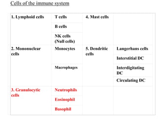
1. 1. Lymphoid cells T cells 4. Mast cells
B cells
NK cells
(Null cells)
2. Mononuclear
cells
Monocytes 5. Dendritic
cells
Langerhans cells
Interstitial DC
Macrophages Interdigitating
DC
Circulating DC
3. Granulocytic
cells
Neutrophils
Eosinophil
Basophil
Cells of the immune system
3. Formation of Granulocytes
Myeloid stem cells (myeloblast)
Promyeloblast
Myelocyte
Metamyelocytes
Granulocytes (N, E and B)
Differentiation
Takes app 14
days
Growth
factors reqd
are
G-CSF
GM-CSF
4. • has a multilobed nucleus (mostly has 2-5 lobes)
• it is often called as polymorphonuclear leukocyte (PMN) (for its
multilobed nucleus)
•are the most abundant white blood cells in humans approximately
108 - 1011 are produced daily)
•they account for approximately 70% of all white blood cells
(leukocytes).
1. Neutrophils
5. •They have granulated cytoplasm- it stains with both acid and
basic dyes
Hematopoiesis in Bone marrow
Neutrophils formed
Released into the peripheral blood and circulate for 7–10 h
After that migrate into the tissues, where they have a life span
of only a few days.
1. Neutrophils
6. Granules present in neutrophils
1. Primary
2. Secondary
3. Tertiary
In the presence of Ag- degranulation occurs
(which is release of contents from granules)
Release of contents
from Granules
Ag
7. Proteins present in the granules
Azurophilic
granules (or
"primary
granules")
Myeloperoxidase
bactericidal/ permeability increasing protein (BPI)
defensins
the serine proteases
neutrophil elastase
cathepsin G
Specific
granules (or
"secondary
granules")
Lactoferrin
cathelicidin
tertiary
granules
cathepsin
gelatinase
10. BPI (bactericidal/ permeability increasing
protein)
is a highly cationic, 55KD protein
Is a cytotoxic protein
Found only in the cells of myeloid series
Has strong affinity for LPS, so has potent
toxicity exclusively towards gram-negative
bacteria
Azurophilic
granules (or
"primary
granules")
Myeloperoxidase
bactericidal/ permeability
increasing protein (BPI)
defensins
the serine proteases
neutrophil elastase
cathepsin G
11. Azurophilic
granules (or
"primary
granules")
Myeloperoxidase
bactericidal/ permeability
increasing protein (BPI)
defensins
the serine proteases
neutrophil elastase
cathepsin G
also known as ELA2 (elastase 2, neutrophil) is a serine
protease
Secreted by neutrophils during inflammation
Hydrolyze elastin (protein present in the extracellular matrix)
Degrades outer membrane protein A (OmpA) of E. coli, and
virulence factors of organisms like Shigella, Salmonella and
Yersinia
Neutrophil elastase (or leukocyte elastase)
12. Cathelicidin (in secondary granules)
Is an antimicrobial peptide
were originally found in neutrophils, but later found to
be present in other cells including epithelial cells and
macrophages
increased in immune cells after activation by bacteria,
viruses, fungi
The human cathelicidin is called as hCAP-18 (Inactive
proprotein)
Specific
granules (or
"secondary
granules")
Lactoferrin
cathelicidin
13. hCAP-18 (Inactive proprotein)
Ag
active peptide (LL-37) (37 amino acids from the C-terminal) is released
exerts antimicrobial activity
Other biological
effects of LL-37
neutralizing the
biological effects of
LPS
promotion of
angiogenesis and
wound healing
angiogenesis is
physiological process
involving the growth
of new blood vessels
from pre-existing
vessels
chemotaxis of
neutrophils,
monocytes and T-
cells
14. Mechanism of killing by neutrophil
-Mainly phagocytosis
-Through garanule release
15. Neurtrophils
are the first to arrive at a site of inflammation
transient increase in the number of circulating
neutrophils, is called leukocytosis- is an
indication of infection
employ both oxygen-dependent and oxygen
independent pathways to generate
antimicrobial substances.
exhibit a larger respiratory burst and express
higher levels of defensins than macrophages
16. Recruitment of Neutrophils into inflamed tissue
Neutrophil extravasation- rolling, adherance---- into tissues and
elimination of pathogen in tissues
Ag
17. Neutrophils are generally the first cell type to bind to
inflamed endothelium and extravasate into the tissues.
Neutrophils first recognize the inflamed endothelium and adhere
strongly enough so that they are not swept away by the flowing
blood.
The bound neutrophils then penetrate the endothelial layer and
migrate into the underlying tissue.
Site of injury
18. The process of neutrophil extravasation are divided into
four sequential steps:
(1) Rolling
(2) activation by chemoattractant
(3) arrest and adhesion and
(4) Transendothelial migration
Site of injury
20. When there is an inflammatory response develops, various
cytokines and other inflammatory mediators act upon the
local blood vessels and induces the increased expression of
endothelial CAMs (cell adhesion molecules).
Endothelium
CAM
This neutrophil is called as activated or inflamed neutrophil
Neutrophil
Ag
21. Rolling (Step 1)
During an inflammatory response,
cytokines and other mediators act
upon the local endothelium and
induces the expression of adhesion
molecules of the selectin family.
Neutrophils attach loosely to the
endothelium by a low-affinity
interaction which involves selectins
and their Ligands.
Selectins are a family of glycoprotein
surface adhesion molecules
The E-selectin (expressed exclusively
on endothelial cells) and and P-
selectin (expressed on platelets and
endothelial cells) bind to mucin like
cell-adhesion molecules on the
neutrophil membrane or with a
sialylated lactosaminoglycan called
sialyl Lewisx
endothelium
22. Rolling:
Though Neutrophil gets attached to endothelium
Due to blood flow, it sometimes gets detached from the
endothelium, but gets attached to the selection present in the
nearby endothelium
This process is reapeated until the neurtophil gets firmly
attached to an endothelium
This is made possible by the rolling of neutrophils
23.
24. Step 2 (Activation)
Chemoattractants on the endothelium
binds to its respective R on Neutrophils
Eg.,
1. Chemoattractive cytokines called as
Chemokines like interleukin 8 (IL-8)
and macrophage inflammatory
protein (MIP)
2. Other Chemoattractants like platelet-
activating factor (PAF), the
complement split products C5a, C3a,
and C5b67 and various N- formyl
peptides produced by the breakdown
of bacterial proteins during an
infection.
Chemoattractants like such as IL-8 then
binds to a G-protein–linked receptor on
the neutrophil, triggering an activating
signal.
G protein linked receptor
25. Step 3
This signal induces a
conformational change in
the integrin molecules,
enabling them to adhere
firmly to Ig-superfamily
molecules on the
endothelium.
G protein linked receptor
26. After attachment to endothelium, the neutrophil migrates
through the vessel wall into the tissues (it is transendothelial
migration)
27. Eosinophils are like neutrophils- ie., are
motile phagocytic cells that can migrate
from the blood into the tissue spaces
play a role in the defense against parasitic
organisms
EOSINOPHILS
28. The granules contain substances like
histamine and proteins like
eosinophil peroxidase,
ribonuclease (RNase),
deoxyribonucleases,
lipase,
plasminogen, and
major basic protein.
These mediators are released by a process called
degranulation following activation of the eosinophil
The secreted contents of eosinophilic granules may
damage the parasite membrane and is also toxic to the
host tissue.
29. Basophils are nonphagocytic granulocytes
They can be stained with basic dyes (eg toulidine blue)
Are least common (represent about 0.01% to 0.3% of
circulating white blood cells)
They function by releasing pharmacologically active substances
from their cytoplasmic granules.
BASOPHILS
30. Nucleus has 2 lobes
they contain anticoagulant heparin
they contain vasodilator histamine (which promotes blood
flow to tissues)
These substances play a major role in certain allergic
responses.
they appear in tissues where allergic reactions are occurring
and probably contribute to the severity of these reactions
31. Basophils
Ig R IgE Ab
Allergens
B cells
Release IgE
Bind to R on Basophils
Degranulation of basophils occurs
32. Degranulation- is extracellular release of the mediators like
1.Histamine
2.Proteoglycans (like heparin and chondroitin)
3.Proteolytic enzymes (elastase, lysophospholipase)
4.Lipid mediators (leukotrienes)
5.Many Cytokines
Cause inflammation
33. Basopenia- is a decrease in B count
Basophilia- is a increase in B count
34. 1. Lymphoid cells T cells 4. Mast cells
B cells
NK cells
(Null cells)
2. Mononuclear
cells
Monocytes 5. Dendritic
cells
Langerhans cells
Interstitial DC
Macrophages Interdigitating
DC
Circulating DC
3. Granulocytic
cells
Neutrophils
Eosinophil
Basophil
Cells of the immune system
35. Mast cells
Myeloid stem cells
Basophil Progenitor
produce either Mast Cells or Basophils
Mast cells and basophils play a central role in inflammatory and
immediate allergic reactions
36. Both mast cells and basophils contain special
cytoplasmic granules which store mediators of
inflammation.
The extracellular release of the mediators is known as
degranulation
Mast cells Basophil
usually do not circulate in the
blood stream
are the smallest circulating
granulocytes. They arise in the
bone marrow, and following
maturation and differentiation,
are released into the blood
circulation
Are present in connective tissues If they are adequately stimulated
they may settle in the tissues
37. MAST CELLS or mastocyte
Mast-cell precursors (in bone marrow)
(undifferentiated cell)
----------------------------------------------------------
enter to blood
Blood undifferentiated cell
---------------------------------------------------------
Tissue enter to tissue
Differentiated cell
38. first described by Paul Ehrlich in 1878
Tissues where mast cells are present
-Present in tissues nearby to blood vessels
1. Skin
2. connective tissues of various organs
3. mucosal epithelial tissue of the respiratory, genitourinary, and
digestive tracts.
4. Mucosa of mouth, conjunctiva and nose
cytoplasmic granules contain
1. histamine and
2. other pharmacologically active substances.
39. Mast cell- plays important role in
1. Allergy and
2. Anaphylaxis (a type I hypersensitivity reaction)
40. 2 types of mediators in mast cells and basophils are
1. Preformed mediators and
2. Newly generated mediators
Preformed mediators,
These are the mediators which are stored in secretory granules
They are secreted upon cell activation by agents (eg., a biogenic
amine, histamine, proteoglycans, heparin, chondroitin sulphates
and a spectrum of neutral proteases).
Newly generated mediators
they are often absent in the resting mast cells
they are produced during IgE-mediated activation
eg of such mediators are arachidonic acid metabolites
(leukotriene C and prostaglandin D) , TNF, IL-4, IL-5 and IL-6.