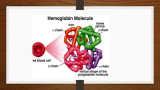
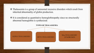
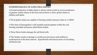
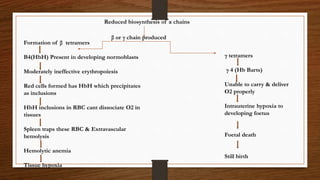
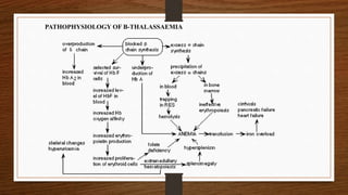
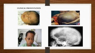
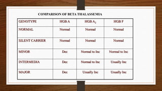
MANEESHA M JOSEPH
MSc MLT (MICROBIOLOGY)
ST.JOHN’S MEDICAL COLLEGE
 Thalassemia is a group of autosomal recessive disorders which result from
inherited abnormality of globin production.
 It is considered as quantitative hemoglobinopathy since no structurally
abnormal hemoglobin is synthesized.
TYPES OF THALASSEMIA
ALPHA THALASSEMIA BETA THALASSEMIA
Misc.THALASSEMIC
SYNDROMES
GENETICS
 Alpha Thalassemia results when there is disturbance in production of α -globin genes.
 Genes are responsible for regulating the synthesis and structure of different globins which are divided
into 2 clusters.
 The α-globin genes are encoded on chromosome 16 and the γ, δ, and β-globin genes are encoded on
chromosome 11
 A normal person carries a linked pair of alpha globin genes, 2 each from maternal and paternal
chromosome.Therefore, alpha thalassemia occurs when there is a disturbance in production of α-globin
from any or all four of the α-globin genes.
 When functional point mutations, frame shift mutations, nonsense mutations, and chain termination
mutations occur within or around the coding sequences of the alpha-globin gene cluster hemoglobin is
impaired.
ALPHA THALASSEMIA
PATHOPHYSIOLOGY OF ALPHA THALASSEMIA
Normal production of alpha chains is absent which results in excess production
of gamma- globin chains in the fetus and newborn or beta- globin chains in
children and adults
The β-globin chains are capable of forming soluble tetramers (beta-4, or HbH)
This form of hemoglobin is still unstable and precipitates within the cell,
forming insoluble inclusions called Heinz bodies
These Heinz bodies damage the red blood cells.
This further results in damage to erythrocyte precursors and ineffective
erythropoiesis in the bone marrow, hypochromia and microcytosis of circulating
red blood cells
Reduced biosynthesis of α chains
β or γ chain produced
Formation of β tetramers
Β4(HbH) Present in developing normoblasts
Moderately ineffective erythropoiesis
Red cells formed has HbH which precipitates
as inclusions
HbH inclusions in RBC cant dissociate O2 in
tissues
Spleen traps these RBC & Extravascular
hemolysis
Hemolytic anemia
Tissue hypoxia
γ tetramers
γ 4 (Hb Barts)
Unable to carry & deliver
O2 properly
Intrauterine hypoxia to
developing foetus
Foetal death
Still birth
Four clinical syndromes present in alpha thalassemia:
 Silent Carrier State
 Alpha Thalassemia Trait (Alpha Thalassemia Minor)
 Hemoglobin H Disease
 Bart's Hydrops Fetalis Syndrome
 Deletion of one alpha gene, leaving three functional alpha genes.
 Alpha/Beta chain ratio nearly normal.
 No hematologic abnormalities present.
 No reliable way to diagnose silent carriers by hematologic methods; Must be done
by genetic mapping.
 May see borderline low MCV (78-80fL)
1) Silent Carrier State
2) Alpha Thalassemia Trait (Alpha Thalassemia Minor)
 Also called Alpha Thalassemia Minor.
 Caused by two missing alpha genes. May be homozygous (-a/-a) or heterozygous(--/aa).
 Exhibits mild microcytic, hypochromic anemia.
 MCV between 70-75 fL.
 May be confused with iron deficiency anemia.
 Although some Bart's hemoglobin (γ4) present at birth, no Bart's hemoglobin present in
adults.
 Second most severe form alpha thalassemia.
 Usually caused by presence of only one gene producing alpha chains (--/-a).
 Results in accumulation of excess unpaired gamma or beta chains. Born with 10-40%
Bart's hemoglobin (γ4). Gradually replaced with Hemoglobin H (β4). In adult, have about
30-50% Hb H.
γ4 β4
 Infants develop progressive anemia and splenomegaly
3) Hemoglobin H Disease
 Smear demonstrates moderate degree of anisopoikilocytosis with
microcytes,hypochromia and target cells
 Reticulocyte preparation demonstrates uniform sized tiny HbH inclusions –Golf ball like
appearance of RBC
 Hb electrophoresis demonstrates fast moving Hb H bands in te range of 5-35%
4) Bart’s Hydrops Fetalis Syndrome
 Most severe form. Incompatible with life. Have no functioning alpha chain genes (--/--).
 Baby born with hydrops fetalis, which is edema and ascites caused by accumulation serous
fluid in fetal tissues as result of severe anemia.
 Predominant hemoglobin is Hemoglobin Bart, along with Hemoglobin Portland and traces of
Hemoglobin H.
 Hemoglobin Bart's has high oxygen affinity so cannot carry oxygen to tissues. Fetus dies in
utero or shortly after birth.
 At birth, see severe hypochromic, microcytic anemia with numerous NRBCs.
Genotype Hb A Hb Bart Hb H
Normal 97-98% 0 0
Silent Carrier 96-98% 0-2% 0
Alpha Thalassemia
Trait
85-95% 5-10% 0
Hemoglobin H Disease Dec 25-40% 2-40%
Hydrops Fetalis 0 80% (with 20%
Hgb Portland)
0-20%
COMPARISON OF ALPHA THALASSEMIAS
BETA THALASSEMIAS
 In this type,there is reduced synthesis of β chains of globin.
 Commonest type of thalassemia
 Β thalassemia is the consequence of various point mutations that causes the suppression of β chain
synthesis.
On the basis of synthetic ability β-genes are designated as
β gene – can synthesize normal amount of β-chain
β+ gene – can synthesize reduced amount of β-chain
β0 gene – cannot synthesize β-chain
Depending on this there are 2 types of thalassemia
1)The absence of beta-globin is referred to as beta-zero (B0) thalassemia. HbA is completely absent Hb F
is increased markedly.
2)Other β gene mutations allow some beta-globin to be produced but in reduced amounts. A reduced
amount of beta-globin is called beta-plus (B+) thalassemia.So some HbA is produced
CLASSICAL SYNDROMES OF BETA THALASSEMIA
Silent carrier state – the mildest form of beta thalassemia.
Beta thalassemia minor - heterozygous disorder resulting in mild hypochromic,
microcytic hemolytic anemia.
Beta thalassemia intermedia - Severity lies between the minor and major.
Beta thalassemia major - homozygous disorder resulting in severe transfusion-
dependent hemolytic anemia.
1)Silent Carrier State for β Thalassemia
Are various heterogenous beta mutations that produce only small decrease in production of beta chains.
Patients have nearly normal beta/alpha chain ratio and no hematologic abnormalities.
Have normal levels of Hb A2.
2) Beta Thalassemia Minor
 Usually presents as mild, asymptomatic hemolytic anemia unless patient in under stress such as
pregnancy, infection, or folic acid deficiency.
 Have one normal beta gene and one mutated beta gene.
 Hemoglobin level in 10-13 g/dL range with normal or slightly elevated RBC count.
 Anemia usually hypochromic and microcytic with slight aniso and poik, including target cells and
elliptocytes; May see basophilic stippling.
 Rarely see hepatomegaly or splenomegaly.
 Have high Hb A2 levels (3.5-8.0%) and normal to slightly elevated Hb F levels.
 Normally require no treatment.
3) Beta Thalassemia Intermedia
 Patients able to maintain minimum hemoglobin (7 g/dL or greater) without
transfusions.
 Expression of disorder falls between thalassemia minor and thalassemia major.
 May be either heterozygous for mutations causing mild decrease in beta chain
production, or may be homozygous causing a more serious reduction in beta chain
production.
 See increase in both Hb A2 production and Hb F production.
 Peripheral blood smear picture similar to thalassemia minor.
 Have varying symptoms of anemia, jaundice, splenomegaly and hepatomegaly.
 Have significant increase in bilirubin levels.
 Anemia usually becomes worse with infections, pregnancy, or folic acid deficiencies.
 May become transfusion dependent as adults.
 Tend to develop iron overloads as result of increased gastrointestinal absorption.
 Usually survive into adulthood.
4)Beta Thalassemia Major (cooley’s anemia)
Homozygous β 0/ β 0 , β +/ β + or double heterozygous of β 0/ β +
Characterized by severe microcytic, hypochromic anemia.
Detected early in childhood:
 Have pallor, variable degree of jaundice, abdominal enlargement, and hepatosplenomegaly.
 Severe anemia causes marked bone changes due to expansion of marrow space for increased
erythropoiesis.
 See characteristic changes in skull, long bones, and hand bones. X ray of skull shows “hair on
end appearence “
 Have protrusion upper teeth and Mongoloid facial features.
 Physical growth and development delayed.
 The skin may show pallor from anemia and jaundice from hyperbilirubinemia.

 Heart examination may reveal findings of cardiac failure and arrhythmia, related to either
severe anemia or iron overload.
 Patients who have received blood transfusions may have hepatomegaly or chronic
hepatitis due to iron overload.
 The gallbladder may contain bilirubin stones formed as a result of the patient's
lifelong hemolytic state.
 Splenomegaly typically is observed as part of the extramedullary hematopoiesis or
as a hypertrophic response related to the extravascular hemolysis.
 In addition to cardiac dysfunction, hepatomegaly, and hepatitis, iron overload can
also cause endocrine dysfunction, especially affecting the pancreas, testes, and
thyroid.
PATHOPHYSIOLOGY OF Β-THALASSAEMIA
CLINICAL PRESENTATION
LABORATORY DIAGNOSIS OF BETA THALASSEMIA MAJOR
1) CBC WITH DIFFERENTIAL
 See decrease in hemoglobin between 4 and 8 gm/dL. , hematocrit, MCV in range
of 50 to 60 fL, MCHC 22-30%
 See slightly decreased MCH 20-28pg
 Have normal or elevated RBC count with a normal red cell volume distribution
(RDW).
 Decrease in MCV very noticeable when compared to decrease in Hb and Hct.
 Elevated RBC count with markedly decreased MCV differentiates thalassemia
from iron deficiency anemia.
 On differential, see microcytic, hypochromic RBCs (except in carrier states).
 See mild to moderate poikilocytosis.
 In more severe cases, see marked number of target cells and elliptocytes.
 Will see polychromasia, basophilic stippling, and NRBCs,occasionally RBC with
Howel Jolly bodies.
2) RETIC COUNT
Reticulocyte count Usually elevated. Degree of elevation depends upon severity of anemia.
3)BONE MARROW
Bone marrow shows erythroid hyperplasia ,erythropoiesis is normoblastic
M:E Ratio reversed to 1:1 to 1:5
Bone marrow iron increased
4)OSMOTIC FRAGILITY
Have decreased osmotic fragility.
Is not very useful fact for diagnosing thalassemia. Is an inexpensive way of screening for carrier
states.
5)BRILLIANT CRESYL BLUE STAIN
Incubation with brilliant cresyl blue stain causes Hemoglobin H to precipitate. Results in
characteristic appearance of multiple discrete inclusions -golf ball appearance of
RBCs. Inclusions smaller than Heinz bodies and are evenly distributed throughout cell.
6)ACID ELUTION STAIN
Based on Kleihauer-Betke procedure. Acid pH will dissolve Hemoglobin A from red
cells. Hemoglobin F is resistant to denaturation and remains in cell. Stain slide with eosin. Normal
adult cells appear as "ghost" cells while cells with Hb F stain varying shades of pink.
7) ROUTINE CHEMISTRY TESTS
 Serum haptoglobins markedly decreased
 Bilirubin increased-unconjugated type
 Urine urobilinogen increased
 Assessment of iron status, total iron binding capacity, and ferritin level important in differentiating
thalassemia from iron deficiency anemia.
8) HEMOGLOBIN ELECTROPHORESIS
 Important role in diagnosing and differentiating various forms of thalassemias.
 Can differentiate among Hb A, Hb A2, and Hb F, as well as detect presence of abnormal
hemoglobins such as Hemoglobin Lepore, hemoglobin Bart's, or Hemoglobin Constant Spring.
 Also aids in detecting combinations of thalassemia and hemoglobinopathies.
 Both HbA and HbF in β + thalassemia and only HbF and Hb A2 <3.5% in β0 thalassemia
9. HPLC
10.Mutation studies
11.Biomagnetic liver spectrometry –Non invasive metod to detect hemosiderosis of liver
MANAGEMENT OF THALASSEMIA MAJOR
1.Blood transfusion-Depending upon severity transfusion may be given every 2-4
weeks.Patients should be tested at regular intervals for transfusion transmitted diseases
2.Iron chelation
Since these patients develop iron overload that damages heart liver & other endocrine
organs start chelation therapy when S.ferritin is >1200μg/L
3.Splenectomy
After 5yrs of age ,as spleen enlarges some patients develop pancytopenia due to
hypersplenism and transfusion requirements increased.splnectomy helps to reduce
severity of pancytopenia
4.Bone marrow transplantation
BMT indicated when HLA matched siblings are available in the family.
OTHER THALASSEMIAS CAUSED BY DEFECTS IN THE BETA-CLUSTER GENES
1.Delta Beta Thalassemia:-
 Group of disorders due either to a gene deletion that removes or inactivates
only delta and beta genes so that only alpha and gamma chains produced.
 Similar to beta thalassemia minor.
 Growth and development nearly normal. Splenomegaly
modest. Peripheral blood picture resembles beta thalassemia
2.Hemoglobin Lepore:-
 Rare class of delta beta thalassemia.
 Caused by gene crossovers between delta locus on one chromosome and
beta locus on second chromosome
 Rare condition characterized by continued synthesis of Hemoglobin F in adult life.
 Do not have usual clinical symptoms of thalassemia.
 Little significance except when combined with other forms of thalassemia or
hemoglobinopathies.
 If combined with sickle cell anemia, produces milder form of disease due to presence of Hb F
 Hb F more resistant to denaturation than Hb A. Can be demonstrated on blood smears using
Kleihauer Betke stain.
3.Hereditary Persistence of Fetal Hemoglobin (HPFH)
4.Beta Thalassemia with Hb S
 Inherit gene for Hb S from one parent and gene for Hb A with beta thalassemia from second
parent.
 Great variety in clinical severity. Production of Hb A ranges from none produced to varying
amounts. If no Hb A produced, see true sickle cell symptoms. If some Hb A produced, have
lessening of sickle cell anemia symptoms
 Is unusual because results in more severe disorder than homozygous E disease.
 Very severe anemia developing in childhood.
 Transfusion therapy required.
5.Beta Thalassemia with Hb E
6.Beta Thalassemia with Hb C
 Shows variability in clinical and hematologic symptoms.
 Usually asymptomatic anemia
GENOTYPE HGB A HGB A2 HGB F
NORMAL Normal Normal Normal
SILENT CARRIER Normal Normal Normal
MINOR Dec Normal to Inc Normal to Inc
INTERMEDIA Dec Normal to Inc Usually Inc
MAJOR Dec Usually Inc Usually Inc
COMPARISON OF BETA THALASSEMIA
REFERENCES
1.Text book of hematology Dacie and Lewis
2.Wintrobes text book of hematolgy
3.Hoffbrand post graduate hematology
2.Text book of hematology Tejindar singh
Thalassemia

Thalassemia

  • 1.
    MANEESHA M JOSEPH MScMLT (MICROBIOLOGY) ST.JOHN’S MEDICAL COLLEGE
  • 3.
     Thalassemia isa group of autosomal recessive disorders which result from inherited abnormality of globin production.  It is considered as quantitative hemoglobinopathy since no structurally abnormal hemoglobin is synthesized. TYPES OF THALASSEMIA ALPHA THALASSEMIA BETA THALASSEMIA Misc.THALASSEMIC SYNDROMES
  • 4.
    GENETICS  Alpha Thalassemiaresults when there is disturbance in production of α -globin genes.  Genes are responsible for regulating the synthesis and structure of different globins which are divided into 2 clusters.  The α-globin genes are encoded on chromosome 16 and the γ, δ, and β-globin genes are encoded on chromosome 11  A normal person carries a linked pair of alpha globin genes, 2 each from maternal and paternal chromosome.Therefore, alpha thalassemia occurs when there is a disturbance in production of α-globin from any or all four of the α-globin genes.  When functional point mutations, frame shift mutations, nonsense mutations, and chain termination mutations occur within or around the coding sequences of the alpha-globin gene cluster hemoglobin is impaired. ALPHA THALASSEMIA
  • 5.
    PATHOPHYSIOLOGY OF ALPHATHALASSEMIA Normal production of alpha chains is absent which results in excess production of gamma- globin chains in the fetus and newborn or beta- globin chains in children and adults The β-globin chains are capable of forming soluble tetramers (beta-4, or HbH) This form of hemoglobin is still unstable and precipitates within the cell, forming insoluble inclusions called Heinz bodies These Heinz bodies damage the red blood cells. This further results in damage to erythrocyte precursors and ineffective erythropoiesis in the bone marrow, hypochromia and microcytosis of circulating red blood cells
  • 6.
    Reduced biosynthesis ofα chains β or γ chain produced Formation of β tetramers Β4(HbH) Present in developing normoblasts Moderately ineffective erythropoiesis Red cells formed has HbH which precipitates as inclusions HbH inclusions in RBC cant dissociate O2 in tissues Spleen traps these RBC & Extravascular hemolysis Hemolytic anemia Tissue hypoxia γ tetramers γ 4 (Hb Barts) Unable to carry & deliver O2 properly Intrauterine hypoxia to developing foetus Foetal death Still birth
  • 7.
    Four clinical syndromespresent in alpha thalassemia:  Silent Carrier State  Alpha Thalassemia Trait (Alpha Thalassemia Minor)  Hemoglobin H Disease  Bart's Hydrops Fetalis Syndrome
  • 8.
     Deletion ofone alpha gene, leaving three functional alpha genes.  Alpha/Beta chain ratio nearly normal.  No hematologic abnormalities present.  No reliable way to diagnose silent carriers by hematologic methods; Must be done by genetic mapping.  May see borderline low MCV (78-80fL) 1) Silent Carrier State 2) Alpha Thalassemia Trait (Alpha Thalassemia Minor)  Also called Alpha Thalassemia Minor.  Caused by two missing alpha genes. May be homozygous (-a/-a) or heterozygous(--/aa).  Exhibits mild microcytic, hypochromic anemia.  MCV between 70-75 fL.  May be confused with iron deficiency anemia.  Although some Bart's hemoglobin (γ4) present at birth, no Bart's hemoglobin present in adults.
  • 9.
     Second mostsevere form alpha thalassemia.  Usually caused by presence of only one gene producing alpha chains (--/-a).  Results in accumulation of excess unpaired gamma or beta chains. Born with 10-40% Bart's hemoglobin (γ4). Gradually replaced with Hemoglobin H (β4). In adult, have about 30-50% Hb H. γ4 β4  Infants develop progressive anemia and splenomegaly 3) Hemoglobin H Disease
  • 10.
     Smear demonstratesmoderate degree of anisopoikilocytosis with microcytes,hypochromia and target cells  Reticulocyte preparation demonstrates uniform sized tiny HbH inclusions –Golf ball like appearance of RBC  Hb electrophoresis demonstrates fast moving Hb H bands in te range of 5-35%
  • 11.
    4) Bart’s HydropsFetalis Syndrome  Most severe form. Incompatible with life. Have no functioning alpha chain genes (--/--).  Baby born with hydrops fetalis, which is edema and ascites caused by accumulation serous fluid in fetal tissues as result of severe anemia.  Predominant hemoglobin is Hemoglobin Bart, along with Hemoglobin Portland and traces of Hemoglobin H.  Hemoglobin Bart's has high oxygen affinity so cannot carry oxygen to tissues. Fetus dies in utero or shortly after birth.  At birth, see severe hypochromic, microcytic anemia with numerous NRBCs.
  • 12.
    Genotype Hb AHb Bart Hb H Normal 97-98% 0 0 Silent Carrier 96-98% 0-2% 0 Alpha Thalassemia Trait 85-95% 5-10% 0 Hemoglobin H Disease Dec 25-40% 2-40% Hydrops Fetalis 0 80% (with 20% Hgb Portland) 0-20% COMPARISON OF ALPHA THALASSEMIAS
  • 13.
    BETA THALASSEMIAS  Inthis type,there is reduced synthesis of β chains of globin.  Commonest type of thalassemia  Β thalassemia is the consequence of various point mutations that causes the suppression of β chain synthesis. On the basis of synthetic ability β-genes are designated as β gene – can synthesize normal amount of β-chain β+ gene – can synthesize reduced amount of β-chain β0 gene – cannot synthesize β-chain Depending on this there are 2 types of thalassemia 1)The absence of beta-globin is referred to as beta-zero (B0) thalassemia. HbA is completely absent Hb F is increased markedly. 2)Other β gene mutations allow some beta-globin to be produced but in reduced amounts. A reduced amount of beta-globin is called beta-plus (B+) thalassemia.So some HbA is produced
  • 14.
    CLASSICAL SYNDROMES OFBETA THALASSEMIA Silent carrier state – the mildest form of beta thalassemia. Beta thalassemia minor - heterozygous disorder resulting in mild hypochromic, microcytic hemolytic anemia. Beta thalassemia intermedia - Severity lies between the minor and major. Beta thalassemia major - homozygous disorder resulting in severe transfusion- dependent hemolytic anemia.
  • 15.
    1)Silent Carrier Statefor β Thalassemia Are various heterogenous beta mutations that produce only small decrease in production of beta chains. Patients have nearly normal beta/alpha chain ratio and no hematologic abnormalities. Have normal levels of Hb A2. 2) Beta Thalassemia Minor  Usually presents as mild, asymptomatic hemolytic anemia unless patient in under stress such as pregnancy, infection, or folic acid deficiency.  Have one normal beta gene and one mutated beta gene.  Hemoglobin level in 10-13 g/dL range with normal or slightly elevated RBC count.  Anemia usually hypochromic and microcytic with slight aniso and poik, including target cells and elliptocytes; May see basophilic stippling.  Rarely see hepatomegaly or splenomegaly.  Have high Hb A2 levels (3.5-8.0%) and normal to slightly elevated Hb F levels.  Normally require no treatment.
  • 16.
    3) Beta ThalassemiaIntermedia  Patients able to maintain minimum hemoglobin (7 g/dL or greater) without transfusions.  Expression of disorder falls between thalassemia minor and thalassemia major.  May be either heterozygous for mutations causing mild decrease in beta chain production, or may be homozygous causing a more serious reduction in beta chain production.  See increase in both Hb A2 production and Hb F production.  Peripheral blood smear picture similar to thalassemia minor.  Have varying symptoms of anemia, jaundice, splenomegaly and hepatomegaly.  Have significant increase in bilirubin levels.  Anemia usually becomes worse with infections, pregnancy, or folic acid deficiencies.  May become transfusion dependent as adults.  Tend to develop iron overloads as result of increased gastrointestinal absorption.  Usually survive into adulthood.
  • 17.
    4)Beta Thalassemia Major(cooley’s anemia) Homozygous β 0/ β 0 , β +/ β + or double heterozygous of β 0/ β + Characterized by severe microcytic, hypochromic anemia. Detected early in childhood:  Have pallor, variable degree of jaundice, abdominal enlargement, and hepatosplenomegaly.  Severe anemia causes marked bone changes due to expansion of marrow space for increased erythropoiesis.  See characteristic changes in skull, long bones, and hand bones. X ray of skull shows “hair on end appearence “  Have protrusion upper teeth and Mongoloid facial features.  Physical growth and development delayed.  The skin may show pallor from anemia and jaundice from hyperbilirubinemia.   Heart examination may reveal findings of cardiac failure and arrhythmia, related to either severe anemia or iron overload.
  • 18.
     Patients whohave received blood transfusions may have hepatomegaly or chronic hepatitis due to iron overload.  The gallbladder may contain bilirubin stones formed as a result of the patient's lifelong hemolytic state.  Splenomegaly typically is observed as part of the extramedullary hematopoiesis or as a hypertrophic response related to the extravascular hemolysis.  In addition to cardiac dysfunction, hepatomegaly, and hepatitis, iron overload can also cause endocrine dysfunction, especially affecting the pancreas, testes, and thyroid.
  • 19.
  • 20.
  • 21.
    LABORATORY DIAGNOSIS OFBETA THALASSEMIA MAJOR 1) CBC WITH DIFFERENTIAL  See decrease in hemoglobin between 4 and 8 gm/dL. , hematocrit, MCV in range of 50 to 60 fL, MCHC 22-30%  See slightly decreased MCH 20-28pg  Have normal or elevated RBC count with a normal red cell volume distribution (RDW).  Decrease in MCV very noticeable when compared to decrease in Hb and Hct.  Elevated RBC count with markedly decreased MCV differentiates thalassemia from iron deficiency anemia.  On differential, see microcytic, hypochromic RBCs (except in carrier states).  See mild to moderate poikilocytosis.  In more severe cases, see marked number of target cells and elliptocytes.  Will see polychromasia, basophilic stippling, and NRBCs,occasionally RBC with Howel Jolly bodies.
  • 23.
    2) RETIC COUNT Reticulocytecount Usually elevated. Degree of elevation depends upon severity of anemia. 3)BONE MARROW Bone marrow shows erythroid hyperplasia ,erythropoiesis is normoblastic M:E Ratio reversed to 1:1 to 1:5 Bone marrow iron increased 4)OSMOTIC FRAGILITY Have decreased osmotic fragility. Is not very useful fact for diagnosing thalassemia. Is an inexpensive way of screening for carrier states. 5)BRILLIANT CRESYL BLUE STAIN Incubation with brilliant cresyl blue stain causes Hemoglobin H to precipitate. Results in characteristic appearance of multiple discrete inclusions -golf ball appearance of RBCs. Inclusions smaller than Heinz bodies and are evenly distributed throughout cell.
  • 24.
    6)ACID ELUTION STAIN Basedon Kleihauer-Betke procedure. Acid pH will dissolve Hemoglobin A from red cells. Hemoglobin F is resistant to denaturation and remains in cell. Stain slide with eosin. Normal adult cells appear as "ghost" cells while cells with Hb F stain varying shades of pink. 7) ROUTINE CHEMISTRY TESTS  Serum haptoglobins markedly decreased  Bilirubin increased-unconjugated type  Urine urobilinogen increased  Assessment of iron status, total iron binding capacity, and ferritin level important in differentiating thalassemia from iron deficiency anemia.
  • 25.
    8) HEMOGLOBIN ELECTROPHORESIS Important role in diagnosing and differentiating various forms of thalassemias.  Can differentiate among Hb A, Hb A2, and Hb F, as well as detect presence of abnormal hemoglobins such as Hemoglobin Lepore, hemoglobin Bart's, or Hemoglobin Constant Spring.  Also aids in detecting combinations of thalassemia and hemoglobinopathies.  Both HbA and HbF in β + thalassemia and only HbF and Hb A2 <3.5% in β0 thalassemia
  • 26.
    9. HPLC 10.Mutation studies 11.Biomagneticliver spectrometry –Non invasive metod to detect hemosiderosis of liver
  • 27.
    MANAGEMENT OF THALASSEMIAMAJOR 1.Blood transfusion-Depending upon severity transfusion may be given every 2-4 weeks.Patients should be tested at regular intervals for transfusion transmitted diseases 2.Iron chelation Since these patients develop iron overload that damages heart liver & other endocrine organs start chelation therapy when S.ferritin is >1200μg/L 3.Splenectomy After 5yrs of age ,as spleen enlarges some patients develop pancytopenia due to hypersplenism and transfusion requirements increased.splnectomy helps to reduce severity of pancytopenia 4.Bone marrow transplantation BMT indicated when HLA matched siblings are available in the family.
  • 28.
    OTHER THALASSEMIAS CAUSEDBY DEFECTS IN THE BETA-CLUSTER GENES 1.Delta Beta Thalassemia:-  Group of disorders due either to a gene deletion that removes or inactivates only delta and beta genes so that only alpha and gamma chains produced.  Similar to beta thalassemia minor.  Growth and development nearly normal. Splenomegaly modest. Peripheral blood picture resembles beta thalassemia 2.Hemoglobin Lepore:-  Rare class of delta beta thalassemia.  Caused by gene crossovers between delta locus on one chromosome and beta locus on second chromosome
  • 29.
     Rare conditioncharacterized by continued synthesis of Hemoglobin F in adult life.  Do not have usual clinical symptoms of thalassemia.  Little significance except when combined with other forms of thalassemia or hemoglobinopathies.  If combined with sickle cell anemia, produces milder form of disease due to presence of Hb F  Hb F more resistant to denaturation than Hb A. Can be demonstrated on blood smears using Kleihauer Betke stain. 3.Hereditary Persistence of Fetal Hemoglobin (HPFH) 4.Beta Thalassemia with Hb S  Inherit gene for Hb S from one parent and gene for Hb A with beta thalassemia from second parent.  Great variety in clinical severity. Production of Hb A ranges from none produced to varying amounts. If no Hb A produced, see true sickle cell symptoms. If some Hb A produced, have lessening of sickle cell anemia symptoms
  • 30.
     Is unusualbecause results in more severe disorder than homozygous E disease.  Very severe anemia developing in childhood.  Transfusion therapy required. 5.Beta Thalassemia with Hb E 6.Beta Thalassemia with Hb C  Shows variability in clinical and hematologic symptoms.  Usually asymptomatic anemia
  • 31.
    GENOTYPE HGB AHGB A2 HGB F NORMAL Normal Normal Normal SILENT CARRIER Normal Normal Normal MINOR Dec Normal to Inc Normal to Inc INTERMEDIA Dec Normal to Inc Usually Inc MAJOR Dec Usually Inc Usually Inc COMPARISON OF BETA THALASSEMIA
  • 32.
    REFERENCES 1.Text book ofhematology Dacie and Lewis 2.Wintrobes text book of hematolgy 3.Hoffbrand post graduate hematology 2.Text book of hematology Tejindar singh