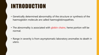
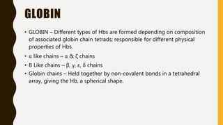
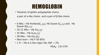
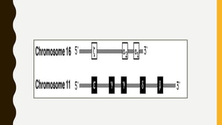
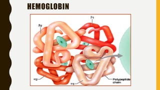
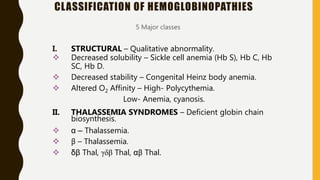
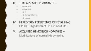
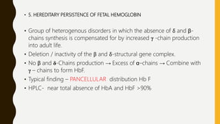
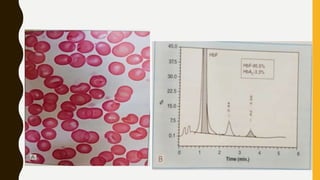
This document provides information on hemoglobinopathies and thalassemias. It begins by defining hemoglobinopathies as genetically determined abnormalities of the hemoglobin molecule, associated with globin chains. It then discusses the different types of globin chains and normal hemoglobin development.
The document classifies hemoglobinopathies into 5 major classes including structural abnormalities, thalassemia syndromes, thalassemic hemoglobin variants, hereditary persistence of fetal hemoglobin, and acquired hemoglobinopathies. It provides detailed information on beta thalassemia syndromes, genetics, clinical features, hematologic findings, bone marrow findings, laboratory tests, and complications/management of thalassemia major. It also briefly discusses
Hemoglobinopathies are a group of inherited disorders involving abnormal hemoglobin. They are classified into quantitative disorders affecting globin chain synthesis (thalassemias) and qualitative disorders altering globin structure (sickle cell disease). Thalassemias include β-thalassemia resulting from reduced β-chain production and α-thalassemia from reduced α-chain production. Sickle cell disease is caused by a mutation substituting valine for glutamic acid on the β-chain, causing hemoglobin S which polymerizes and sickles red blood cells under low oxygen conditions.
This document discusses thalassemia, specifically alpha and beta thalassemia. It describes the genetics, pathophysiology, clinical presentations, and laboratory diagnosis of the different types. The main points are:
1) Thalassemia results from inherited abnormalities in globin chain production, causing excess unpaired chains. Alpha thalassemia affects alpha chain production, while beta thalassemia affects beta chain production.
2) There are different clinical syndromes for each type depending on the severity of the genetic mutation, ranging from silent carriers to severe anemia requiring transfusions.
3) Laboratory testing helps diagnose and classify the specific type of thalassemia based on hemoglobin electrophoresis
Dr. Neela Ferdoushi presented on thalassemias and hemoglobinopathies. Thalassemias are genetic disorders caused by mutations that decrease the synthesis of the alpha or beta globin chains, resulting in imbalanced globin chain production and anemia. The clinical severity depends on the specific gene mutations inherited. Untreated homozygous forms can be fatal in childhood. Heterozygous carriers may be asymptomatic or have mild anemia. Laboratory tests can help identify abnormalities in hemoglobin type or amounts through hemograms and hemoglobin electrophoresis. Management involves blood transfusions and iron chelation therapy for severe forms.
The document discusses the thalassemias, a group of inherited blood disorders caused by defects in hemoglobin synthesis. There are two main types: alpha thalassemia results from reduced alpha globin chain production, while beta thalassemia is caused by reduced beta globin chains. Symptoms range from none to severe anemia requiring blood transfusions, depending on the number of defective genes. Thalassemias are most common in people from Mediterranean, African, and Southeast Asian descent and are diagnosed based on blood tests showing microcytic anemia and abnormalities in hemoglobin electrophoresis and red blood cell indices.
1. Thalassemia is caused by a defective production of the globin portion of hemoglobin, resulting in an imbalance between alpha and beta globin chains.
2. There are two main types - alpha thalassemia affects alpha chain production while beta thalassemia affects beta chain production.
3. Beta thalassemia major is the most severe form, characterized by severe anemia starting in early childhood requiring lifelong regular blood transfusions and iron chelation therapy to prevent iron overload.
Thalassemia is a genetic blood disorder caused by mutations in the genes that control globin production. There are two main types - alpha thalassemia affects alpha globin genes, while beta thalassemia affects beta globin genes. Thalassemia severity depends on the number of affected genes, ranging from no symptoms to severe anemia requiring chronic blood transfusions. The thalassemia gene is maintained in populations where malaria is common due to heterozygote resistance to the disease.
Thalassemia is a genetic blood disorder characterized by reduced or absent globin chain synthesis. The main types are alpha and beta thalassemia. Beta thalassemia major occurs when both beta globin genes are affected and results in severe anemia requiring lifelong blood transfusions. Thalassemia intermedia has a milder phenotype between thalassemia major and minor. Thalassemia minor is the carrier state with minimal symptoms. Diagnosis involves blood tests showing microcytic hypochromic anemia and abnormalities on hemoglobin electrophoresis or HPLC. Bone marrow aspiration demonstrates erythroid hyperplasia. Management ranges from transfusions for major to monitoring and prevention of complications for minor
1. The document discusses the structure and function of normal hemoglobin and describes various hemoglobin abnormalities that can be detected using high-performance liquid chromatography (HPLC).
2. HPLC separates hemoglobin variants by differences in retention time on the column, allowing detection of abnormalities like thalassemias and hemoglobinopathies.
3. Key things to check on an HPLC analysis include the baseline, peak shape/profile, order of peaks, and hemoglobin A2 retention time to identify potential hemoglobin disorders.
This document discusses hemoglobin, its structure and types. It describes that hemoglobin is made up of heme and globin proteins, with each molecule containing two alpha globin chains and two non-alpha chains. The main types discussed are HbA, HbA2, HbF. Hemoglobinopathies and thalassemias are disorders involving abnormalities in hemoglobin structure or globin gene expression. Specific disorders covered include sickle cell anemia, sickle cell trait, hemoglobin H disease, Bart's hydrops fetalis, hereditary persistence of fetal hemoglobin and hemoglobin E.
This document discusses the laboratory diagnosis of hemoglobinopathies. It covers the different types of hemoglobin variants including structural variants that cause reduced solubility or instability. It describes various laboratory techniques for detection such as peripheral blood smear examination, hemoglobin electrophoresis using cellulose acetate or agarose gel, and automated high-performance liquid chromatography. Diagnostic tests for specific variants like HbS and unstable hemoglobins are also outlined. Guidelines for investigation of suspected thalassemia including blood counts, HbA2 quantification and HbF analysis are provided.
Thalassemia is an inherited blood disorder caused by reduced or absent production of hemoglobin subunits (globin chains). There are two main types: alpha thalassemia involves a deficiency of alpha globin chains, while beta thalassemia involves a deficiency of beta globin chains. The severity of symptoms depends on how many globin genes are affected. Alpha thalassemia ranges from silent carrier state with no symptoms to hydrops fetalis, which is fatal if untreated.
1) Hemoglobinopathies are inherited disorders affecting hemoglobin structure or production, ranging from asymptomatic to fatal. The most common types are sickle cell disease and thalassemias.
2) Thalassemias are caused by deficient production of globin chains, leading to imbalanced globin synthesis and red blood cell damage. Beta thalassemias result from low beta chain production while alpha thalassemias involve alpha chains.
3) Clinical features vary by specific disorder from mild anemia to transfusion-dependent anemia and organ damage. Management involves treatment of complications, transfusions, chelation therapy, and in severe cases, stem cell transplant.
The document discusses haemoglobin disorders and haemoglobinopathies. It provides details on the molecular basis, inheritance patterns, clinical presentation and diagnosis of conditions like thalassaemia, sickle cell disease and other haemoglobin variants. Key points include that haemoglobin disorders are globally common due to ancestral mutations, are usually inherited in an autosomal recessive pattern, and can be diagnosed through blood tests, family history and molecular genetic analysis. Screening programs have helped identify carriers and provide prenatal diagnosis services.
Hemoglobinopathies and thalassemia are genetic blood disorders that result in abnormal hemoglobin. Hemoglobinopathies are caused by mutations in the globin chains of hemoglobin molecules, while thalassemias are caused by reduced or absent globin chain production. Sickle cell disease is a hemoglobinopathy caused by a mutation in the beta globin chain that results in sickle-shaped red blood cells. Thalassemias include alpha and beta thalassemia, which are characterized by decreased alpha or beta globin chain production leading to anemia. Management involves blood transfusions, iron chelation therapy, and in some cases stem cell transplantation.
This document summarizes different types of hemoglobinopathies and thalassemias. It describes normal adult hemoglobins and the two major disorders - qualitative hemoglobinopathies caused by structural mutations like sickle cell anemia, and quantitative hemoglobinopathies caused by reduced globin chain synthesis like thalassemias. Sickle cell disease results from a glutamic acid to valine substitution and causes polymerization of deoxygenated hemoglobin. Thalassemias are caused by alpha or beta globin chain deficiency. Beta thalassemia major involves homozygosity for beta thalassemia genes and requires frequent blood transfusions. Alpha thalassemia ranges from silent carrier state to Bart's hydrops
This patient is married to a man from Saudi Arabia whose family members require regular blood transfusions. She is concerned about the risk of genetic disorders causing anemia in her potential children. The document discusses thalassemia, a genetic blood disorder caused by defects in hemoglobin production. It explains the genetics and inheritance of alpha and beta thalassemia. The advisor would recommend confirming the blood conditions of the parents, performing lab work and genetic testing, and providing genetic counseling to help the couple understand the risks and make informed reproductive decisions.
challenges in interpreting abnormal hemoglobin study- the key is to correlate with patient age, ethnicity,RBC indices & morphology findings. Two tier approach for correct characterization of abnormal hemoglobins of HPLC &/or capillary electrophoresis.
The hemoglobinopathies are diseases characterized by abnormalities in hemoglobin synthesis. Qualitative abnormalities result from changes to the amino acid structure, as in sickle cell anemia. Quantitative abnormalities (thalassemias) involve normal amino acid sequences but impaired globin chain production. Examples include alpha-thalassemia reducing alpha chain production and beta-thalassemia reducing beta chain production. Other hemoglobin variants like HbC and HbE involve single amino acid substitutions and are associated with mild anemias.
Hemoglobinopathies are genetic disorders that result in abnormalities in the structure or synthesis of hemoglobin. There are two main types: qualitative abnormalities that change the structure of the globin chains, and quantitative abnormalities that reduce globin chain production (thalassemias). Thalassemias are caused by mutations that decrease or eliminate alpha or beta globin chain synthesis. This leads to imbalanced globin chain production and severe anemia. The most severe form is beta thalassemia major, which requires lifelong blood transfusions if untreated. Beta thalassemia trait is a milder form with microcytic anemia and elevated hemoglobin A2 levels.
The document discusses thalassemia, a genetic blood disorder. It defines thalassemia and describes its prevalence worldwide. There are two main types - alpha and beta thalassemia - depending on which globin chain is deficient. Symptoms range from mild to severe anemia. The disorder is diagnosed through blood tests and analysis of red blood cells. Treatment involves blood transfusions and medication.
This document summarizes different types of erythrocyte inclusions seen on supravital and Wright staining, along with their composition and associated disease states. It describes inclusions such as basophilic stippling composed of RNA seen in iron deficiency, Howell-Jolly bodies which are nuclear fragments seen in megaloblastic anemia, Heinz bodies containing denatured hemoglobin associated with G6PD deficiency, and siderotic granules containing iron found in sideroblastic anemia.
Hemoglobinopathies are disorders that affect the structure, function, or production of hemoglobin. The document discusses normal hemoglobin structure and composition, the genes that encode the globin chains, fetal hemoglobin development, classification of hemoglobinopathies including structural abnormalities and thalassemias, and details on sickle cell disease which results from a single nucleotide change causing valine to replace glutamic acid in the beta globin chain.
This document summarizes the molecular basis of hemoglobin disorders and sickle cell disease. It discusses hemoglobin genes, structure, and variants. Key points include: hemoglobin is a tetramer of alpha and non-alpha chains; sickle cell disease is caused by a single point mutation resulting in hemoglobin S; hemoglobin S polymerizes under low oxygen conditions, causing red blood cells to sickle. Clinical manifestations of sickle cell disease include anemia, pain crises, organ damage. Treatment focuses on pain management, transfusions, and hydroxyurea to raise fetal hemoglobin levels. New therapies aim to correct the genetic defect through gene therapy or induced pluripotent stem cells.
This a series of notes on hematology useful for undergraduate and postgraduate medical and paramedical students. Notes are prepared from standard texts and are easy to reproduce in exams.
Thalassemia is a group of hemolytic anemias caused by inherited abnormalities in globin chain production. There are two main types: alpha and beta thalassemia. Beta thalassemia major presents in infants with severe anemia, jaundice, hepatosplenomegaly, and failure to thrive. Diagnosis involves blood tests showing microcytic hypochromic anemia and hemoglobin electrophoresis. Treatment requires lifelong blood transfusions combined with iron chelation therapy to prevent complications from iron overload.
Thalassemia is caused by defective production of the globin portion of hemoglobin, resulting in an imbalance of globin chain production. There are two main types: alpha thalassemia involves a defect in alpha chain synthesis, while beta thalassemia involves a defect in beta chain synthesis. Beta thalassemia major is the most severe form, causing severe anemia starting in infancy that requires lifelong blood transfusions and iron chelation therapy. Beta thalassemia intermedia is milder, while beta thalassemia minor causes few or no symptoms. Alpha thalassemia can range from the lethal hydrops fetalis form to asymptomatic alpha thalassemia trait. Diagnosis involves blood
This document discusses thalassemia and sickle cell disease. It begins by describing the different types of hemoglobin present during development and in adults. It then covers the two main types of hemoglobinopathies - qualitative involving abnormal hemoglobin structure like sickle cell, and quantitative involving reduced hemoglobin production like thalassemia. The document goes into detail about the genetic causes and clinical manifestations of alpha and beta thalassemia as well as sickle cell disease. Laboratory findings and treatments are also summarized.
Thalassemia is a genetic blood disorder caused by reduced or absent globin chain production, classified as alpha or beta thalassemia. Alpha thalassemia involves alpha chain defects and beta thalassemia involves beta chain defects. Symptoms range from none in trait carriers to severe anemia requiring transfusions in major forms. Prevention involves genetic screening and counseling for at-risk populations.
1. Thalassemias are genetic blood disorders caused by mutations in the globin genes that result in reduced or absent globin chain production and imbalanced hemoglobin synthesis.
2. There are two main types: alpha-thalassemia affects alpha chain production and beta-thalassemia affects beta chain production.
3. The clinical severity of thalassemias depends on the number of defective globin genes and ranges from asymptomatic carriers to severe anemias requiring lifelong blood transfusions. Laboratory tests can identify the type and severity through hemoglobin analysis and peripheral blood smears.
Hemoglobin is composed of four globin chains that each bind to a heme group. The genes that encode for the different types of globin chains are located on chromosomes 11 and 16. Mutations in these genes can result in disorders called thalassemias, where there is imbalanced production of the globin chains. In β-thalassemia major, there is little to no production of the β-globin chains. This causes unpaired α-globin chains to precipitate and destroy red blood cell precursors in the bone marrow. Patients with β-thalassemia major suffer severe anemia starting in infancy and require regular blood transfusions to survive, but this leads to iron overload in tissues
1. Thalassemia is a heterogeneous group of inherited disorders characterized by decreased synthesis of the alpha or beta globin chains of hemoglobin A, leading to ineffective erythropoiesis and hemolysis.
2. Beta-thalassemias result from reduced or absent beta chain synthesis and include beta-thalassemia major (severe transfusion dependent anemia), beta-thalassemia intermedia (severe anemia but not transfusion dependent), and beta-thalassemia minor (asymptomatic with mild anemia).
3. Laboratory findings in beta-thalassemia major include severe anemia, peripheral blood changes, increased HbF and HbA2 on electroph
This document discusses hemolytic anemia, defined as anemias resulting from increased red blood cell destruction coupled with an intact bone marrow response. It is classified as congenital/hereditary or acquired. Specific hereditary causes include thalassemia, sickle cell anemia, and G6PD deficiency. Thalassemia results from a defect in globin chain synthesis, and causes severe anemia requiring frequent blood transfusions and iron chelation therapy. Sickle cell anemia is caused by a mutation producing abnormal hemoglobin S, which can polymerize and deform red blood cells, causing pain crises. Hereditary spherocytosis is due to a membrane defect impairing the sodium pump and deforming red
Halima, an 11-year-old girl with consanguineous parents, presented with not growing well, gradual pallor, and abdominal distension for 7 years. On examination, she was severely pale with facial dysmorphism and hepatosplenomegaly. Her history included repeated blood transfusions. She was diagnosed with hereditary hemolytic anemia. The seminar discussed thalassemia, including the types of thalassemia, clinical features, investigations, complications, and management with a focus on blood transfusions and chelation therapy.
Hemolytic anemia results from increased red blood cell destruction coupled with the bone marrow's ability to increase red blood cell production in response. There are two types: congenital/hereditary forms caused by genetic defects, and acquired forms caused by external factors. Common hereditary types include thalassemia from hemoglobin defects, sickle cell anemia from abnormal hemoglobin, and G6PD deficiency from enzyme defects. Symptoms include anemia, jaundice, splenomegaly. Diagnosis involves blood tests showing markers of hemolysis like low haptoglobin, increased LDH and bilirubin. Treatment depends on the underlying cause but may include blood transfusions, iron chelation therapy,
Based on the FBC results provided:
- The low MCV and Hb along with normal iron studies is consistent with thalassaemia trait
- The elevated HbA2 and HbF levels provide further support for beta thalassaemia minor
- The absence of HbH inclusions makes alpha thalassaemia less likely
- The findings of basophilic stippling, though nonspecific, can occur in beta thalassaemia minor
- Therefore, the most likely diagnosis is beta thalassaemia minor based on the haematological and biochemical investigation results.
This document discusses hematological disorders including hemolytic anemias and congenital red blood cell disorders. It provides details on laboratory tests used to evaluate anemias, such as blood smears, hemoglobin electrophoresis, and enzyme assays. Specific disorders covered include thalassemias, sickle cell disease, hereditary spherocytosis, elliptocytosis, and G6PD deficiency. Thalassemias result from decreased alpha or beta globin chain production and are classified based on severity from hydrops fetalis to thalassemia minor. Sickle cell disease is caused by a mutation in the beta globin gene.
This document discusses hematological disorders including hemolytic anemias and congenital red blood cell disorders. It provides details on laboratory tests used to evaluate anemias, such as a complete blood count, blood smear, and hemoglobin electrophoresis. Specific disorders covered include thalassemias, sickle cell disease, glucose-6-phosphate dehydrogenase deficiency, hereditary spherocytosis, and hereditary elliptocytosis. The roles of decreased red blood cell production and increased red blood cell destruction in causing anemia are explained.
Thalassemia and sideroblastic anemia are inherited blood disorders characterized by reduced hemoglobin synthesis. Thalassemia results from a genetic mutation affecting either the alpha or beta globin chains, leading to imbalanced globin chain production and anemia. Sideroblastic anemia features iron accumulation in erythroblast mitochondria, causing ineffective red blood cell production. The document defines and compares the genetic causes and clinical manifestations of different types of thalassemia and sideroblastic anemia. Laboratory findings include abnormal hemoglobin levels, red blood cell morphology, and iron studies.
This document discusses hemolytic anemias, which are genetically determined blood disorders caused by defects in red blood cells that lead to their premature destruction. The document categorizes hemolytic anemias based on whether the cause is intrinsic or extrinsic to red blood cells, and also based on the site of hemolysis. Specific examples of hemolytic anemias are discussed in detail, including thalassemia, sickle cell anemia, hereditary spherocytosis, and G6PD deficiency. The clinical features, laboratory findings, management, and treatment options are summarized for each condition.
This document discusses various hemoglobinopathies that are prevalent in India. It begins by describing normal hemoglobin and its subunits. It then classifies different types of hemoglobinopathies including structural abnormalities like sickle cell disease, thalassemias due to defective globin chain synthesis, and acquired hemoglobinopathies. Sickle cell disease results from a genetic mutation causing hemoglobin S polymerization and red blood cell sickling. Thalassemias involve reduced alpha or beta globin chain production. The document provides detailed information on the genetics, pathophysiology, clinical manifestations, diagnosis, and management of these common hemoglobin disorders.
1. Thalassemias are inherited blood disorders caused by reduced or absent globin chain synthesis leading to imbalanced hemoglobin production and anemia.
2. Beta thalassemias result from beta globin chain deficiency and include thalassemia major, intermedia, and minor. Thalassemia major is the severe transfusion-dependent form.
3. Laboratory findings in thalassemia major include severe anemia, microcytosis, hypochromia, and elevated fetal hemoglobin levels on electrophoresis. Complications arise from extramedullary hematopoiesis and iron overload from transfusions.
Thalassemia is a blood disorder caused by reduced or absent globin chain production, leading to anemia. It is classified as alpha or beta thalassemia depending on the deficient chain. Common in areas like Southeast Asia and the Mediterranean. Diagnosis involves blood tests showing hypochromic microcytic anemia and globin gene testing. Symptoms range from mild to severe depending on the number of defective genes. The most severe form is beta thalassemia major requiring lifelong blood transfusions and iron chelation therapy. Complications include anemia, jaundice, bone changes, iron overload affecting organs, and growth issues.
1) Microcytic hypochromic anemia is characterized by small, pale red blood cells and can be caused by iron deficiency, thalassemia, sideroblastic anemia, or other conditions.
2) Iron deficiency anemia is the most common cause and results from inadequate iron intake or absorption. It disrupts hemoglobin synthesis and cellular proliferation.
3) Thalassemia is an inherited disorder of hemoglobin production that can range from mild to severe. Thalassemia major requires regular blood transfusions and causes severe anemia from ineffective erythropoiesis and hemolysis.
Telepatholgy is the practice of pathology at a distance . there are three types: static image based, dynamic/ real time/dynamic robotic, and virtual or whole slide imaging. uses of telepathology, advantages and problems.
Efficacy of liquid based cytology versus conventional smearsAnamika Dev
This study compares the efficacy of liquid-based cytology (LBC) to conventional smear methods for fine needle aspiration cytology samples. The study analyzed 110 cases of various lesions collected over 2 years. Results showed that LBC produced more clear backgrounds and better preserved and dispersed cells compared to conventional smears. LBC allowed for improved diagnosis of lesions like ductal carcinoma, lymph node metastases, and bone lesions. However, LBC required more processing and had some artifacts that could complicate interpretation. Overall, LBC improved sample quality and reduced inadequate sampling compared to conventional smears.
This document discusses autoantibodies and methods for detecting anti-nuclear antibodies (ANA). ANAs are antibodies directed against nuclear and cytoplasmic antigens and are associated with various autoimmune diseases. The three main methods for detecting ANAs are indirect immunofluorescence assay using HEp-2 cells, ELISA, and multiplex bead immunoassays. The immunofluorescence assay is commonly used for initial screening due to its ability to detect multiple antigen patterns but has limitations. ELISA and bead assays allow detection of specific autoantibodies and have improved sensitivity and specificity compared to immunofluorescence.
Semen analysis WHO 2010 BY DR ANAMIKA DEVAnamika Dev
The document provides information about semen analysis, including the typical components and fractions of semen, the structures and sizes of sperm cells, indications for semen analysis, the process of sample collection and handling, and the steps involved in an initial semen analysis. Key points include that semen is made up of contributions from various glands, liquefies within 15-30 minutes, and an analysis involves examining the sample under a microscope to assess characteristics like motility, viscosity, and presence of other cells.
This document outlines the World Health Organization's 2016 classification of central nervous system tumors. It lists 17 categories of CNS tumors and provides brief descriptions of some of the tumor types within categories 1, 2, and 10, including diffuse astrocytic and oligodendroglial tumors, other astrocytic tumors characterized by more circumscribed growth and specific genetic alterations, and the various subtypes of meningiomas ranging from low to high grade.
This document discusses various nutritional disorders including obesity, kwashiorkor, marasmus, anorexia nervosa, and bulimia nervosa. It notes that kwashiorkor is a childhood protein-energy deficiency characterized by edema, while marasmus is caused by overall lack of calories seen as "skin and bone." Obesity results from calorie intake exceeding utilization and is measured by body mass index. The body regulates energy balance through hormones like leptin from fat cells and ghrelin from the stomach.
This document discusses various bacterial infections caused by different species of bacteria. It describes infections such as furuncles, carbuncles, hydradenitis suppurativa, osteomyelitis, toxic shock syndrome, food poisoning, scalded skin syndrome, opportunistic infections, urinary tract infections, and infections caused by different types of streptococci including Streptococcus pyogenes, Streptococcus agalactiae, Corynebacterium diphtheriae, Bacillus anthracis, and Nocardia asteroides. It provides details on symptoms, pathogenesis, and clinical presentation of these infections.
Local Advanced Lung Cancer: Artificial Intelligence, Synergetics, Complex Sys...Oleg Kshivets
Overall life span (LS) was 1671.7±1721.6 days and cumulative 5YS reached 62.4%, 10 years – 50.4%, 20 years – 44.6%. 94 LCP lived more than 5 years without cancer (LS=2958.6±1723.6 days), 22 – more than 10 years (LS=5571±1841.8 days). 67 LCP died because of LC (LS=471.9±344 days). AT significantly improved 5YS (68% vs. 53.7%) (P=0.028 by log-rank test). Cox modeling displayed that 5YS of LCP significantly depended on: N0-N12, T3-4, blood cell circuit, cell ratio factors (ratio between cancer cells-CC and blood cells subpopulations), LC cell dynamics, recalcification time, heparin tolerance, prothrombin index, protein, AT, procedure type (P=0.000-0.031). Neural networks, genetic algorithm selection and bootstrap simulation revealed relationships between 5YS and N0-12 (rank=1), thrombocytes/CC (rank=2), segmented neutrophils/CC (3), eosinophils/CC (4), erythrocytes/CC (5), healthy cells/CC (6), lymphocytes/CC (7), stick neutrophils/CC (8), leucocytes/CC (9), monocytes/CC (10). Correct prediction of 5YS was 100% by neural networks computing (error=0.000; area under ROC curve=1.0).
Muktapishti is a traditional Ayurvedic preparation made from Shoditha Mukta (Purified Pearl), is believed to help regulate thyroid function and reduce symptoms of hyperthyroidism due to its cooling and balancing properties. Clinical evidence on its efficacy remains limited, necessitating further research to validate its therapeutic benefits.
Recomendações da OMS sobre cuidados maternos e neonatais para uma experiência pós-natal positiva.
Em consonância com os ODS – Objetivos do Desenvolvimento Sustentável e a Estratégia Global para a Saúde das Mulheres, Crianças e Adolescentes, e aplicando uma abordagem baseada nos direitos humanos, os esforços de cuidados pós-natais devem expandir-se para além da cobertura e da simples sobrevivência, de modo a incluir cuidados de qualidade.
Estas diretrizes visam melhorar a qualidade dos cuidados pós-natais essenciais e de rotina prestados às mulheres e aos recém-nascidos, com o objetivo final de melhorar a saúde e o bem-estar materno e neonatal.
Uma “experiência pós-natal positiva” é um resultado importante para todas as mulheres que dão à luz e para os seus recém-nascidos, estabelecendo as bases para a melhoria da saúde e do bem-estar a curto e longo prazo. Uma experiência pós-natal positiva é definida como aquela em que as mulheres, pessoas que gestam, os recém-nascidos, os casais, os pais, os cuidadores e as famílias recebem informação consistente, garantia e apoio de profissionais de saúde motivados; e onde um sistema de saúde flexível e com recursos reconheça as necessidades das mulheres e dos bebês e respeite o seu contexto cultural.
Estas diretrizes consolidadas apresentam algumas recomendações novas e já bem fundamentadas sobre cuidados pós-natais de rotina para mulheres e neonatos que recebem cuidados no pós-parto em unidades de saúde ou na comunidade, independentemente dos recursos disponíveis.
É fornecido um conjunto abrangente de recomendações para cuidados durante o período puerperal, com ênfase nos cuidados essenciais que todas as mulheres e recém-nascidos devem receber, e com a devida atenção à qualidade dos cuidados; isto é, a entrega e a experiência do cuidado recebido. Estas diretrizes atualizam e ampliam as recomendações da OMS de 2014 sobre cuidados pós-natais da mãe e do recém-nascido e complementam as atuais diretrizes da OMS sobre a gestão de complicações pós-natais.
O estabelecimento da amamentação e o manejo das principais intercorrências é contemplada.
Recomendamos muito.
Vamos discutir essas recomendações no nosso curso de pós-graduação em Aleitamento no Instituto Ciclos.
Esta publicação só está disponível em inglês até o momento.
Prof. Marcus Renato de Carvalho
www.agostodourado.com
Does Over-Masturbation Contribute to Chronic Prostatitis.pptxwalterHu5
In some case, your chronic prostatitis may be related to over-masturbation. Generally, natural medicine Diuretic and Anti-inflammatory Pill can help mee get a cure.
Histololgy of Female Reproductive System.pptxAyeshaZaid1
Dive into an in-depth exploration of the histological structure of female reproductive system with this comprehensive lecture. Presented by Dr. Ayesha Irfan, Assistant Professor of Anatomy, this presentation covers the Gross anatomy and functional histology of the female reproductive organs. Ideal for students, educators, and anyone interested in medical science, this lecture provides clear explanations, detailed diagrams, and valuable insights into female reproductive system. Enhance your knowledge and understanding of this essential aspect of human biology.
Rasamanikya is a excellent preparation in the field of Rasashastra, it is used in various Kushtha Roga, Shwasa, Vicharchika, Bhagandara, Vatarakta, and Phiranga Roga. In this article Preparation& Comparative analytical profile for both Formulationon i.e Rasamanikya prepared by Kushmanda swarasa & Churnodhaka Shodita Haratala. The study aims to provide insights into the comparative efficacy and analytical aspects of these formulations for enhanced therapeutic outcomes.
These lecture slides, by Dr Sidra Arshad, offer a quick overview of the physiological basis of a normal electrocardiogram.
Learning objectives:
1. Define an electrocardiogram (ECG) and electrocardiography
2. Describe how dipoles generated by the heart produce the waveforms of the ECG
3. Describe the components of a normal electrocardiogram of a typical bipolar lead (limb II)
4. Differentiate between intervals and segments
5. Enlist some common indications for obtaining an ECG
6. Describe the flow of current around the heart during the cardiac cycle
7. Discuss the placement and polarity of the leads of electrocardiograph
8. Describe the normal electrocardiograms recorded from the limb leads and explain the physiological basis of the different records that are obtained
9. Define mean electrical vector (axis) of the heart and give the normal range
10. Define the mean QRS vector
11. Describe the axes of leads (hexagonal reference system)
12. Comprehend the vectorial analysis of the normal ECG
13. Determine the mean electrical axis of the ventricular QRS and appreciate the mean axis deviation
14. Explain the concepts of current of injury, J point, and their significance
Study Resources:
1. Chapter 11, Guyton and Hall Textbook of Medical Physiology, 14th edition
2. Chapter 9, Human Physiology - From Cells to Systems, Lauralee Sherwood, 9th edition
3. Chapter 29, Ganong’s Review of Medical Physiology, 26th edition
4. Electrocardiogram, StatPearls - https://www.ncbi.nlm.nih.gov/books/NBK549803/
5. ECG in Medical Practice by ABM Abdullah, 4th edition
6. Chapter 3, Cardiology Explained, https://www.ncbi.nlm.nih.gov/books/NBK2214/
7. ECG Basics, http://www.nataliescasebook.com/tag/e-c-g-basics
2. INTRODUCTION
• Genetically determined abnormality of the structure or synthesis of the
haemoglobin molecule are called haemoglobinopathies.
• The abnormality is associated with globin chains, heme portion will be
normal.
• Range in severity is from asymptomatic laboratory anomalies to death in
utero.
3. GLOBIN
• GLOBIN – Different types of Hbs are formed depending on composition
of associated globin chain tetrads; responsible for different physical
properties of Hbs.
• α like chains – α & ζ chains
• Β Like chains – β, γ, ε, δ chains
• Globin chains – Held together by non-covalent bonds in a tetrahedral
array, giving the Hb, a spherical shape.
4. HEMOGLOBIN
• Tetramer of globin polypeptide chains:
a pair of α-like chains and a pair of β-like chains
• 6 Wks – Hb Portland(ζ 2γ2), Hb Gower I(ζ 2ε2) and Hb
Gower II(α2 ε2).
• 10-11 Wks – Hb F(α2 γ2)
• 35 Wks – Hb A2(α2 δ2)
• 38 Wks – Hb A(α2 β2)
• New born – Hb F-50-85%
• 1 Yr – Hb A is the major Hb, HbF < 2%,
HbA2 - 1.8-3.5%
8. III. THALASSEMIC Hb VARIANTS –
– HbS/β-Thal.
– HbS/α-Thal.
– Hb E.
– Hb Constant Spring.
– Hb Lepore.
IV. HEREDITARY PERSISTENCE OF FETAL Hb-(
HPFH) – High levels of Hb F in adult life.
V. ACQUIRED HEMOGLOBINOPATHIES –
Modifications of normal Hb by toxins.
10. “Thalassa” – (Gk.) Sea.(referring to Mediterranean)
Grp of congenital anemias that have deficient synthesis of one or more
of the globin subunits of the normal Hb.
Thomas Cooley identified it.
It is quantitative hemoglobinopathy since no structurally abnormal
hemoglobin is synthesized.
11. CLASSIFICATION OF THALASSEMIAS
Normally α : β - 1:1
Degree of imbalance is related to the clinical expression.
A. Impaired α–chain synthesis – α-Thal
B. Impaired β–chain synthesis – β-Thal
C. Misc. Thalassemic syndromes –HbS Thal, HbD Thal, HbE Thal, δβ-
13. ß THALASSEMIA SYNDROMES
• Thalassemia Major – Most severe form, homozygous state
• Thalassemia Intermedia – Double heterozygous state, milder form
• Thalassemia Minor/Trait(BTT) – Heterozygous state, asymptomatic with little or no
anemia
• Thalassemia Minima – Clinically undetectable
14. GENETICS
• Autosomal Recessive disorders
• 2 β- Thalassemia genes (βThal)–
• β˚ - No production of β-Chains.
• β+ - Some but still subnormal production.
• Any combination of normal β-genes and βThal genes are possible –
various phenotypes.
• Point mutation on globin gene cluster; i.e, single nucleotide substitution
– Splicing mutation – Most common cause of β+ Thalassemia
– Promoter region mutation - β+ Thalassemia
– Chain terminator mutation - β0 Thalassemia
15. BETA -THALASSEMIA MAJOR
• Also K/A Cooleys anemia
• Homozygous form of β0 / β0 or β+ / β+ or double heterozygous β0 /β+
• Infants well at birth
• Later by 6 months , develop moderate to severe anemia, failure to thrive,
hepatosplenomegaly and bone changes
• Transfusion dependent
16.
17. CLINICAL FEATURES
Irritable, pale infant, failure to thrive, diarrhea, fever
and enlarged abdomen.
Severe anemia → Cardiac failure.
Chronic Hemolysis → Gout, Gall stones, Icterus
Thrombotic complications.
Massive Splenomegaly.
Flattened nose, wide set eyes, frontal bossing of the skull, prominent
cheek bones, and overgrowth of zygomatic bones– “Chipmunk Facies” /
thalassemic facies/ mongoloid facies.
Enlarged marrow cavities d/t hyperplastic marrow.
Hair-on-end appearance on X-Ray skull.
18.
19. HEMATOLOGIC FINDINGS
• Anemia – moderate to severe when first diagnosed ; 3-8gm%
• MCV – 50-70fl
• MCH -12-20pg
• MCHC - 22-32%
• PERIPHERAL SMEAR-
– RBCs are microcytic and hypochromia is marked and red cells are thin
– Moderate to marked degree of aniso-poikilocytosis
– Many target cells. Central puddle of Hb may be eccentric
– Basophilic stippling – constant feature
– Nucleated RBCs (mainly late normoblasts )- 5 - 40/100 WBC
– Presence of tear drop , elliptical, fragmented red cells and occasional red cell with Howel Jolly
body
• Aggregates of free α chains can be seen by phase contrast microscopy on supravital
staining with methyl violet
• Reticulocyte count <2 % because of ineffective erythropoiesis & does not correspond to
severity of anemia
20.
21. IRON STATUS
• S. ferritin >1000 µg/L ( 50-150)
• Transferrin saturation ↑’d 55-90% (30-35%)
• Total iron binding capacity ↓ ‘d 250-300 µg% (320-360)
• S. Iron is increased
22. BONE MARROW
• Markedly hypercellular
• Erythroid hyperplasia is marked
• M:E ratio is reversed to 1:1 to 1:2
• Erythropoiesis is normoblastic
• Some normoblasts demonstrates features of
dyserythropoiesis like irregular nuclear and
cytoplasmic borders.
• Normoblasts demonstrate pink inclusions of
free α chains and basophilic stippling in
intermediate and late normoblasts
• Few gaucher like cells may be seen
• Myelopoiesis and megakaryopoiesis - normal
• Abundance of iron ( Prussian blue stain)
23. LABORATORY TESTS FOR DIAGNOSIS
1. Acid elution test / kleihauer’s cytochemical method
• Hb F levels are high 30%-90%; being higher in β0 Thalassemia than in β+ Thalassemia
• In the red cells, Hb F can be demonstrated by acid elution test
• Procedure : blood smear fixed with ethyl alcohol and incubated in prewarmed citric
acid phosphate buffer solution for 5 mins and then stained with hematoxylin and
erythrosine B
• Result:
– Red cells containing Hb F stained pink (resistant to acid elution)
– Red cells containing Hb A appear as GHOST cells
24.
25. 2. Hb Electrophoresis
– Done on starch agarose/ cellulose acetate membrane
– Shows bands of both Hb A and Hb F in β+ Thalassemia
– In β0 Thalassemia – Hb F >90%
3. HPLC
4. Globin Chain synthesis analysis
– Due to lack of synthesis of ß chains,α:ß ratio is altered to 2-30:1 (normal 1:1).
– In transfused patients – Globin chain synthesis analysis is done by separating peripheral
blood reticulocytes
– Done by incubation of red cells with a radioactive tracer such as H-leucine
5. Mutation studies by DNA analysis, DNA Scanning and DNA Sequencing
28. THALASSEMIA INTERMEDIA
• Double heterozygote for mild β+ Thalassemia alleles
• It’s a clinical spectrum intermediate between thalassemia major and minor
• Pts are anemic but not transfusion dependent
• Hematological features are of moderate severity
• Hb- 7-10gm/dl
• Hb F – 10-30%
29. THALASSEMIA MINOR/ ß THALASSEMIA TRAIT
(BTT)
• Heterozygotes for thalassemia gene
• Carriers are asymptomatic
• Women during pregnancy are diagnosed as trait cases in thalassemia screening
programmes.
• Hematologic findings
– Hb- 10-12gm/dl
– RBC count > 5.2 million /cumm
– MCV -65-80fl
– MCH – 20-25pg
– MCHC is nearly normal – 29-33%
– Hb A -90-93%
– HbA2 – 3.6-8% (normal < 3.5 %)
30.
31. • If Hb A2 is 3.3 -3.7% iron status to be studied because it can be BTT with associated
IDA
• Peripheral smear – mild degree of aniso-poikilocytosis with microcytosis, hypochromic
, few target cells and low RDW compared to IDA
• Serum iron and serum ferritin are normal to increased
• NESTROF test
– Naked eye single tube red cell osmotic fragility test
– Method : 5 ml of 0.35% saline solution is taken in two test tube. To the tubes is added 0.02
ml of blood of a normal person( control) and patients blood ( test )
– After half an hour put a white paper with dark black line behind both tubes
– In control tube , black line is clearly seen.
– In test tube, line is not clearly seen in positive cases since microcytic hypochromic red cells
of thalassemia are more resistant to lysis than normal normocytic normochromic red cells
– Used in screening programmes
– If +ve do HbA2 estimation to detect BTT
– False positives : IDA, Hb E thalassemia , Hb D thalassemia
32.
33. THALASSEMIA VS IRON DEFICIENCY
Differential diagnosis of BTT – iron deficiency anemia
BTT – high RBC count , target cells, and stippled cells, HbA2
> 3.5%
IDA – RDW increased and MCHC is low, Hb A2 – 1-3.5%
Thal Minor Fe Def Anemia
RDW N ↑
RBC N or ↑ ↓↓
MCV ↓↓ ↓
MCH ↓↓ ↓
MCHC ↓ ↓↓
HbA2
↑ N
FEP N ↑
34. Management of Thalassemia Major
• Regular blood transfusions at an early age – to prevent
hyperstimulation of the bone marrow.
• “Supertransfusion Regimen”
• Bone marrow transplantation
• Cord blood transplantation
• In-utero Stem cell transplantation
• Gene Therapy – to correct defects at molecular level.
• HbF reactivation - 5-Azacytidine, Hydroxyurea, Cytarabine,
Vinblastine, Butyrates.
• Splenectomy – when excessive destruction of RBCs.
35. Complications of Treatment
• Transfusion Related–
• Infections – HBV, HCV, HGV.
• Hemosiderosis –Each unit BT contains 200mg Iron.
PATHOGENESIS –tissue damage d/t- Iron-induced peroxidative injury
to the phospholipids of lysosomes and mitochondria; free radical
formation.
Myocardial Iron Toxicity
Hepatic fibrosis and cirrhosis.
Endocrinal
Pulmonary insufficiency
CHELATION – Desferrioxamine(DFO) , Deferiprone(DPO) and
Deferasirox
36. • BM Transplantation Related
• Chronic Graft-Vs-Host Disease.
• Short stature and fertility related problems.
37. PREVENTION OF THALASSEMIA
MAJOR
• All mothers during first antenatal check up screened for Hb, MCV, MCH, MCHC, PS,
and NESTROF test.
• Pts with MCV<70, MCH <23 and +ve NESTROF test Assess HbA2 level of mother ,if
3.6-8% , then evaluate father also.
• If positive for trait if < 12 weeks – chorionic villous biopsy sampling
12-20 weeks – amniotic fluid sampling
• Then PCR analysis on fetal DNA is done to detect point mutations
38. MISC. THALASSEMIC SYNDROMES
1. HbS/β-Thalassemia(βS/βThal)
• Double heterozygous state of HbS & β thalassemia.
• Severity depends upon inheritance of β-Thal gene i.e β˚ or β+
CLINICAL FEATURES
•Hepatosplenomegaly.
•Lymphadenopathy.
•Vaso-occlusive crisis
•Mild episodes of skeletal pain and fever
39. LABORATORY FEATURES –
Similar to SCA to Heterozygous Thal
Microcytic hypochromic anemia (Clue to the presence of Thalassemia)
MCV, MCH, MCHC
Hb – 5-10g/dL
PS – Anisocytosis, Poikilocytosis, Target cells, basophilic stippling
Increase in HbF and Hb S levels
• Hb Electrophoresis –
- In HbS/β˚Thal – HbA – Absent
HbS – 70%
HbA2 & HbF – Increased
- In HbS/β+Thal – HbA – 50%
HbS – 50%
HbA2 & HbF - Increased
40. 2. Hb E THALASSEMIA
– Patient inherits ßthal gene from one parent and HbE gene from another parent
– HPLC and Hb electrophoresis HbA , HbF and HbE inß+/E
– No HbA in ß 0 /E
3. Hb D THALASSEMIA
– HbA , HbF and HbD
4. δß THALASSEMIA
– Reduced or absent production of both δ and ß chains and an increase in γ chain synthesis
– 2 types – δß + & (δß) 0
– Δß + production of Hb Lepore has normal α chains and δß chains instead of ß chain
– HbF 10-20%
– HPLC – shows humps on downward slope
41.
42. • 5. HEREDITARY PERSISTENCE OF FETAL HEMOGLOBIN
• Group of heterogenous disorders in which the absence of δ and β-
chains synthesis is compensated for by increased γ -chain production
into adult life.
• Deletion / inactivity of the β and δ-structural gene complex.
• No β and δ-Chains production → Excess of α–chains → Combine with
γ – chains to form HbF.
• Typical finding – PANCELLULAR distribution Hb F
• HPLC- near total absence of HbA and HbF >90%
43.
44.
45. A. ALPHA THALASSEMIA
Normal Hb α-Chain Production
• α chains of globin are not / partly synthesized.
•most frequently by deletions of DNA that involve one or
more α-genes.
•Less common causes are point mutations and presence of
an abnormal α-gene.
46.
47.
48. HYDROPS FETALIS/ HB BARTS
• Deletion of all the four α genes
• Intra-uterine death; if born , dies in 2 hours
• Baby is pale and bloated
• Placenta is edematous
• Moderate to massive hepatomegaly
• Barts has high affinity for oxygen, therefore oxygen does not dissociate from γ4 severe tissue hypoxia and
fetal death
• Hb Electrophoresis
• Hb Bart’s – 80-90%
• Hb H & Hb Portland – 10-20%
• TREATMENT
• No effective therapy.
• IU transfusions, In-utero stem cell transplants attempted unsuccessfully.
• Early termination of at-risk pregnancy.
49. Hb H DISEASE–(α-Thal-1/α-Thal-2)Or(–,–/–,α)
•Most frequent in South-East Asia.
•Excess of β-Chains form tetramers(β4) – Hb H.
β
ββ
β
β
ββ
β
Hb H
PATHOPHYSIOLOGY
•HbH – An unstable thermolabile protein with high O2
affinity.
•Form intracellular inclusions Membrane Damage
50. • Shortened RBC life span → Chronic Hemolytic Anemia.
• Ineffective Erythropoesis – Not very severe.
CLINICAL FEATURES –
• Mild to severe anemia.
• Worsening of anemia during pregnancy, infections, oxidant
drugs intake.
• Splenomegaly.
• Respiratory infections, leg ulcers, gall stones, Jaundice
• Moderate skeletal changes.
LABORATORY FEATURES
• Hb – 6-10 g/dL.
• Reticulocytosis – 5-10%
• MCV, MCH, MCHC – Reduced
• Erythrocytosis.
51. • PS – Microcytic Hypochromic BP.
– Variable poikilocytosis, anisocytosis.
HbH inclusions when stained with supravital stain.
Brilliant cresyl blue;40X
– Blue globules, Many per cell, require time for formation
– Gives a golf ball appearance
• BM – Erythroid hyperplasia with normoblasts having scant
supply of Hb.
•Hb Electrophoresis –
•HbH – Up to 40%
•HbA2 – Decreased •HbF - Normal
52. • α-THALASSEMIA TRAIT
Common in Mediterranean Area, West Africa and in South-East
Asia.
Asymptomatic or mild anemia.
Hb – 10-12 g/dL.
MCV – 60-70 fL, MCH – 20-25 pg.
PS – Microcytosis, Hypochromia
– Occasionally HbH inclusions
– Target cells, Basophilic stippling
53. SILENT CARRIER – (α-Thal-2/Normal)
• One α-gene is deleted.
• Adequate normal Hb synthesis.
• Definitive diagnosis by Gene Mapping.
• Asymptomatic
54. REFERENCES
• Tejinder singh. Atlas and text of hematology. 3rd edition.
• Kumar, Abbas, Fausto. Robbins and Cotran Pathologic Basis of Disease.9th ed.
• Internet sources