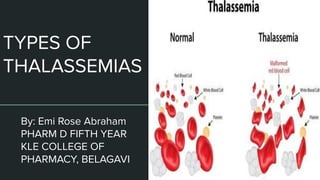
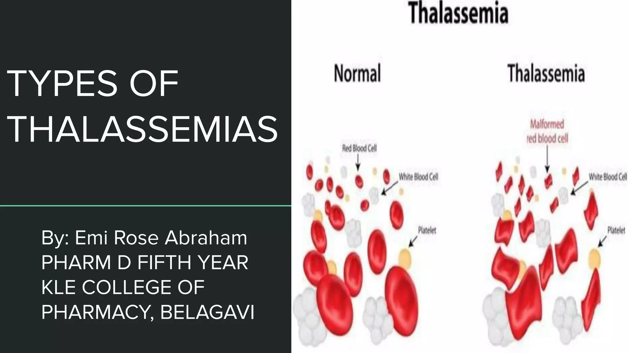
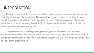
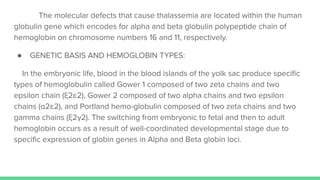
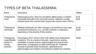
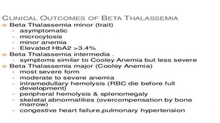
2. INTRODUCTION:
The hereditary disorders of the hemoglobin molecule are among the commonest of
clinically serious genetic conditions .They are of two general types: those in which a
mutation interferes with the amount of protein produced (thalassemias), and those that
result in a structural change of the hemoglobin molecule, leading to the production of a
variant protein (hemoglobinopathies).
Thalassemias are a heterogenous group of genetic disorders, transmitted as
autosomal recessive inheritance, in which the rate of hemoglobin production is partially or
completely suppressed due to the reduced rate of synthesis of α- or β-chain, the two chains
of adult hemo-globin (Hb A).
3. The molecular defects that cause thalassemia are located within the human
globulin gene which encodes for alpha and beta globulin polypeptide chain of
hemoglobin on chromosome numbers 16 and 11, respectively.
● GENETIC BASIS AND HEMOGLOBIN TYPES:
In the embryonic life, blood in the blood islands of the yolk sac produce specific
types of hemoglobulin called Gower 1 composed of two zeta chains and two
epsilon chain (ξ2ε2), Gower 2 composed of two alpha chains and two epsilon
chains (α2ε2), and Portland hemo-globulin composed of two zeta chains and two
gamma chains (ξ2γ2). The switching from embryonic to fetal and then to adult
hemoglobin occurs as a result of well-coordinated developmental stage due to
specific expression of globin genes in Alpha and Beta globin loci.
4.
5. TYPES OF THALASSEMIAS:
There are two main types, alpha thalassemia and beta thalassemia.The
severity of alpha and beta thalassemia depends on how many of the four genes for
alpha globin or two genes for beta globin are missing.
1. ALPHA THALASSEMIA:
Alpha-thalassemia is a form of thalassemia involving the genes HBA1 and
HBA2.In alpha-thalassemia, there is a quantitative decrease in the amount of alpha
chains, resulting in fewer normal hemoglobin molecules. Furthermore,
alpha-thalassemia leads to the production of unstable beta globin molecules which
cause increased red blood cell destruction
6. PATHOPHYSIOLOGY OF ALPHA THALASSEMIA:
The mechanism sees that α thalassemias results in decreased alpha-globin
production, therefore fewer alpha-globin chains are produced, resulting in an
excess of β chains in adults and excess γ chains in newborns. The excess β chains
form unstable tetramers called hemoglobin H or HbH of four beta chains. The
excess γ chains form tetramers which are poor carriers of O2 since their affinity for
O2 is too high, so it is not dissociated in the periphery. Homozygote α0
thalassaemias, where numerous γ4 but no α-globins occur at all (referred to as Hb
Barts), often result in death soon after birth.
7. SIGNS AND SYMPTOMS:
Common Uncommon
● Anemia: People with alpha-thalassemia can
experience both a decrease in the production
of red blood cells (RBC) and an increase in
the destruction of RBCs which can result in
other symptoms such as fatigue and
dizziness.
● Enlarged spleen
● Gallstones
● Delayed growth
● Jaundice
● Pronounced forehead: This occurs due to the
expansion of the bone marrow in the frontal
bone in the skull.The bone marrow expands
because more blood cells are being produced
in order to compensate for the anemia.
● Extramedullary hematopoesis (blood
formation outside of the bone marrow)
● Hypertension (in pregnancy)
8. TYPES OF ALPHA THALASSEMIA:
The severity of the α-thalassemias is correlated with the number of affected
α-globin alleles: the greater, the more severe will be the manifestations of the
disease. When noting the genotype, an "α" indicates a functional alpha chain, and
'-' a pathological one.
Alleles affected Description Genotype
One This is known as alpha thalassemia
silent and with this type, the effect
on hemoglobin synthesis is minimal.
Three α-globin genes are enough to
permit normal hemoglobin
production, and no clinical
symptoms present. It occurs due to
a deletion or non-deletion mutation
- α/α α
9. Two The condition is called alpha thalassemia trait; two α
genes permit nearly normal production of red blood cells,
but a mild microcytic hypochromic anemia is seen. The
disease in this form can be mistaken for iron-deficiency
anemia and treated inappropriately with iron.
Alpha-thalassemia trait can exist in two forms:
Alpha-thal-1 (- -/α α): involves cis deletion of both alpha
genes on the same chromosome. Higher incidence in
people of Asian descent when compared with the general
population.
Alpha-thal-2 (- α/- α): involves trans deletion of alpha
genes; this occurs on different (homologous)
chromosomes. Higher incidence in people of African
descent when compared with the general population
- -/α α
or
- α/- α
10. Three This condition is called hemoglobin H disease; two unstable
hemoglobins are present in the blood; hemoglobin Barts (tetrameric γ
chains) and hemoglobin H (tetrameric β chains). Both of these
unstable hemoglobins have a higher affinity for oxygen than normal
hemoglobin.A microcytic hypochromic anemia with target cells and
Heinz bodies (precipitated HbH) on the peripheral blood smear can
occur, as well as hepatosplenomegaly. The disease is noticed in
childhood or in early adult life; anemia and hepatosplenomegaly are
noted.
- -/- α
Four This is known as alpha thalassemia major; these fetuses are
edematous, have little circulating hemoglobin, and the hemoglobin that
is present is all tetrameric γ chains. When all four alleles are affected,
the fetus likely will not survive gestation without in utero intervention;
most infants with alpha-thalassemia major are stillborn with hydrops
fetalis. Fetuses treated with intrauterine transfusions throughout
pregnancy starting at an early gestational age can survive to birth with
acceptable morbidity. After birth, the treatment options include bone
marrow transplantation or continued chronic transfusions
- -/- -
11.
12. 2. BETA THALASSEMIA:
They are forms of thalassemia caused by reduced or absent synthesis of the beta
chains of hemoglobin that result in variable outcomes ranging from severe anemia
to clinically asymptomatic individuals.
● PATHOPHYSIOLOGY:
Beta thalassemias occur due to malfunctions in the hemoglobin subunit beta
or HBB. The severity of the disease depends on the nature of the mutation. HBB
blockage over time leads to decreased beta-chain synthesis. The body's inability to
construct new beta-chains leads to the underproduction of HbA.Reductions in HbA
available overall to fill the red blood cells in turn leads to microcytic anemia.
Microcytic anemia ultimately develops in respect to inadequate HBB protein for
sufficient red blood cell functioning.Due to this factor, the patient may require blood
transfusions to make up for the blockage in the beta-chains.Repeated blood
transfusions cause severe problems associated with iron overload
13. TYPES OF BETA THALASSEMIA:
Name Description Alleles
Thalassemia
minor
Heterozygous form: Only one of β globin alleles bears a mutation.
Individuals will suffer from microcytic anemia. Detection usually
involves lower than normal mean corpuscular volume value (<80 fL)
β+/β
βo/β
Thalassemia
intermedia
Affected individuals can often manage a normal life but may need
occasional transfusions, e.g., at times of illness or pregnancy,
depending on the severity of their anemia.
β+/β+
βo/β+
Thalassemia
major(Mediter
ranean or
Cooley
Anemia)
Homozygous form: Occurs when both alleles have thalassemia
mutations. This is a severe microcytic, hypochromic anemia.
Untreated, it causes anemia, splenomegaly and severe bone
deformities. It progresses to death before age 20. Treatment
consists of periodic blood transfusion; splenectomy for
splenomegaly and chelation of transfusion-related iron overload.
βo/βo
14.
15.
16. Reference:
1. THALASSAEMIA INTERNATIONAL FEDERATION, GUIDELINES FOR THE
MANAGEMENT OF NON TRANSFUSION DEPENDENT THALASSAEMIA (NTDT)
2ND EDITION.
2. NATIONAL HEALTH MISSION GUIDELINES ON HEMOGLOBINOPATHIES IN
INDIA, 2016.
3. Michael Angastiniotis and Stephan Lobitz ,Thalassemias: An Overview, 20
March 2019
4. Isam Jaber Al-Zwaini, Introductory Chapter: Thalassemia - An Overview, 2018