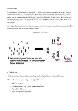
Kurdistan Regional Government – Iraq
Ministry of Higher Education and Scientific Research
Duhok polytechnic university
Shekhan technical college of health
Report aboutThalassemia
Shekhan technical college of health
Medical Lab. Technology
Academic Debate
( 2015 – 2016 )
Content :-
 Introduction
 Thalassemia
 Hemoglobinopathies
 Incidence of thalassemia in Thailand
 Mode of inheritance
 How to name thalassemia ?
 Common types of thalassemia
 Alpha Thalassemia
 Symbolism Alpha Thalassemia
 Classification & Terminology Alpha Thalassemia
 Types of a-thalassemia
 Compound heterozygotes
 Comparison of α Thalassemias
 Beta thalassemias (β thalassemias)
 Types of β Thalassemia
 β Thalassemia Symptoms Include
 Diagnosis of Beta Thalassemia
 Prevention
 What Is The Treatment For Thalassemia
 Conclusion
 References
Introduction :-
Thalassemia is an inherited disorder of autosomal recessive gene disorder caused by impaired synthesis of one or
more globin chains. The impairment alters production of hemoglobin (Hb) (Ridolfi et al., 2002). Thalassemia
causes varying degrees of anemia, which can range from significant to life threatening. People of Mediterranean,
Middle Eastern, African, and Southeast Asian descent are at higher risk of carrying the genes for thalassemia
(Weatherall, 1997). These hereditary anemias are caused by mutations that decrease hemoglobin synthesis and
red cell survival. These hereditary anemia caused by decreased or absent production of one type of globin chain
either ┙ or ┚ globin chain.
These hematologic disorders range from asymptomatic to severe anemia that can cause significant morbidity and
mortality. It was first recognized clinically in 1925 by Dr. Thomas Cooley who described a syndrome of anemia
with microcytic erythrocytes. Then it was called Cooley’s anemia. Later Wipple and Bradford renamed this
disease as “Thalassemia.” Because it was found in the region of the Mediterranean Sea (thalasa is an old Greek
word for sea) (Cooley, 1946). Thalassemias can cause significant problems because these are inherited disorders,
newborn screening and prenatal diagnosis are important in management of patients. This topic will review the
clinical features of thalassemia while focusing on pathophysiology, clinical features, complication, management,
screening and diagnosis.
Thalassemia
 Syndromes arising form decreased rate or absence of globin chain synthesis.
 The resulting imbalance-globin chain synthesis takes place, giving rise to the excess amount of
the normally synthesized globin chain.
F.1
Hemoglobinopathies :-
 The syndrome arising from the synthesis of abnormal haemoglobin or hemoglobin variants.
 Rate of globin chain synthesis are theoritically normal.
 Abnormal hemoglobins have different properties from the normal ones.
Hemoglobinopathies
F.2
Incidence of thalassemia in Thailand
 a-thalassemia : 20-30%
 b-thalassemia : 3-9%
 Hb E : 8-70 % (very high in E-sarn (
 Hb Constant Spring : 1-6%
 Thalassemia disease : 1%
Mode of inheritance
 Autosomal recessive
 Heterozygote or double heterozygote are not affected.
 Homozygote or compound heterozygote are affected.
How to name thalassemia?
Named after globin chain that is abnormally synthesized!!!!
 Reduced or absent a-globin chain : a-thalassemia
 Reduced or absent b-globin chain : b-thalassemia
 Reduced or absent g-globin chain : g-thalassemia
 Reduced or absent d-globin chain : d-thalassemia
 Reduced or absent gdb-globin chains : gdb-thalassemia
F.3
Common types of thalassemia :-
1. a-thalassemia
2. b-thalassemia
ALPHA THALASSEMIAS
Absence of α chains will result in increase/ excess of g globin chains during fetal life and excess β globin chains
later in postnatal life.
Severity of disease depends on number of genes affected.
Symbolism Alpha Thalassemia
)/(Indicates division between genes inherited from both parents :aa/aa (Normal)
 Each chromosome 16 carries 2 genes. Therefore the total complement of a genes in an individual is 4.
(-) Indicates a gene deletion: - a/aa
 alpha+ Thalassemia (one gene deletion)
 3 functional working genes.
 Called alpha thal 2.
(-) Indicates a gene deletion: ---/ aa
 Alpha 0 Thalassemia (two gene deletion) in the same chromosome.
 2functional working genes.
 Called alpha thal 1.
Symbolism Other Thalassemia
Superscript T denotes nonfunctioning (mutated gene, not deletion) gene :
 Alpha T
Classification & Terminology Alpha Thalassemia :-
Normal alpha alpha/alpha alpha
Silent carrier -alpha/alpha alpha
Minor -alpha/-alpha
--/ alpha alpha
Hb H disease --/- alpha
Barts hydrops fetalis --/--
α Thalassemia
The most common cause of α thalassemia is due to α gene/s deletions.
The most likely mechanism for α gene deletion is due to homologous pairing between α1 and α2 and
recombination. This results in loss of α gene. Other causes of α thalassemias are deletions in the locus control
regions (HS40) or chain termination mutations (nonsense mutations) .
Types of a-thalassemia :-
a-thalassemia-1 or ao-thalassemia (--)
F.4
a-thalassemia-2 or a+-thalassemia (-a)
F.5
Compound heterozygotes :-
Hb H disease ( --/-a)
F.6
Hb Bart’s hydrops fetalis syndrome (--/--)
F.7
α Thalassemia
As said, the genetic basis of α thal is mostly deletions: If you have 4 functional α genes, then you are normal.
With 3 functional α genes, you are a silent carrier . With 2 functional α genes you have α thalassemia trait which
is clinically benign, but there is mild microcytic anemia .With only one functional α chain, you have severe
hemolytic anemia with primarily HbH, composed of 4 β chains (β4). This is clinically severe. In the absence of α
chain in the fetus, the gamma forms a tetramer of globin chains, and is called Hb Bart’s. Both Hb-H and Hb-
Barts are high affinity Hbs, thus neither of them is capable of releasing oxygen to the tissues, also these
hemoglobins are fast moving hemoglobins in Hb electrophoresis at alkaline pH .
Infants with severe α Thalassemia (zero functional alpha genes) and Hb Barts suffer from severe intrauterine
hypoxia and are born with massive generalized fluid accumulation, a condition known as hydrops fetalis or also
called erythroblastosis fetalis.
in α Thalassemia
Is usually caused by deletion of 1 or more of the 4 α globin genes on chromosome 16. And Severity of disease
depends on number of the deleted α genes.and Absence of α chains will result in increase/ excess of g chains
during fetal life and excess β chains later in life; Causes hemoglobins like Hb Bart's (g4) or HbH (β4), to form
which are physiologically useless (very high affinity) . and Like β thalassemia the excess globin chains causes
the problem.
But: Alpha chain accumulation and deposition are more toxic than beta chain accumulation and deposition. Thus
beta thalassemia is more severe than alpha thalassemia.
α Thalassemia: Hb-H Disease
α Thalassemia
Predominant cause of alpha thalassemias is large number of gene deletions in the α-globin genes .
There are four clinical syndromes present in alpha thalassemia -:
 Silent Carrier State
 Alpha Thalassemia Trait (Alpha Thalassemia Minor (
 Hemoglobin H Disease
 Bart's Hydrops Fetalis Syndrome
Silent Carrier α Thalassemia
 -α/αα
 One alpha gene deletion, 3 intact alpha genes.
 Healthy persons.
 Normal Hb and Hct
 No treatment
 Can only be detected by DNA studies.
Comparison of α Thalassemias
Phenotype Hb A Hb Barts Hb H
Normal 97-98% 0 0
Silent Carrier 96-98% %0-2
(At birth)
0
α Thalassemia Trait 85-95% %2-8
(At birth)
<2%
Hb H Disease Dec <10 %
(At birth)
5-40%
Hydrops Fetalis 0 70-80% (with
20% Hb
Portland)
0-20%
Table number 1
Beta thalassemias (β thalassemias) :-
They are the most important types of thalassemias because they are so common and usually produce severe
anemia in their homozygous and compound heterozygous states (compound= when combined with other
hemoglobinopathies or thalassemias(
Beta thalassemias are autosomal inherited disorders of b globin synthesis. In most, globin structure is normal but
the rate of production is reduced because of decrease in transcription of DNA, abnormal processing of pre-
mRNA, or decreased translation of mRNA leading to decreased Hb-A production (A=Adult).
Usually and mostly they are caused by gene mutations in the b gene in chromosome 11, although deletions do
occur. Hundreds of mutations possible in the b globin gene, therefore b thalassemia is more diverse disease in its
presentation (the presentation differs between people depending on the type of mutation.) This results in excess
alpha chains, because they cannot find their counterparts (the beta chains) to bind to.
Excess alpha chains
The classes of mutations that underlie β-thalassaemia :-
F.8
Types of βThalassemia
Three common types of b Thalassemia:
 Thalassemia major : transfusion dependent , Anaemia is sever ( Hb about 5 g/dl ) , Starts within first year
of life.
 Thalassemia intermedia : moderate anaemia ( Hb >7 g/dl ) , Infrequent or on need for transfusions , Late
presentation and long survival.
 Thalassemia minor : mild or no anaemia , Minimal red cell morphological changes , increased Hb-A2 ,
also called thalassemia trait or carrier.
F.9
β Thalassemia Symptoms Include :-
 Fatigue
 Weakness
 Pale appearance
 Yellow discoloration of skin (jaundice)
 Facial bone deformities
 Slow growth
 Abdominal swelling
 Dark urine
Diagnosis of Beta Thalassemia :-
 Blood smear : you can seen
1. microcytic (low MCV (
2. Hypochromic.
3. Vary in size (anisocytosis (
4. Vary in shape (poikilocytosis (
5. Be nucleated (normal, mature RBCs do not have a nucleus(
6. target cells
 Complete blood count ( CBC ) : If the MCV is low , thalassemia should be considered.
 Iron : ferritin, unsaturated iron binding capacity (UIBC), total iron binding capacity (TIBC), andpercent
saturation of transferrin.
 Reticulocyte count
 Genetic testing
 Prenatal testing
 Chorionic villus sampling
 Amniocentesis
Prevention :-
In most cases, thalassemia Cannot be prevented.
If you have Thalassemia, or if you carry a thalassemia gene, consider talking with a genetic counselor for
guidance before you have or father a child .
What Is The Treatment For Thalassemia ?
 Thalassemia patients would require frequent blood transfusions depending upon the severity of the
disease. They would also require medication to reduce the iron overload.
 Spleenectomy is done electively to reduce the destruction of blood cells, if the spleen is very much
enlarged.
 Recent treatment modalities include the use of cord blood cell of the sibling which aim at curing
Thalessemia.
Conclusion :-
 It is the most common genetic disorder on worldwide basis .
 Children with thalassemia have shorter RBC life, more HbF, & the RBCS are more sensitive to the
oxidative stress .
 Thalassemia have main three type (THALASSEMIA INTERMEDIA and THALASSEMIA MINIMA &
MINOR and THALASSEMIA TRAI )
 A Thalassemia is caused by mutations in the DNA of cells that make haemoglobin.
 The mutations associated with thalassemia are passed from parents to children.
References :-
 Mayo Clinic. "Thalassemia". Mayo Clinic. Retrieved 17 October 2014.
 Robbins Basic Pathology, Page No:428
 Weatherall, David J. "Ch. 47: The Thalassemias: Disorders of Globin Synthesis". In Lichtman MA, Kipps
TJ, Seligsohn U, Kaushansky K, Prchal, JT. Williams Hematology (8e ed.).
 http://www.cdc.gov/ncbddd/thalassemia/
 http://www.healthline.com/health/thalassemia
 https://en.wikipedia.org/wiki/Thalassemia
 Pediatric Thalassemia~treatment at eMedicine
 Harrison's Principles of Internal Medicine (17th ed.). McGraw-Hill medical. September 2008. p. 776.
ISBN 0-07-164114-9.

Thalassemia

  • 2.
    Kurdistan Regional Government– Iraq Ministry of Higher Education and Scientific Research Duhok polytechnic university Shekhan technical college of health Report aboutThalassemia Shekhan technical college of health Medical Lab. Technology Academic Debate ( 2015 – 2016 )
  • 3.
    Content :-  Introduction Thalassemia  Hemoglobinopathies  Incidence of thalassemia in Thailand  Mode of inheritance  How to name thalassemia ?  Common types of thalassemia  Alpha Thalassemia  Symbolism Alpha Thalassemia  Classification & Terminology Alpha Thalassemia  Types of a-thalassemia  Compound heterozygotes  Comparison of α Thalassemias  Beta thalassemias (β thalassemias)  Types of β Thalassemia  β Thalassemia Symptoms Include  Diagnosis of Beta Thalassemia  Prevention  What Is The Treatment For Thalassemia  Conclusion  References
  • 4.
    Introduction :- Thalassemia isan inherited disorder of autosomal recessive gene disorder caused by impaired synthesis of one or more globin chains. The impairment alters production of hemoglobin (Hb) (Ridolfi et al., 2002). Thalassemia causes varying degrees of anemia, which can range from significant to life threatening. People of Mediterranean, Middle Eastern, African, and Southeast Asian descent are at higher risk of carrying the genes for thalassemia (Weatherall, 1997). These hereditary anemias are caused by mutations that decrease hemoglobin synthesis and red cell survival. These hereditary anemia caused by decreased or absent production of one type of globin chain either ┙ or ┚ globin chain. These hematologic disorders range from asymptomatic to severe anemia that can cause significant morbidity and mortality. It was first recognized clinically in 1925 by Dr. Thomas Cooley who described a syndrome of anemia with microcytic erythrocytes. Then it was called Cooley’s anemia. Later Wipple and Bradford renamed this disease as “Thalassemia.” Because it was found in the region of the Mediterranean Sea (thalasa is an old Greek word for sea) (Cooley, 1946). Thalassemias can cause significant problems because these are inherited disorders, newborn screening and prenatal diagnosis are important in management of patients. This topic will review the clinical features of thalassemia while focusing on pathophysiology, clinical features, complication, management, screening and diagnosis. Thalassemia  Syndromes arising form decreased rate or absence of globin chain synthesis.  The resulting imbalance-globin chain synthesis takes place, giving rise to the excess amount of the normally synthesized globin chain. F.1
  • 5.
    Hemoglobinopathies :-  Thesyndrome arising from the synthesis of abnormal haemoglobin or hemoglobin variants.  Rate of globin chain synthesis are theoritically normal.  Abnormal hemoglobins have different properties from the normal ones. Hemoglobinopathies F.2 Incidence of thalassemia in Thailand  a-thalassemia : 20-30%  b-thalassemia : 3-9%  Hb E : 8-70 % (very high in E-sarn (  Hb Constant Spring : 1-6%  Thalassemia disease : 1% Mode of inheritance  Autosomal recessive  Heterozygote or double heterozygote are not affected.  Homozygote or compound heterozygote are affected. How to name thalassemia? Named after globin chain that is abnormally synthesized!!!!  Reduced or absent a-globin chain : a-thalassemia  Reduced or absent b-globin chain : b-thalassemia  Reduced or absent g-globin chain : g-thalassemia  Reduced or absent d-globin chain : d-thalassemia  Reduced or absent gdb-globin chains : gdb-thalassemia
  • 6.
    F.3 Common types ofthalassemia :- 1. a-thalassemia 2. b-thalassemia ALPHA THALASSEMIAS Absence of α chains will result in increase/ excess of g globin chains during fetal life and excess β globin chains later in postnatal life. Severity of disease depends on number of genes affected. Symbolism Alpha Thalassemia )/(Indicates division between genes inherited from both parents :aa/aa (Normal)  Each chromosome 16 carries 2 genes. Therefore the total complement of a genes in an individual is 4. (-) Indicates a gene deletion: - a/aa  alpha+ Thalassemia (one gene deletion)  3 functional working genes.  Called alpha thal 2. (-) Indicates a gene deletion: ---/ aa  Alpha 0 Thalassemia (two gene deletion) in the same chromosome.  2functional working genes.  Called alpha thal 1. Symbolism Other Thalassemia Superscript T denotes nonfunctioning (mutated gene, not deletion) gene :  Alpha T
  • 7.
    Classification & TerminologyAlpha Thalassemia :- Normal alpha alpha/alpha alpha Silent carrier -alpha/alpha alpha Minor -alpha/-alpha --/ alpha alpha Hb H disease --/- alpha Barts hydrops fetalis --/-- α Thalassemia The most common cause of α thalassemia is due to α gene/s deletions. The most likely mechanism for α gene deletion is due to homologous pairing between α1 and α2 and recombination. This results in loss of α gene. Other causes of α thalassemias are deletions in the locus control regions (HS40) or chain termination mutations (nonsense mutations) . Types of a-thalassemia :- a-thalassemia-1 or ao-thalassemia (--) F.4 a-thalassemia-2 or a+-thalassemia (-a) F.5
  • 8.
    Compound heterozygotes :- HbH disease ( --/-a) F.6 Hb Bart’s hydrops fetalis syndrome (--/--) F.7 α Thalassemia As said, the genetic basis of α thal is mostly deletions: If you have 4 functional α genes, then you are normal. With 3 functional α genes, you are a silent carrier . With 2 functional α genes you have α thalassemia trait which is clinically benign, but there is mild microcytic anemia .With only one functional α chain, you have severe hemolytic anemia with primarily HbH, composed of 4 β chains (β4). This is clinically severe. In the absence of α chain in the fetus, the gamma forms a tetramer of globin chains, and is called Hb Bart’s. Both Hb-H and Hb- Barts are high affinity Hbs, thus neither of them is capable of releasing oxygen to the tissues, also these hemoglobins are fast moving hemoglobins in Hb electrophoresis at alkaline pH . Infants with severe α Thalassemia (zero functional alpha genes) and Hb Barts suffer from severe intrauterine hypoxia and are born with massive generalized fluid accumulation, a condition known as hydrops fetalis or also called erythroblastosis fetalis.
  • 9.
    in α Thalassemia Isusually caused by deletion of 1 or more of the 4 α globin genes on chromosome 16. And Severity of disease depends on number of the deleted α genes.and Absence of α chains will result in increase/ excess of g chains during fetal life and excess β chains later in life; Causes hemoglobins like Hb Bart's (g4) or HbH (β4), to form which are physiologically useless (very high affinity) . and Like β thalassemia the excess globin chains causes the problem. But: Alpha chain accumulation and deposition are more toxic than beta chain accumulation and deposition. Thus beta thalassemia is more severe than alpha thalassemia. α Thalassemia: Hb-H Disease α Thalassemia Predominant cause of alpha thalassemias is large number of gene deletions in the α-globin genes . There are four clinical syndromes present in alpha thalassemia -:  Silent Carrier State  Alpha Thalassemia Trait (Alpha Thalassemia Minor (  Hemoglobin H Disease  Bart's Hydrops Fetalis Syndrome
  • 10.
    Silent Carrier αThalassemia  -α/αα  One alpha gene deletion, 3 intact alpha genes.  Healthy persons.  Normal Hb and Hct  No treatment  Can only be detected by DNA studies. Comparison of α Thalassemias Phenotype Hb A Hb Barts Hb H Normal 97-98% 0 0 Silent Carrier 96-98% %0-2 (At birth) 0 α Thalassemia Trait 85-95% %2-8 (At birth) <2% Hb H Disease Dec <10 % (At birth) 5-40% Hydrops Fetalis 0 70-80% (with 20% Hb Portland) 0-20% Table number 1 Beta thalassemias (β thalassemias) :- They are the most important types of thalassemias because they are so common and usually produce severe anemia in their homozygous and compound heterozygous states (compound= when combined with other hemoglobinopathies or thalassemias( Beta thalassemias are autosomal inherited disorders of b globin synthesis. In most, globin structure is normal but the rate of production is reduced because of decrease in transcription of DNA, abnormal processing of pre- mRNA, or decreased translation of mRNA leading to decreased Hb-A production (A=Adult). Usually and mostly they are caused by gene mutations in the b gene in chromosome 11, although deletions do occur. Hundreds of mutations possible in the b globin gene, therefore b thalassemia is more diverse disease in its presentation (the presentation differs between people depending on the type of mutation.) This results in excess alpha chains, because they cannot find their counterparts (the beta chains) to bind to.
  • 11.
    Excess alpha chains Theclasses of mutations that underlie β-thalassaemia :- F.8 Types of βThalassemia Three common types of b Thalassemia:  Thalassemia major : transfusion dependent , Anaemia is sever ( Hb about 5 g/dl ) , Starts within first year of life.  Thalassemia intermedia : moderate anaemia ( Hb >7 g/dl ) , Infrequent or on need for transfusions , Late presentation and long survival.  Thalassemia minor : mild or no anaemia , Minimal red cell morphological changes , increased Hb-A2 , also called thalassemia trait or carrier.
  • 12.
    F.9 β Thalassemia SymptomsInclude :-  Fatigue  Weakness  Pale appearance  Yellow discoloration of skin (jaundice)  Facial bone deformities  Slow growth  Abdominal swelling  Dark urine Diagnosis of Beta Thalassemia :-  Blood smear : you can seen 1. microcytic (low MCV ( 2. Hypochromic. 3. Vary in size (anisocytosis ( 4. Vary in shape (poikilocytosis ( 5. Be nucleated (normal, mature RBCs do not have a nucleus( 6. target cells  Complete blood count ( CBC ) : If the MCV is low , thalassemia should be considered.  Iron : ferritin, unsaturated iron binding capacity (UIBC), total iron binding capacity (TIBC), andpercent saturation of transferrin.  Reticulocyte count  Genetic testing  Prenatal testing  Chorionic villus sampling  Amniocentesis
  • 13.
    Prevention :- In mostcases, thalassemia Cannot be prevented. If you have Thalassemia, or if you carry a thalassemia gene, consider talking with a genetic counselor for guidance before you have or father a child . What Is The Treatment For Thalassemia ?  Thalassemia patients would require frequent blood transfusions depending upon the severity of the disease. They would also require medication to reduce the iron overload.  Spleenectomy is done electively to reduce the destruction of blood cells, if the spleen is very much enlarged.  Recent treatment modalities include the use of cord blood cell of the sibling which aim at curing Thalessemia. Conclusion :-  It is the most common genetic disorder on worldwide basis .  Children with thalassemia have shorter RBC life, more HbF, & the RBCS are more sensitive to the oxidative stress .  Thalassemia have main three type (THALASSEMIA INTERMEDIA and THALASSEMIA MINIMA & MINOR and THALASSEMIA TRAI )  A Thalassemia is caused by mutations in the DNA of cells that make haemoglobin.  The mutations associated with thalassemia are passed from parents to children.
  • 14.
    References :-  MayoClinic. "Thalassemia". Mayo Clinic. Retrieved 17 October 2014.  Robbins Basic Pathology, Page No:428  Weatherall, David J. "Ch. 47: The Thalassemias: Disorders of Globin Synthesis". In Lichtman MA, Kipps TJ, Seligsohn U, Kaushansky K, Prchal, JT. Williams Hematology (8e ed.).  http://www.cdc.gov/ncbddd/thalassemia/  http://www.healthline.com/health/thalassemia  https://en.wikipedia.org/wiki/Thalassemia  Pediatric Thalassemia~treatment at eMedicine  Harrison's Principles of Internal Medicine (17th ed.). McGraw-Hill medical. September 2008. p. 776. ISBN 0-07-164114-9.