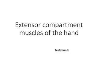
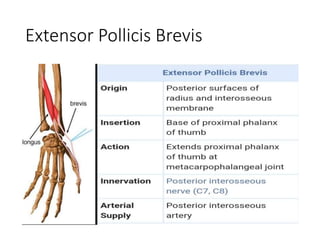
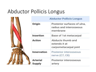
Can read freely here
https://sethiortho.blogspot.com/
Thoracic outlet syndrome
Neurovascular symptoms in the upper extremities due to pressure on the nerves and vessels in the thoracic outlet area
The specific structures compressed are usually the nerves of the branchial plexus and occasionally the subclavian artery or subclavian vein
Anatomy
Thoracic outlet
Entrance/ Exit region of the upper limb
The thoracic outlet is defined as the interval from the supraclavicular fossa to the axilla that passes between the clavicle and the first rib
Anatomy - Scalane triangle
Anatomy of the costoclavicular space
Pectoralis minor space
Located inferior to the coracoid process
anterior to the second through fourth ribs
posterior to the pectoralis minor muscle
The cords of the brachial plexus
Axillary artery
Axillary vein.
Soft-tissue Causes (70%)
Scalene muscle
Variations in insertion
Hypertrophy
Accessory scalenus minimus muscle
Anomalous ligaments or bands
Soft-tissue tumors
Osseous Causes
Cervical rib
Prominent C7 transverse process
Displacement or callus from first rib fracture
Malunited clavicle or first rib fracture
AC or SC joint injury or dislocation
Osseous tumor
Poor posture
Drooping the shoulders
Holding the head in a forward position
Repetitive activity
Athletes and swimmers
Neurogenic TOS
Compression – scalene triangle and costoclavicular space
May be associated with normal anatomy
Traction of the lowest trunk of the brachial plexus
Often in association with arterial TOS
Features of Lower brachial plexus compression - Common
Female predominance
Appearance of Amedio Modigliani painting
Complains of pain and paresthesia extending from the shoulder /down the ulnar aspect of the arm into the medial two fingers
Neurogenic TOS
Upper brachial plexus compression C5,C6 and C7
Less common
Compression mainly occurs in scalene triangle
Symptoms
Unilateral occipito-frontal headache
Facial or jaw pain
The Gilliatt-Sumner hand
A characteristic finding of neurogenic TOS, is described as atrophy of the abductor pollicis brevis and, to a lesser degree, the hypothenar musculature and the interossei.
Venous TOS
Causes
Hypertrophy of the subclavius muscle,
Chondroma formation
Clinical presentation
Most patients are sportsmen, musicians or manual workers undertaking repetitive arm movements.
The condition occurs more commonly in the dominant limb
Male predominance
Clinical presentation
Acute presentation -
Swollen and tensed upper limb
Upper limb aching pain
blueish- purple arm due to venous engorgement
Collateral veins may be visible
Feeling of heaviness that is worse after activity
Symptoms are precipitated by working with the arms elevated and are relieved by dependency, a pathognomonic feature of vTOS.
Arterial TOS
Rare but has more devastating consequences
Caused by
Intermittent subclavian arterial compression - Costoclavicular compression with normal anatomy.
Flu Vaccine Alert in Bangalore Karnatakaaddon Scans
As flu season approaches, health officials in Bangalore, Karnataka, are urging residents to get their flu vaccinations. The seasonal flu, while common, can lead to severe health complications, particularly for vulnerable populations such as young children, the elderly, and those with underlying health conditions.
Dr. Vidisha Kumari, a leading epidemiologist in Bangalore, emphasizes the importance of getting vaccinated. "The flu vaccine is our best defense against the influenza virus. It not only protects individuals but also helps prevent the spread of the virus in our communities," he says.
This year, the flu season is expected to coincide with a potential increase in other respiratory illnesses. The Karnataka Health Department has launched an awareness campaign highlighting the significance of flu vaccinations. They have set up multiple vaccination centers across Bangalore, making it convenient for residents to receive their shots.
To encourage widespread vaccination, the government is also collaborating with local schools, workplaces, and community centers to facilitate vaccination drives. Special attention is being given to ensuring that the vaccine is accessible to all, including marginalized communities who may have limited access to healthcare.
Residents are reminded that the flu vaccine is safe and effective. Common side effects are mild and may include soreness at the injection site, mild fever, or muscle aches. These side effects are generally short-lived and far less severe than the flu itself.
Healthcare providers are also stressing the importance of continuing COVID-19 precautions. Wearing masks, practicing good hand hygiene, and maintaining social distancing are still crucial, especially in crowded places.
Protect yourself and your loved ones by getting vaccinated. Together, we can help keep Bangalore healthy and safe this flu season. For more information on vaccination centers and schedules, residents can visit the Karnataka Health Department’s official website or follow their social media pages.
Stay informed, stay safe, and get your flu shot today!
ARTIFICIAL INTELLIGENCE IN HEALTHCARE.pdfAnujkumaranit
Artificial intelligence (AI) refers to the simulation of human intelligence processes by machines, especially computer systems. It encompasses tasks such as learning, reasoning, problem-solving, perception, and language understanding. AI technologies are revolutionizing various fields, from healthcare to finance, by enabling machines to perform tasks that typically require human intelligence.
Title: Sense of Smell
Presenter: Dr. Faiza, Assistant Professor of Physiology
Qualifications:
MBBS (Best Graduate, AIMC Lahore)
FCPS Physiology
ICMT, CHPE, DHPE (STMU)
MPH (GC University, Faisalabad)
MBA (Virtual University of Pakistan)
Learning Objectives:
Describe the primary categories of smells and the concept of odor blindness.
Explain the structure and location of the olfactory membrane and mucosa, including the types and roles of cells involved in olfaction.
Describe the pathway and mechanisms of olfactory signal transmission from the olfactory receptors to the brain.
Illustrate the biochemical cascade triggered by odorant binding to olfactory receptors, including the role of G-proteins and second messengers in generating an action potential.
Identify different types of olfactory disorders such as anosmia, hyposmia, hyperosmia, and dysosmia, including their potential causes.
Key Topics:
Olfactory Genes:
3% of the human genome accounts for olfactory genes.
400 genes for odorant receptors.
Olfactory Membrane:
Located in the superior part of the nasal cavity.
Medially: Folds downward along the superior septum.
Laterally: Folds over the superior turbinate and upper surface of the middle turbinate.
Total surface area: 5-10 square centimeters.
Olfactory Mucosa:
Olfactory Cells: Bipolar nerve cells derived from the CNS (100 million), with 4-25 olfactory cilia per cell.
Sustentacular Cells: Produce mucus and maintain ionic and molecular environment.
Basal Cells: Replace worn-out olfactory cells with an average lifespan of 1-2 months.
Bowman’s Gland: Secretes mucus.
Stimulation of Olfactory Cells:
Odorant dissolves in mucus and attaches to receptors on olfactory cilia.
Involves a cascade effect through G-proteins and second messengers, leading to depolarization and action potential generation in the olfactory nerve.
Quality of a Good Odorant:
Small (3-20 Carbon atoms), volatile, water-soluble, and lipid-soluble.
Facilitated by odorant-binding proteins in mucus.
Membrane Potential and Action Potential:
Resting membrane potential: -55mV.
Action potential frequency in the olfactory nerve increases with odorant strength.
Adaptation Towards the Sense of Smell:
Rapid adaptation within the first second, with further slow adaptation.
Psychological adaptation greater than receptor adaptation, involving feedback inhibition from the central nervous system.
Primary Sensations of Smell:
Camphoraceous, Musky, Floral, Pepperminty, Ethereal, Pungent, Putrid.
Odor Detection Threshold:
Examples: Hydrogen sulfide (0.0005 ppm), Methyl-mercaptan (0.002 ppm).
Some toxic substances are odorless at lethal concentrations.
Characteristics of Smell:
Odor blindness for single substances due to lack of appropriate receptor protein.
Behavioral and emotional influences of smell.
Transmission of Olfactory Signals:
From olfactory cells to glomeruli in the olfactory bulb, involving lateral inhibition.
Primitive, less old, and new olfactory systems with different path
Prix Galien International 2024 Forum ProgramLevi Shapiro
June 20, 2024, Prix Galien International and Jerusalem Ethics Forum in ROME. Detailed agenda including panels:
- ADVANCES IN CARDIOLOGY: A NEW PARADIGM IS COMING
- WOMEN’S HEALTH: FERTILITY PRESERVATION
- WHAT’S NEW IN THE TREATMENT OF INFECTIOUS,
ONCOLOGICAL AND INFLAMMATORY SKIN DISEASES?
- ARTIFICIAL INTELLIGENCE AND ETHICS
- GENE THERAPY
- BEYOND BORDERS: GLOBAL INITIATIVES FOR DEMOCRATIZING LIFE SCIENCE TECHNOLOGIES AND PROMOTING ACCESS TO HEALTHCARE
- ETHICAL CHALLENGES IN LIFE SCIENCES
- Prix Galien International Awards Ceremony
The prostate is an exocrine gland of the male mammalian reproductive system
It is a walnut-sized gland that forms part of the male reproductive system and is located in front of the rectum and just below the urinary bladder
Function is to store and secrete a clear, slightly alkaline fluid that constitutes 10-30% of the volume of the seminal fluid that along with the spermatozoa, constitutes semen
A healthy human prostate measures (4cm-vertical, by 3cm-horizontal, 2cm ant-post ).
It surrounds the urethra just below the urinary bladder. It has anterior, median, posterior and two lateral lobes
It’s work is regulated by androgens which are responsible for male sex characteristics
Generalised disease of the prostate due to hormonal derangement which leads to non malignant enlargement of the gland (increase in the number of epithelial cells and stromal tissue)to cause compression of the urethra leading to symptoms (LUTS
These lecture slides, by Dr Sidra Arshad, offer a quick overview of physiological basis of a normal electrocardiogram.
Learning objectives:
1. Define an electrocardiogram (ECG) and electrocardiography
2. Describe how dipoles generated by the heart produce the waveforms of the ECG
3. Describe the components of a normal electrocardiogram of a typical bipolar leads (limb II)
4. Differentiate between intervals and segments
5. Enlist some common indications for obtaining an ECG
Study Resources:
1. Chapter 11, Guyton and Hall Textbook of Medical Physiology, 14th edition
2. Chapter 9, Human Physiology - From Cells to Systems, Lauralee Sherwood, 9th edition
3. Chapter 29, Ganong’s Review of Medical Physiology, 26th edition
4. Electrocardiogram, StatPearls - https://www.ncbi.nlm.nih.gov/books/NBK549803/
5. ECG in Medical Practice by ABM Abdullah, 4th edition
6. ECG Basics, http://www.nataliescasebook.com/tag/e-c-g-basics
Ozempic: Preoperative Management of Patients on GLP-1 Receptor Agonists Saeid Safari
Preoperative Management of Patients on GLP-1 Receptor Agonists like Ozempic and Semiglutide
ASA GUIDELINE
NYSORA Guideline
2 Case Reports of Gastric Ultrasound
5. Associated Pathology
• De Quervain's tenosynovitis is a stenosing
tenosynovial inflammation of the 1st dorsal
compartment.
• Diagnosis is made clinically with radial sided wrist
pain made worse with the Finkelstein maneuver.
• Treatment is generally conservative with thumb
spica braces, injections and in refractory cases, 1st
dorsal compartment surgical release.
•
•
6. Epidemiology
• Incidence
• very common
• ~1 per 1000 annually
• Demographics
• woman > men , 30 - 50 years old
• Anatomic location
• most commonly in the dominant wrist
• Risk factors
• overuse
• golfers and racquet sports
• post-traumatic
• postpartum
•
7. Etiology
• Pathophysiology
• pathoanatomy
• thickening and swelling of extensor
retinaculum causes increased tendon friction
• NOT considered an inflammatory process
• may be related to accumulation of
mucopolysaccharides
•
•
8. Presentation
• Symptoms
• gradual onset
• radial sided wrist pain
pain exacerbated by gripping and raising objects with wrist in
neutral
Physical exam
Inspection
tenderness over 1st dorsal compartment at level of radial
styloid
motion
usually normal wrist motion
pain with resisted radial deviation
neurovascular exam
normal
9. provocative tests
• Finkelstein maneuver
• On grasping the patient’s thumb and quickly
abducting the hand ulnarward, the pain over the
styloid tip is painful
• more indicative of EPB > APL tendon pathology
• Eichhoff maneuver
• ulnar deviated wrist while patient clenches thumb
in fist, followed by relief of pain once the thumb is
extended even if the wrist remains ulnar deviated
•
•
10. Imaging
• Radiographs
• recommended views-- AP, lateral views of wrist
• indications
• radiographs usually not indicated
• findings
• may be used to rule out
• - basilar arthritis of the thumb
• - carpal arthritis
•
•
13. Treatment
• Nonoperative
• rest, NSAIDS, thumb spica splint, steroid iinjectin
• indications--- first line of treatment
• technique
• NSAIDS, rest and immobilisation usually first step
• steroid injections into first dorsal compartment usually
second step
• outcomes
• overall corticosteriods found to be superior to splinting
• concomitant splinting and/or NSAIDs after steriods
injection does not improve outcomes
•
•
14. Trt...ctnd
• Operative
surgical release of 1st dorsal compartment
indications
severe symptoms
usually consider after 6 months of failed
nonoperative management
technique
radial based incision proximal to the wrist
protect the superficial radial sensory nerve
15. Try.. ctd..
• Techniques
Surgical release of 1st dorsal compartment
approach
transverse incision with release on dorsal side of 1st
compartment to prevent volar subluxation of the tendon
EPB is more dorsal than APL
has variable anatomy with APL usually having at
least 2 tendon slips and its own fibro-osseous
compartment
a distinct EPB sheath is often encountered dorsally
16. Complications
• Sensory branch of radial nerve injury
• Neuroma formation
• Failure to decompress with recurrence
• may be caused by failure to recognize and
decompress EPB or APL lying in separate
subsheath/compartment
• Complex regional pain syndrome
• Prognosis
• Most cases resolve with non-operative management
• High recurrence rate
•
•