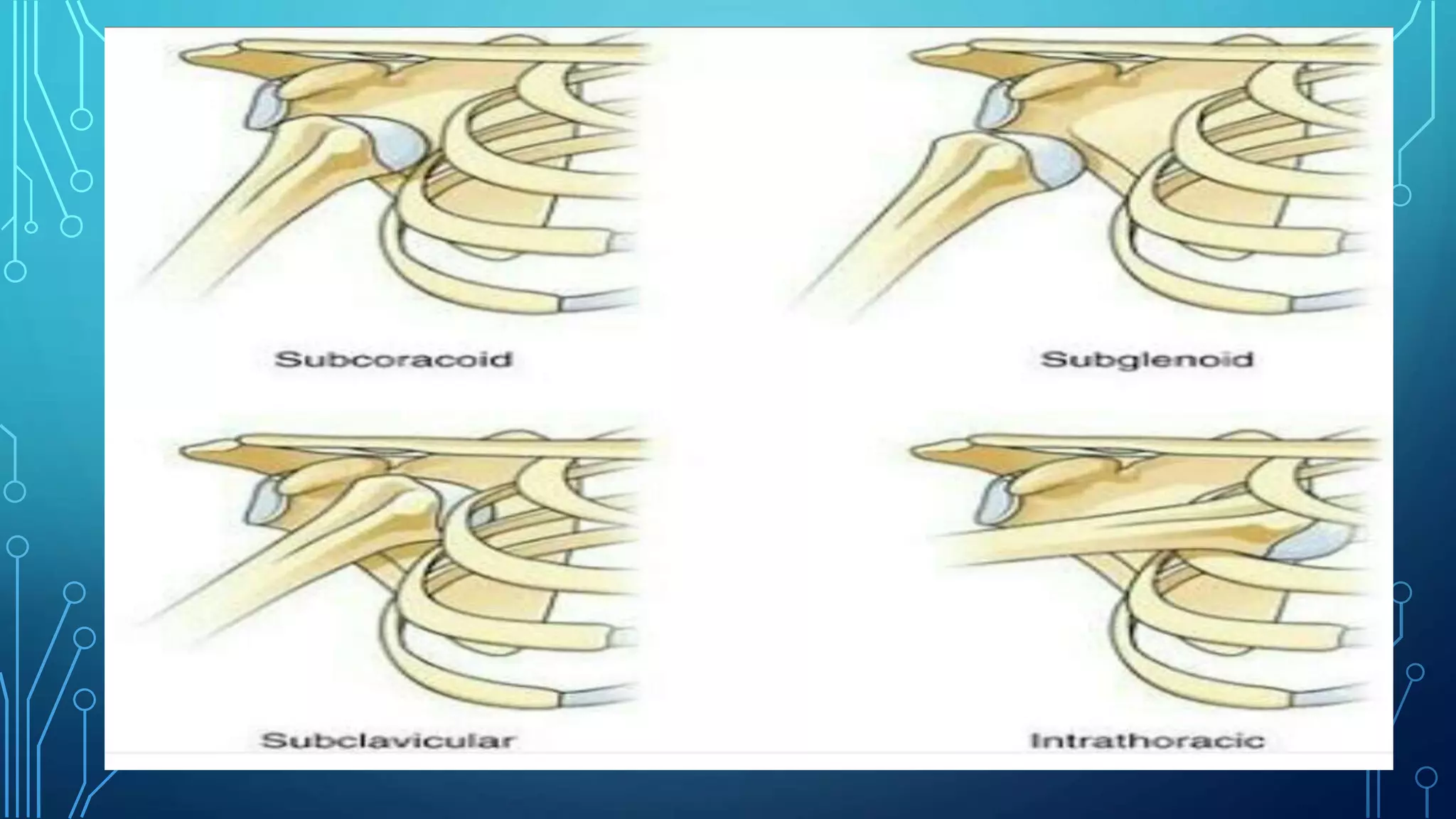
SHOULDER DISLOCATION
WITH PHYSIOTHERAPY
MANAGEMENT
INTRODUCTION
•This is the commonest joint in human body
to dislocate.
•More comon in adults.
•Anterior dislocation is much more common.
TYPES OF SHOULDER DISLOCATION
•Aanterior
•Posterior
•Luxatio erecta( erect dislocation)
ANTERIOR DISLOCATION
•The most common mode of injury is
fall on out stretched hand with limb in
lateral rotation.
•In this position the head of the
humerus is thrust against the
thightened Anterior capsule which
gets torn or avulsed from bone .
•Lateral rotation of the arm is particularly
important in causing this injury rather than
abduction.
•In this type of dislocation the position of
head of humerus may slip to one of the
following:
1. Subcoracoid
2. Subglenoid
3. Subcvicular
•Subcoracoid:
•Most common type of dislocation.
•The dislocated humeral head comes to lie
anteriorly below the coracoid process.
SUBCAROCOID DISLOCATION
•Subglenoid:
•The head of the humerus passes through
the lower part of the capsule and remain
beneath the glenoid cavity.
•Subclavicular:
•Rarely, the head of humerus occupies a
position just below the clavicle.
SUBGLENOID DISLOCATION XRAY:
RECURRENT ANTERIOR DISLOCATION OF
SHOULDER
•It occurs by repeated dislocation of Shoulder jt.
Following one episode of acute dislocation.
•occurs due to failure of healing of torn avulsed
capsule anteriorly because of inadequate
treatment during first episode.
•The repeated dislocations usually require less
violence and therefore the subsequent
dislocations occur when the arm is externally
rotated And abducted during the routine
movemens of playing and dressing.
•There are two types of lesions which may occur
during anterior dislocation of the shoulder:
1. bankart’s lesion :
2.Hill-sachs lesion :
•1. Bankart’s lesion
The avulsion of the glenoid labrum and
anterior capsule creates a pouch anterior to the
neak of scapula into which the humeral head
slips with every dislocation .
•2 . hill- sachs lesion:
•It is deffect in the posterolateral quadrant of
the head of tbe humerus .
•It caused by the anterior edge of the scapula
when the dialocated head impiges against
HILL-SACHS LESION:
HILL-SACHS LESION X-RAY:
POSTERIOR DISLOCATION
•Less common
•Caused by direct blow on the front of the
shoulder with the arm in internal rotation,
e.g.during electroconvulsive therapy or
epileptic attack or severe electrial shock.
•This injury is often missed even on x-
ray,so careful examination of x-ray is
needed.
•Loss of external rotation.
•CT scan may be diagnostic.
POSTERIOR DISLOCATION X-RAY:
LAXATIO ERECTA
•It occurs in rare cases.
•Occurs when limb is strongly abducted
•E.g. Holding a branch of a tree with arm
in wide abduction while falling down
From tree.
•As a result of injury the head of humerus
is pushed down underneath the glenoid
CONTINUED.....
Fixed in wide abduction-elevation almost
by the side of the head.
LUXATIO ERECTA X-RAY:
DIAGNOSIS:
◆Presenting complaints:
•The pt.enters the casulty with
his shoulder abducted and the
elbow supported wirh
opposite hand.
• Also pain is present.
• inability to move the Shoulder.
• there may be a history of similar episodes in
the past.
◆ On examination:
•The pt. Keeps his arm abducted.
•the normal contour of the shoulder jt. Is lost,
and it
becomes flattened.
•on carefully inspection , fullness below the
clavicle Is noticeable, due to the displaced head.
It is felt by rotating the arm.
•The pt. Resists any attempted movt. Of
abduction and external rotation. This is
called apprehension sign.
•Dugas test:
Pt’s position: sitting. And instruct him to
touch the opposite Shoulder and bring the
elbow to the chest wall.
Positive test:inability to touch the opposite
Shoulder, because of pain . Which indicates
•Dugas test:
•Hamilton ruler test :
Because of flattni g of tbe Shoulder, it is
possible to place a ruler on the lateral side
of the arm.
The ruler touches the acromion and lateral
Condyle of humerus simultaneously.
• x-ray are also used for diagnosis.
NORMAL SIDE AFFECTED
SIDE
◆TREATMENT:
•It has three phases;
∆ Reduction
∆ Immobilization phase
∆ Mobilization phase
◆ Reduction:
•It is done under sedation or general
anesthesia.
•There are two techniques of reduction of
Shoulder dislocation:
1. Kocher’s monoeurve:
2. Hippocrates monoeurve:
KOCHER’S MONOEURVE:
•This is the most commonly used
method.
1. Traction- with elbow flexed to a
right angle steady traction is applied
along the long axis of the humerus.
2. External rotation of the arm
3. adduction
1 2
3 4
•Hippocrates manoeuvre:
•In this method surgeon applies a firm and
steady pull in the semi-abducted arm.
•He keeps his foot in the axilla against tbe
chest wall.
•Head of the humerus is levered back into
position using the foot as a fulcrum.
◆ Imobilisation:
•In chest arm bandage.
•For 3 weeks.
◆ mobilization:
•After bandage is removed , pt. can move
the Shoulder.
COMPLICATIONS:
•Fracture of the greater tuberosity or
surgical neak of humerus.
•Supraspinatus tendinitis.
•Rotator cuff injury .
•Injury to the axillary nerve during
reduction.
•Recurrent anterior dislocation of Shoulder.
SURGICAL OPERATION:
◆Putti-platt operation:
•Procedure: in this procedure the
subscapularis muscle is divided along with
the anterior capsule and it is sutured back
by overlapping(double breasting) both the
divided edges of the subscapularis muscle
along with rhe capsule.
•capsuloraphy
•Indication: it is indicated for pts with
unidirectional anterior shoulder instability.
•Evaluation of outcomes and biomechanics
suggests that this procedure is rarely
indicated.
•Contraindication:it only repairs anterior
instability.
•Glenohumeral arthritis .
•Any limitation in external rotation
•Technical consideration:
•Athletes and laborers requiring normal
range of motion in external rotation And
are limited with this procedure.
•This procedure also limits the use for
throwers and overhead athletes .
•The applicability of this procedure is
reduced because of loss of external
rotation .
•Bankart’s operation:
•Procedure:the goal of procedure is to
reattach and thighten the torn labrum and
ligament of the jt.
•This is technically demanding procedure.
•It becomes simpler with the use of special
fixation device called anchors.
•Bristow’s operation:
•Procedure:in this procedure the coracoid
process along with its attached muscles,
is osteomized at the base and fixed to
lower half of the anterior margin of tbe
glenoid.
•Musxles attached to the coracoid provide
dynamic anterior support rto the head of
humerus.
•Arthroscopic benkart repair:
•Now days repair of benkart’s lesion is done
arthroscopically.
•Minimal invasive surgery.
•Definite advantage.
•The post operative morbidity is less and
rehabilitation is faster.
• lower complication rate than open repair
procedure.
PHYSIOTHERAPY MANAGEMENT
•Basic objective:
•To regain full range of active movements
of Shoulder.
•Early return of movements of abduction
and external rotation.
∆ During immobilization:first 3 weeks
•only wrist and finger movements are possible
because the arm is strapped to the trunk in
position of adduction and internal rotation.
•So exercises at this phase are:
* Strong resistive movement at wrist and finger.
* isometric contractions can safely be instituted
to the deltoid, biceps, and triceps.
∆ Mobilization:after 3 weeks
•After Removal of strapping the limb is
supported in a sling.
•Elbow should be mobilized to the full extent by
removing the sling intermittently.
•Then mobilization of the shoulder flexion-
extension should be initiated as a small range
pendular swinging movt. In forward stoop
position.
CONTINUED....
• Initiation of Shoulder abduction and
external rotation:
• as this two are instrumental in causing
redislocation they have to be initiated with
atmost care and adequate stabilization at
GH jt.
•Initial aim should be relaxed passive
abduction up to 45 degrees.this is done in
supine wirh arm in internal rotation.
•External rotation Should also be initiated
in supine with arm adducted by the side
of the body, and pt have to do external
rotation up to 45 degree.
•Relaxed passive movts. To the Shoulder
should be carried out to the full or near
normal range at the earliest, to avoid
adhesive capsulitis.
•Self assisted relaxed movts. With wand in
•Once good passive range is attained, regimen
of strengthening is begun.
•Self-resisted isometric and slow isotonic
movt. Should be taught as a home treatment
programme. Dumbells could be used as a
resistive device.
•90% of full range is achieved by 6-8 weeks
following dislocation.
•Heavy resistive exercise, passive stretching,
and forced external rotation and abduction
are safe after12 weeks.
•It may be difficult in some pts. To achieve
the terminal range of abduction- elevation
and external rotation this could be
painful and needs to be facilitated by a
suitable thermotherapy adjunct.
•But majority of pts. get full function by 12
weeks following injury.
•Physiotherapy also play very important
role in preventing recurrent anterior
Preventive regime of Physiotherapy:
Principle objective of physiotherapautic
management:
1. To strenghen the ligaments and
muscles crossing the shoulder jt. to
iptimum level.
2. To regain full passive ROM of all the
movts.
∆Strenghning procedure: to be successful,
it needs several repitations.
•The exercise should be taught in standing
or sitting So they can be conveniently
performed several times.
•Weighted dumbells or weight belts may
also be used as resistive device.
∆ To achieve and maintain full range
passive motion:
The arc of movts. Of abduction-elevation,
flexion-elevation and external rotation
need to be done gradually and carefully.
•Extra care should be taken during the
terminal range of elevation and external
rotation.
•As adequate stabilization of Shoulder
girdle facilitates relaxation of gh jt, pt.
Should be advised to get some assistance
at home while performing these movts.
•Surgically managed pts:the regime of
physiotherapy for the pts. treated with
•The main difference is the secured safety
of performming movments
•It needs hard effort to achieve active
terminal range in external rotation and
elevation.
•Usually extreme level of external rotation
remain deficient.
•Strong and functional shoulder can be
achieved with in 10 to 12weeks.
REFRENCE:
• Eessentials of orthopedics and applied physiotherapy:
jayant joshi;second edition
• Essential of orthopedics:maheshwari and mahaskar;5th
edition
THANK YOU
Shoulder dislocation with physiotherapy management

Shoulder dislocation with physiotherapy management

  • 1.
  • 2.
    INTRODUCTION •This is thecommonest joint in human body to dislocate. •More comon in adults. •Anterior dislocation is much more common.
  • 3.
    TYPES OF SHOULDERDISLOCATION •Aanterior •Posterior •Luxatio erecta( erect dislocation)
  • 4.
    ANTERIOR DISLOCATION •The mostcommon mode of injury is fall on out stretched hand with limb in lateral rotation. •In this position the head of the humerus is thrust against the thightened Anterior capsule which gets torn or avulsed from bone .
  • 5.
    •Lateral rotation ofthe arm is particularly important in causing this injury rather than abduction. •In this type of dislocation the position of head of humerus may slip to one of the following: 1. Subcoracoid 2. Subglenoid 3. Subcvicular
  • 6.
    •Subcoracoid: •Most common typeof dislocation. •The dislocated humeral head comes to lie anteriorly below the coracoid process.
  • 7.
  • 8.
    •Subglenoid: •The head ofthe humerus passes through the lower part of the capsule and remain beneath the glenoid cavity. •Subclavicular: •Rarely, the head of humerus occupies a position just below the clavicle.
  • 9.
  • 10.
    RECURRENT ANTERIOR DISLOCATIONOF SHOULDER •It occurs by repeated dislocation of Shoulder jt. Following one episode of acute dislocation. •occurs due to failure of healing of torn avulsed capsule anteriorly because of inadequate treatment during first episode. •The repeated dislocations usually require less violence and therefore the subsequent dislocations occur when the arm is externally rotated And abducted during the routine movemens of playing and dressing.
  • 12.
    •There are twotypes of lesions which may occur during anterior dislocation of the shoulder: 1. bankart’s lesion : 2.Hill-sachs lesion :
  • 13.
    •1. Bankart’s lesion Theavulsion of the glenoid labrum and anterior capsule creates a pouch anterior to the neak of scapula into which the humeral head slips with every dislocation . •2 . hill- sachs lesion: •It is deffect in the posterolateral quadrant of the head of tbe humerus . •It caused by the anterior edge of the scapula when the dialocated head impiges against
  • 15.
  • 16.
  • 17.
    POSTERIOR DISLOCATION •Less common •Causedby direct blow on the front of the shoulder with the arm in internal rotation, e.g.during electroconvulsive therapy or epileptic attack or severe electrial shock.
  • 18.
    •This injury isoften missed even on x- ray,so careful examination of x-ray is needed. •Loss of external rotation. •CT scan may be diagnostic.
  • 19.
  • 20.
    LAXATIO ERECTA •It occursin rare cases. •Occurs when limb is strongly abducted •E.g. Holding a branch of a tree with arm in wide abduction while falling down From tree. •As a result of injury the head of humerus is pushed down underneath the glenoid
  • 21.
    CONTINUED..... Fixed in wideabduction-elevation almost by the side of the head.
  • 22.
  • 23.
    DIAGNOSIS: ◆Presenting complaints: •The pt.entersthe casulty with his shoulder abducted and the elbow supported wirh opposite hand.
  • 24.
    • Also painis present. • inability to move the Shoulder. • there may be a history of similar episodes in the past.
  • 25.
    ◆ On examination: •Thept. Keeps his arm abducted. •the normal contour of the shoulder jt. Is lost, and it becomes flattened. •on carefully inspection , fullness below the clavicle Is noticeable, due to the displaced head. It is felt by rotating the arm.
  • 26.
    •The pt. Resistsany attempted movt. Of abduction and external rotation. This is called apprehension sign. •Dugas test: Pt’s position: sitting. And instruct him to touch the opposite Shoulder and bring the elbow to the chest wall. Positive test:inability to touch the opposite Shoulder, because of pain . Which indicates
  • 27.
  • 28.
    •Hamilton ruler test: Because of flattni g of tbe Shoulder, it is possible to place a ruler on the lateral side of the arm. The ruler touches the acromion and lateral Condyle of humerus simultaneously. • x-ray are also used for diagnosis.
  • 29.
  • 30.
    ◆TREATMENT: •It has threephases; ∆ Reduction ∆ Immobilization phase ∆ Mobilization phase
  • 31.
    ◆ Reduction: •It isdone under sedation or general anesthesia. •There are two techniques of reduction of Shoulder dislocation: 1. Kocher’s monoeurve: 2. Hippocrates monoeurve:
  • 32.
    KOCHER’S MONOEURVE: •This isthe most commonly used method. 1. Traction- with elbow flexed to a right angle steady traction is applied along the long axis of the humerus. 2. External rotation of the arm 3. adduction
  • 33.
  • 34.
    •Hippocrates manoeuvre: •In thismethod surgeon applies a firm and steady pull in the semi-abducted arm. •He keeps his foot in the axilla against tbe chest wall. •Head of the humerus is levered back into position using the foot as a fulcrum.
  • 36.
    ◆ Imobilisation: •In chestarm bandage. •For 3 weeks. ◆ mobilization: •After bandage is removed , pt. can move the Shoulder.
  • 37.
    COMPLICATIONS: •Fracture of thegreater tuberosity or surgical neak of humerus. •Supraspinatus tendinitis. •Rotator cuff injury . •Injury to the axillary nerve during reduction. •Recurrent anterior dislocation of Shoulder.
  • 38.
    SURGICAL OPERATION: ◆Putti-platt operation: •Procedure:in this procedure the subscapularis muscle is divided along with the anterior capsule and it is sutured back by overlapping(double breasting) both the divided edges of the subscapularis muscle along with rhe capsule. •capsuloraphy
  • 39.
    •Indication: it isindicated for pts with unidirectional anterior shoulder instability. •Evaluation of outcomes and biomechanics suggests that this procedure is rarely indicated. •Contraindication:it only repairs anterior instability. •Glenohumeral arthritis . •Any limitation in external rotation
  • 40.
    •Technical consideration: •Athletes andlaborers requiring normal range of motion in external rotation And are limited with this procedure. •This procedure also limits the use for throwers and overhead athletes . •The applicability of this procedure is reduced because of loss of external rotation .
  • 41.
    •Bankart’s operation: •Procedure:the goalof procedure is to reattach and thighten the torn labrum and ligament of the jt. •This is technically demanding procedure. •It becomes simpler with the use of special fixation device called anchors.
  • 43.
    •Bristow’s operation: •Procedure:in thisprocedure the coracoid process along with its attached muscles, is osteomized at the base and fixed to lower half of the anterior margin of tbe glenoid. •Musxles attached to the coracoid provide dynamic anterior support rto the head of humerus.
  • 45.
    •Arthroscopic benkart repair: •Nowdays repair of benkart’s lesion is done arthroscopically. •Minimal invasive surgery. •Definite advantage. •The post operative morbidity is less and rehabilitation is faster. • lower complication rate than open repair procedure.
  • 47.
    PHYSIOTHERAPY MANAGEMENT •Basic objective: •Toregain full range of active movements of Shoulder. •Early return of movements of abduction and external rotation.
  • 48.
    ∆ During immobilization:first3 weeks •only wrist and finger movements are possible because the arm is strapped to the trunk in position of adduction and internal rotation. •So exercises at this phase are: * Strong resistive movement at wrist and finger. * isometric contractions can safely be instituted to the deltoid, biceps, and triceps.
  • 49.
    ∆ Mobilization:after 3weeks •After Removal of strapping the limb is supported in a sling. •Elbow should be mobilized to the full extent by removing the sling intermittently. •Then mobilization of the shoulder flexion- extension should be initiated as a small range pendular swinging movt. In forward stoop position.
  • 50.
    CONTINUED.... • Initiation ofShoulder abduction and external rotation: • as this two are instrumental in causing redislocation they have to be initiated with atmost care and adequate stabilization at GH jt. •Initial aim should be relaxed passive abduction up to 45 degrees.this is done in supine wirh arm in internal rotation.
  • 51.
    •External rotation Shouldalso be initiated in supine with arm adducted by the side of the body, and pt have to do external rotation up to 45 degree. •Relaxed passive movts. To the Shoulder should be carried out to the full or near normal range at the earliest, to avoid adhesive capsulitis. •Self assisted relaxed movts. With wand in
  • 52.
    •Once good passiverange is attained, regimen of strengthening is begun. •Self-resisted isometric and slow isotonic movt. Should be taught as a home treatment programme. Dumbells could be used as a resistive device. •90% of full range is achieved by 6-8 weeks following dislocation. •Heavy resistive exercise, passive stretching, and forced external rotation and abduction are safe after12 weeks.
  • 53.
    •It may bedifficult in some pts. To achieve the terminal range of abduction- elevation and external rotation this could be painful and needs to be facilitated by a suitable thermotherapy adjunct. •But majority of pts. get full function by 12 weeks following injury. •Physiotherapy also play very important role in preventing recurrent anterior
  • 54.
    Preventive regime ofPhysiotherapy: Principle objective of physiotherapautic management: 1. To strenghen the ligaments and muscles crossing the shoulder jt. to iptimum level. 2. To regain full passive ROM of all the movts. ∆Strenghning procedure: to be successful, it needs several repitations.
  • 55.
    •The exercise shouldbe taught in standing or sitting So they can be conveniently performed several times. •Weighted dumbells or weight belts may also be used as resistive device. ∆ To achieve and maintain full range passive motion: The arc of movts. Of abduction-elevation, flexion-elevation and external rotation need to be done gradually and carefully.
  • 56.
    •Extra care shouldbe taken during the terminal range of elevation and external rotation. •As adequate stabilization of Shoulder girdle facilitates relaxation of gh jt, pt. Should be advised to get some assistance at home while performing these movts. •Surgically managed pts:the regime of physiotherapy for the pts. treated with
  • 57.
    •The main differenceis the secured safety of performming movments •It needs hard effort to achieve active terminal range in external rotation and elevation. •Usually extreme level of external rotation remain deficient. •Strong and functional shoulder can be achieved with in 10 to 12weeks.
  • 58.
    REFRENCE: • Eessentials oforthopedics and applied physiotherapy: jayant joshi;second edition • Essential of orthopedics:maheshwari and mahaskar;5th edition
  • 59.