Soft tissue conditions in
Orthopedics
Facilitator: Dr. Subarna Adhikari
Lecturer, KISTMCTH
Topics
• Bursitis
• Tenosynovitis
• Dupuytren’s Contracture
• Tennis Elbow
• Golfer’s Elbow
• De-Quervain’s Tenosynovitis
• Trigger finger
• Ganglion
• Carpal tunnel syndrome
• Frozen Shoulder
• Plantar Fascitis
• Fibrositis
• Painful-arc Syndrome
• Meralgia paresthetica
• Fibromyalgia
Exams: ★★★★☆
Clinical practice: ★★★★★
Bursitis
• Bursa: a small fluid-filled sac or saclike cavity situated
in places in tissues where friction would otherwise
occur
• Types: adventitious, subcutaneous, synovial, and sub-
muscular.
• Adventitious: non-native, repeated stress;
example Students' elbow and bunion.
• Housemaid’s knee, Clergyman’s knee, Student’s
elbow, Weaver’s Bottom, Tailor’s ankle, Bunion:
(Entrance Preparation)
Irritative Bursistis
• Excessive pressure or friction ☞ Inflammation☞ Effusion
• Prolonged inflammation☞Thickened sac☞ Erosion of adjacent bone
• Treatment:
• Analgesics, rest, removal of causative factor
• Intralesional steroid injection
• Excision of bursa
Infective bursitis
• Uncommon; pyogenic or tubercular; prepatellar or trochanteric
• Surgical drainage
Tenosynovitis
• Unaccustomed overuse/ spontaneous
• Synovial inflammation ☞ Secondary thickening of sheath ☞ Stenosis
of compartment ☞ further compromise
• Inflammation/infection; flexor/extensor tendon sheath of the hand
• Irritative: rest, analgesics, splinting, US therapy, intralesional steroid
(RUPTURE!)
• Infective: Pyogenic or tubercular
Trigger Finger (Digital tenovaginosis)
• Congenital or acquired
• Stenosing tenovaginosis of fibrous digital sheath
• Flexor tendon thickened at the entrance to its sheath (A1 pulley;
passes the sheath on forced extension with a snap (‘triggering’)
• Diabetes, Rheumatoid arthritis
• Clinical: thumb, ring finger and middle finger most commonly affected
• Click on flexion; PIP remains flexed as the hand is unclenched, sudden
straightening with a snap, tender nodule on flexor aspect of MCP
Infantile Trigger Thumb
• Baby cannot extend the thumb tip (IP joint), noticed by parents
• Often delayed diagnosis
• D/D congenitally clasped thumb (both MCP and IP incolved)
Treatment:
• Early cases: corticosteroid injection into
the tendon sheath
• Recurrent cases: Second injection
• Refractory cases: Operation; A1 pulley
release
• Infantile cases: wait until the child is
three years old for spontaneous
recovery ☞ Pulley release
• Cases with rheumatoid arthritis:
Preserve pulley, flexor synovectomy
De Quervain’s disease
• Reactive thickening of sheath around Extensor policis brevis and
Abductor Policis Longus: First extensor compartment
• Overuse/Spontaneous
• Middle age women/Pregnancy/Postpartum
• C/F Pain on wrist, H/O unaccustomed activity: eg wringing out clothes
• Tenderness over radial styloid process, sometimes swelling
• Finkelstein’s test
• Hitchhiker’s sign
• Differential Diagnosis: arthritis at the base of thumb,
Scaphoid non-union, Intersection syndrome
• (Intersection syndrome: extensor tenosynovitis due to friction
between first and second extensor compartment tendons)
• Treatment: Splintage, analgesics, Ultrasound therapy
• Resistant cases: operation: slitting thickened tendon sheath
• Case: 28 years old with a 7 months old child presented with a
history of wrist pain, usually on lifting the child. What is your
diagnosis?
Dupuytren’s contracture
• Nodular hypertrophy of the superficial palmar
fascia (Palmar aponeurosis)
• Autosomal dominant
• Common in European population
• Predisposition with: epileptics receiving phenytoin,
diabetes, smoking, alcoholic cirrhosis, AIDS, PTB
• Palmar fascia: connects palmar aponeurosis with
fibrous digital sheaths, four slips
• Ledderhose’s (soles), Peyronnie’s (penis)
• Fibrous tissue in palmar fascia and digital sheaths: Initial proliferative
phase ☞ Contracture ☞ Flexion deformities of MCP and PIP
• Digital nerve: displaced or enveloped
• Clinical: Middle aged man with nodular thickening in palm; usually
bilateral, palm is puckered, nodular and thick
• D/D: traumatic contracture, trigger finger, clawing
• Treatment: Partial or complete fasciectomy (excision of affected fascia)
Ganglion cysts
• 95% of swelling of the hand, benign; 60% of benign swelling, ganglion
cysts, 60% of ganglion cysts, dorsal surface
• Dorsal wrist ganglions: scapholunate ligament
• Palmar wrist ganglions: arise from scapho-lunate or scapho-trapezoid
ligament
• Leakage of synovial fluid from
a joint or tendon sheath
• Clinical Features:
• Female> Male; Young adult
• Painless/slightly painful well defined cystic lump, transilluminant
• Carpal tunnel ganglion: features of compression of median nerve
• Within bone; scaphoid or lunate: pain
• Treatment indications:
• Aesthetic: less invasive methods
• Pain, limitation of movement, nerve palsies:
surgical removal
Treatment
• Old traditional treatment: hit it with a book, since the cysts can burst
when struck
• Patient reassurance and no treatment
• Manual compression until bursting
• Aspiration with/without steroid injection:
recurrence (upto 60%)
• Open excision/ Arthroscopic excision:
recurrence (upto 40%)
Tennis elbow (lateral epicondalgia)
• Bony attachment of common
extensor tendon over lateral
epicondyle of humerus
• Repeated forceful wrist
extension: tennis players/non-
players
• Pathology: tears,
fibrocartilaginous metaplasia,
microscopic calcification, painful
vascular reaction
Clinical Features
• M=F, over 30-40 years of age
• Prolonged history of pain, usually after unaccustomed activities
• Localized to lateral epicondyle, may spread widely
• Aggravated by movements like pouring tea, turning door handle, shaking
hands, lifting with forearm pronated
• Elbow: normal on inspection with full range of flexion and extension,
tenderness at lateral epicondyle
• Cozen’s test: resisted dorsiflexion of wrist
• Acute passive flexion of wrist with forearm pronated
• Xray: Normal/ Calcification of tendon
• Differential diagnosis: consider radial nerve entrapment in resistant
cases (radial tunnel syndrome)
• Treatment:
• 90% cases resolve spontaneously within 6-12 months
• Identify and restrict activities causing pain
• NSAIDs
• Counterforce bracing and wrist splint
• Non-surgical Treatment:
• Intralesional steroid and local anesthetic injection
• Ultrasound therapy
• Autologous blood patching
• Platelet rich plasma injection
• Hyaluronate injections
• Operative treatment:
• Removal of diseased tendon and reattachment of healthy part
• Physiotherapy
• Strength training and stretching exercises, occupational therapy
Golfer’s elbow (Medial epicondylitis)
• Less common than tennis elbow
• Pronator origin affected
• Associated ulnar neuropathy may be present
• Treatment: similar to tennis elbow
• D/D medial collateral ligament of elbow injury
Carpal Tunnel syndrome
Boundaries:
• Palmar: flexor
retinaculum
• Radial: scaphoid tubercle
and the ridge of
trapezium
• Ulnar: pisiform and hook
of hamate
• Dorsal: Capitate,
trapezoid and trapezium
Contents:
• A total of nine flexor tendons
• flexor digitorum profundus (four tendons)
• flexor digitorum superficialis (four tendons)
• flexor pollicis longus (one tendon)
• flexor carpi radialis (one tendon) passes through the retinaculum
• A single nerve the median nerve between tendons of flexor digitorum
profundus and flexor digitorum superficialis
Clinical features
• Female>Male, 40-5- years of age
• Pregnancy, rheumatoid disease, myxedema
• Pain and paresthesia over lateral three and a half digits
• Worsening at night, waking up with pain, tingling, numbness
• Hanging the hand/ shaking may relieve pain
• Clumsiness of fine movements
• Wasting of thenar eminence
• Nerve conduction studies
• D/D cervical radiculopathy, thoracic outlet syndrome
Splintage
• Wrist splints
• Steroid injection into the carpal tunnel
• Open surgical release of transverse carpal ligament
• Physiotherapy: weight reduction aerobic training,
occupational therapy
Painful arc syndrome
• Rotator cuff: Supraspinatus, Infraspinatus
Teres Minor, Subscapularis
• Pass under the coracoacromial arch
• Function: Stabilize the head of humerus
when there
is abduction due to contraction of deltoid
muscle
• The Subacromial space contains:
• Belly and tendon of the supraspinatus muscle
• Long head of the biceps muscle
• Subacromial bursa
• Abductors of shoulder???
• Coronal sections through the shoulder: irritation of subdeltoid bursa
and supraspinatus tendon by repeated impingement under the
coracoacromial arch during abduction. (a) Joint at rest. (b) In
abduction.
1 Supraspinatus Muscle
2 Acromioclavicular Joint
3 Subdeltoid Bursa
4 Deltoid Muscle
5 Supraspinatus Tendon
6 Synovial Joint
Painful arc Syndrome
• Clinical examination may reveal tenderness along the acromion
• D/D: Frozen shoulder, Biceps tendinitis
• Clinical syndrome in which there is pain in the
shoulder and upper arm during mid-range of
abduction
• Causes:
• Subacute supraspinatus tendinitis
• Calcification of supraspinatus tendon
• Subacromial bursitis
• Greater tuberosity fracture
Treatment:
• Based on cause
• Ultrasound therapy
• NSAIDs
• Injection of corticosteroids in subacromial space
• Excision of anterior prominent part of acromion (acromioplasty):
open or arthroscopic; arthroscopic rotator cuff repair
• Physiotherapy
Frozen shoulder
• Adhesive capsulitis/ Periarthritis shoulder
• Clinically diagnosed
• Characterized by progressive pain and stiffness of the shoulder which
usually resolves spontaneously after about 18 months
• Cause: Unknown
• Associated with
• Diabetes, Dupuytren’s disease, Hyperlipidaemia, Hyperthyroidism
• Cardiac disease, Hemiplegia
• Occasionally after recovery from neurosurgery
Clinical features
• 40-60 years; History of trivial trauma
• Aching in the arm and shoulder, prevents sleeping on affected side
• Three characteristic phases
• Freezing phase: pain gradually increases over several months
• Frozen phase: pain decreases over 6-12 months, stiffness increases
• Thawing phase: Movement gradually regained, may not be as before
• Examination: wasting of muscles, loss of movement in all directions
(active and passive)
• Xrays: Normal
• D/D : infection/post-traumatic stiffness/post-immobilization
stiffness/reflex sympathetic dystrophy
Treatment:
• Conservative treatment
• Pain relief, prevent further stiffening
• Pendulum exercises
• Intra-articular steroid injection
• Joint distension with large volume of normal saline injection (50-200
ml)
• Manipulation under general anesthesia
• External rotation ☞ Abduction ☞Flexion
• Intraarticular injection at the end
• Careful to prevent fracture surgical neck humerus in the elderly osteoporotic
patient
Surgical Treatment
• Prolonged disabling restriction of movement which fails to respond to
conservative therapy
• Arthroscopic capsular release, and release of adhesions
Plantar fascitis
• Common cause of heel pain
• Self-limiting: 18-36 months
• Plantar fascia: originates from calcaneum,
divides into four slips, inserts into
metatarsal heads
• Stiffening and thickening with age
• Micro-tears and thickening of fascia
Clinical features
• Gradual onset, without history of injury
• Maybe a history of sudden increase in activity, change of footwear
• Worse when getting up in the morning, going downstairs, getting up
after rest
• Pain maybe sharp/persistent background ache
Examination
• Localozed tenderness medial aspect beneath the heel, sometimes
midfoot
• Xray: rule out stress fracture, spur on undersurface of calcaneum
Treatment
• Relative rest and NSAIDs
• Footwear modification
• Plantar fascia stretching exercises
• Cushioning of heel pad
• Ultrasound therapy
• Operative treatment: Limited fasciotomy
Differential diagnosis
• Painful fat pad
• Nerve entrapment
Meralgia Paresthetica
• Tingling, burning, numbness over skin
supplied by lateral cutaneous nerve of
thigh
• Entrapment in fascia lata just medial
to anterior superior iliac spine
• Treatment: analgesics, local steroid
injection, decompression of nerve
Fibromyalgia
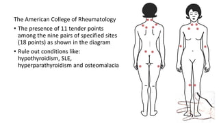
• Disorder of chronic, widespread pain and tenderness
• Typically presents in young or middle-aged women but can affect patients
of either sex and at any age
Signs and symptoms:
• Persistent (≥ 3 mo) widespread pain (pain/tenderness on both sides of the
body, above and below the waist, and includes the axial spine [usually the
paraspinus, scapular, and trapezius muscles])
• Stiffness
• Fatigue; disrupted and unrefreshing sleep
• Cognitive difficulties
• Multiple other unexplained symptoms, anxiety and/or depression, and
functional impairment of activities of daily living (ADLs)
The American College of Rheumatology
• The presence of 11 tender points
among the nine pairs of specified sites
(18 points) as shown in the diagram
• Rule out conditions like:
hypothyroidism, SLE,
hyperparathyroidism and osteomalacia
Treatment
Nonpharmacotherapy
• Diet (eg, promote good nutrition, vitamin supplementation, bone
health, weight loss)
• Stress management
• Aerobic exercise (eg, low-impact aerobics, walking, water aerobics,
stationary bicycle)
• Sleep therapy (eg, education/instruction on sleep hygiene)
• Psychologic/behavioral therapy (eg, cognitive-behavioral, operant-
behavioral)
Pharmacotherapy
• Combine pharmacologic and nonpharmacologic therapy in the
treatment of fibromyalgia. Aggressively treat comorbid depression.
• Analgesics (eg, tramadol)
• Antianxiety agents (eg, alprazolam, clonazepam, zolpidem, zaleplon,
Trazodone, buspirone, temazepam, sodium oxybate)
• Skeletal muscle relaxants (eg, cyclobenzaprine)
• Antidepressants (eg, amitriptyline, duloxetine, milnacipran,
venlafaxine, desvenlafaxine)
• Anticonvulsants (eg, pregabalin, gabapentin, tiagabine)
• Alpha 2 agonists (eg, clonidine)
Polydactyly
• Most common congenital digital anomaly
of the hand and foot.
• Isolated: usually autosomal dominant/random
• Associated with other birth defects autosomal recessive.
• Preaxial polydactyly: the most common type, duplication of the first digit
or ray
• Central polydactyly: duplication of the second, third, or fourth digit or
ray.
• Postaxial polydactyly: fifth digit or ray.
• Radial, central, and ulnar vs tibial, central, and fibular
• Look for other
congenital anomalies
and association with
genetic syndromes
• X-ray: skeletal elements
• 3 years: aware of the
anomaly.
• 7 years: close scrutiny
of peers at school ☞
emotional stress
Treatment: Early surgical
removal
Syndactyly
• Greek συν- "together“; δακτυλος "finger"
• Condition wherein two or more digits are fused
together
• Complete or incomplete
• Complete: the skin is joined all the way to the tip of the
finger
• Incomplete: the skin is only joined part of the
distance to the fingertip
• Simple or complex
• Simple: adjacent fingers or toes are joined by soft tissue
• Complex: the bones of adjacent digits are fused
Treatment
• Surgical Release : syndactyly that prevents full range of motion (not
cosmetic)
• In complex syndactyly, separation of the conjoint fingers may make
the 2 individualized digits nonfunctional; only 1 set of tendons and 1
neurovascular pedicle may be present
• Elective surgical procedures at 5 or 6 months of age
Thank you!
https://drive.google.com/open?id=0BxYO2YbzRfGgWjB5TW0zdUdDeUk
http://www.slideshare.net/justsubun/lecture-kist

Lecture kist

  • 1.
    Soft tissue conditionsin Orthopedics Facilitator: Dr. Subarna Adhikari Lecturer, KISTMCTH
  • 2.
    Topics • Bursitis • Tenosynovitis •Dupuytren’s Contracture • Tennis Elbow • Golfer’s Elbow • De-Quervain’s Tenosynovitis • Trigger finger • Ganglion • Carpal tunnel syndrome • Frozen Shoulder • Plantar Fascitis • Fibrositis • Painful-arc Syndrome • Meralgia paresthetica • Fibromyalgia Exams: ★★★★☆ Clinical practice: ★★★★★
  • 3.
    Bursitis • Bursa: asmall fluid-filled sac or saclike cavity situated in places in tissues where friction would otherwise occur • Types: adventitious, subcutaneous, synovial, and sub- muscular. • Adventitious: non-native, repeated stress; example Students' elbow and bunion. • Housemaid’s knee, Clergyman’s knee, Student’s elbow, Weaver’s Bottom, Tailor’s ankle, Bunion: (Entrance Preparation)
  • 4.
    Irritative Bursistis • Excessivepressure or friction ☞ Inflammation☞ Effusion • Prolonged inflammation☞Thickened sac☞ Erosion of adjacent bone • Treatment: • Analgesics, rest, removal of causative factor • Intralesional steroid injection • Excision of bursa Infective bursitis • Uncommon; pyogenic or tubercular; prepatellar or trochanteric • Surgical drainage
  • 5.
    Tenosynovitis • Unaccustomed overuse/spontaneous • Synovial inflammation ☞ Secondary thickening of sheath ☞ Stenosis of compartment ☞ further compromise • Inflammation/infection; flexor/extensor tendon sheath of the hand • Irritative: rest, analgesics, splinting, US therapy, intralesional steroid (RUPTURE!) • Infective: Pyogenic or tubercular
  • 6.
    Trigger Finger (Digitaltenovaginosis) • Congenital or acquired • Stenosing tenovaginosis of fibrous digital sheath • Flexor tendon thickened at the entrance to its sheath (A1 pulley; passes the sheath on forced extension with a snap (‘triggering’) • Diabetes, Rheumatoid arthritis • Clinical: thumb, ring finger and middle finger most commonly affected • Click on flexion; PIP remains flexed as the hand is unclenched, sudden straightening with a snap, tender nodule on flexor aspect of MCP
  • 7.
    Infantile Trigger Thumb •Baby cannot extend the thumb tip (IP joint), noticed by parents • Often delayed diagnosis • D/D congenitally clasped thumb (both MCP and IP incolved)
  • 8.
    Treatment: • Early cases:corticosteroid injection into the tendon sheath • Recurrent cases: Second injection • Refractory cases: Operation; A1 pulley release • Infantile cases: wait until the child is three years old for spontaneous recovery ☞ Pulley release • Cases with rheumatoid arthritis: Preserve pulley, flexor synovectomy
  • 9.
    De Quervain’s disease •Reactive thickening of sheath around Extensor policis brevis and Abductor Policis Longus: First extensor compartment • Overuse/Spontaneous • Middle age women/Pregnancy/Postpartum • C/F Pain on wrist, H/O unaccustomed activity: eg wringing out clothes • Tenderness over radial styloid process, sometimes swelling • Finkelstein’s test • Hitchhiker’s sign
  • 11.
    • Differential Diagnosis:arthritis at the base of thumb, Scaphoid non-union, Intersection syndrome • (Intersection syndrome: extensor tenosynovitis due to friction between first and second extensor compartment tendons) • Treatment: Splintage, analgesics, Ultrasound therapy • Resistant cases: operation: slitting thickened tendon sheath • Case: 28 years old with a 7 months old child presented with a history of wrist pain, usually on lifting the child. What is your diagnosis?
  • 12.
    Dupuytren’s contracture • Nodularhypertrophy of the superficial palmar fascia (Palmar aponeurosis) • Autosomal dominant • Common in European population • Predisposition with: epileptics receiving phenytoin, diabetes, smoking, alcoholic cirrhosis, AIDS, PTB • Palmar fascia: connects palmar aponeurosis with fibrous digital sheaths, four slips • Ledderhose’s (soles), Peyronnie’s (penis)
  • 13.
    • Fibrous tissuein palmar fascia and digital sheaths: Initial proliferative phase ☞ Contracture ☞ Flexion deformities of MCP and PIP • Digital nerve: displaced or enveloped • Clinical: Middle aged man with nodular thickening in palm; usually bilateral, palm is puckered, nodular and thick • D/D: traumatic contracture, trigger finger, clawing • Treatment: Partial or complete fasciectomy (excision of affected fascia)
  • 14.
    Ganglion cysts • 95%of swelling of the hand, benign; 60% of benign swelling, ganglion cysts, 60% of ganglion cysts, dorsal surface • Dorsal wrist ganglions: scapholunate ligament • Palmar wrist ganglions: arise from scapho-lunate or scapho-trapezoid ligament • Leakage of synovial fluid from a joint or tendon sheath
  • 15.
    • Clinical Features: •Female> Male; Young adult • Painless/slightly painful well defined cystic lump, transilluminant • Carpal tunnel ganglion: features of compression of median nerve • Within bone; scaphoid or lunate: pain • Treatment indications: • Aesthetic: less invasive methods • Pain, limitation of movement, nerve palsies: surgical removal
  • 16.
    Treatment • Old traditionaltreatment: hit it with a book, since the cysts can burst when struck • Patient reassurance and no treatment • Manual compression until bursting • Aspiration with/without steroid injection: recurrence (upto 60%) • Open excision/ Arthroscopic excision: recurrence (upto 40%)
  • 17.
    Tennis elbow (lateralepicondalgia) • Bony attachment of common extensor tendon over lateral epicondyle of humerus • Repeated forceful wrist extension: tennis players/non- players • Pathology: tears, fibrocartilaginous metaplasia, microscopic calcification, painful vascular reaction
  • 18.
    Clinical Features • M=F,over 30-40 years of age • Prolonged history of pain, usually after unaccustomed activities • Localized to lateral epicondyle, may spread widely • Aggravated by movements like pouring tea, turning door handle, shaking hands, lifting with forearm pronated • Elbow: normal on inspection with full range of flexion and extension, tenderness at lateral epicondyle • Cozen’s test: resisted dorsiflexion of wrist • Acute passive flexion of wrist with forearm pronated • Xray: Normal/ Calcification of tendon
  • 19.
    • Differential diagnosis:consider radial nerve entrapment in resistant cases (radial tunnel syndrome) • Treatment: • 90% cases resolve spontaneously within 6-12 months • Identify and restrict activities causing pain • NSAIDs • Counterforce bracing and wrist splint
  • 20.
    • Non-surgical Treatment: •Intralesional steroid and local anesthetic injection • Ultrasound therapy • Autologous blood patching • Platelet rich plasma injection • Hyaluronate injections • Operative treatment: • Removal of diseased tendon and reattachment of healthy part • Physiotherapy • Strength training and stretching exercises, occupational therapy
  • 21.
    Golfer’s elbow (Medialepicondylitis) • Less common than tennis elbow • Pronator origin affected • Associated ulnar neuropathy may be present • Treatment: similar to tennis elbow • D/D medial collateral ligament of elbow injury
  • 22.
    Carpal Tunnel syndrome Boundaries: •Palmar: flexor retinaculum • Radial: scaphoid tubercle and the ridge of trapezium • Ulnar: pisiform and hook of hamate • Dorsal: Capitate, trapezoid and trapezium
  • 23.
    Contents: • A totalof nine flexor tendons • flexor digitorum profundus (four tendons) • flexor digitorum superficialis (four tendons) • flexor pollicis longus (one tendon) • flexor carpi radialis (one tendon) passes through the retinaculum • A single nerve the median nerve between tendons of flexor digitorum profundus and flexor digitorum superficialis
  • 24.
    Clinical features • Female>Male,40-5- years of age • Pregnancy, rheumatoid disease, myxedema • Pain and paresthesia over lateral three and a half digits • Worsening at night, waking up with pain, tingling, numbness • Hanging the hand/ shaking may relieve pain • Clumsiness of fine movements • Wasting of thenar eminence • Nerve conduction studies • D/D cervical radiculopathy, thoracic outlet syndrome
  • 26.
    Splintage • Wrist splints •Steroid injection into the carpal tunnel • Open surgical release of transverse carpal ligament • Physiotherapy: weight reduction aerobic training, occupational therapy
  • 27.
    Painful arc syndrome •Rotator cuff: Supraspinatus, Infraspinatus Teres Minor, Subscapularis • Pass under the coracoacromial arch • Function: Stabilize the head of humerus when there is abduction due to contraction of deltoid muscle • The Subacromial space contains: • Belly and tendon of the supraspinatus muscle • Long head of the biceps muscle • Subacromial bursa • Abductors of shoulder???
  • 28.
    • Coronal sectionsthrough the shoulder: irritation of subdeltoid bursa and supraspinatus tendon by repeated impingement under the coracoacromial arch during abduction. (a) Joint at rest. (b) In abduction. 1 Supraspinatus Muscle 2 Acromioclavicular Joint 3 Subdeltoid Bursa 4 Deltoid Muscle 5 Supraspinatus Tendon 6 Synovial Joint
  • 29.
    Painful arc Syndrome •Clinical examination may reveal tenderness along the acromion • D/D: Frozen shoulder, Biceps tendinitis • Clinical syndrome in which there is pain in the shoulder and upper arm during mid-range of abduction • Causes: • Subacute supraspinatus tendinitis • Calcification of supraspinatus tendon • Subacromial bursitis • Greater tuberosity fracture
  • 30.
    Treatment: • Based oncause • Ultrasound therapy • NSAIDs • Injection of corticosteroids in subacromial space • Excision of anterior prominent part of acromion (acromioplasty): open or arthroscopic; arthroscopic rotator cuff repair • Physiotherapy
  • 31.
    Frozen shoulder • Adhesivecapsulitis/ Periarthritis shoulder • Clinically diagnosed • Characterized by progressive pain and stiffness of the shoulder which usually resolves spontaneously after about 18 months • Cause: Unknown • Associated with • Diabetes, Dupuytren’s disease, Hyperlipidaemia, Hyperthyroidism • Cardiac disease, Hemiplegia • Occasionally after recovery from neurosurgery
  • 32.
    Clinical features • 40-60years; History of trivial trauma • Aching in the arm and shoulder, prevents sleeping on affected side • Three characteristic phases • Freezing phase: pain gradually increases over several months • Frozen phase: pain decreases over 6-12 months, stiffness increases • Thawing phase: Movement gradually regained, may not be as before • Examination: wasting of muscles, loss of movement in all directions (active and passive) • Xrays: Normal • D/D : infection/post-traumatic stiffness/post-immobilization stiffness/reflex sympathetic dystrophy
  • 34.
    Treatment: • Conservative treatment •Pain relief, prevent further stiffening • Pendulum exercises • Intra-articular steroid injection • Joint distension with large volume of normal saline injection (50-200 ml) • Manipulation under general anesthesia • External rotation ☞ Abduction ☞Flexion • Intraarticular injection at the end • Careful to prevent fracture surgical neck humerus in the elderly osteoporotic patient
  • 35.
    Surgical Treatment • Prolongeddisabling restriction of movement which fails to respond to conservative therapy • Arthroscopic capsular release, and release of adhesions
  • 36.
    Plantar fascitis • Commoncause of heel pain • Self-limiting: 18-36 months • Plantar fascia: originates from calcaneum, divides into four slips, inserts into metatarsal heads • Stiffening and thickening with age • Micro-tears and thickening of fascia
  • 37.
    Clinical features • Gradualonset, without history of injury • Maybe a history of sudden increase in activity, change of footwear • Worse when getting up in the morning, going downstairs, getting up after rest • Pain maybe sharp/persistent background ache Examination • Localozed tenderness medial aspect beneath the heel, sometimes midfoot • Xray: rule out stress fracture, spur on undersurface of calcaneum
  • 38.
    Treatment • Relative restand NSAIDs • Footwear modification • Plantar fascia stretching exercises • Cushioning of heel pad • Ultrasound therapy • Operative treatment: Limited fasciotomy Differential diagnosis • Painful fat pad • Nerve entrapment
  • 39.
    Meralgia Paresthetica • Tingling,burning, numbness over skin supplied by lateral cutaneous nerve of thigh • Entrapment in fascia lata just medial to anterior superior iliac spine • Treatment: analgesics, local steroid injection, decompression of nerve
  • 40.
    Fibromyalgia • Disorder ofchronic, widespread pain and tenderness • Typically presents in young or middle-aged women but can affect patients of either sex and at any age Signs and symptoms: • Persistent (≥ 3 mo) widespread pain (pain/tenderness on both sides of the body, above and below the waist, and includes the axial spine [usually the paraspinus, scapular, and trapezius muscles]) • Stiffness • Fatigue; disrupted and unrefreshing sleep • Cognitive difficulties • Multiple other unexplained symptoms, anxiety and/or depression, and functional impairment of activities of daily living (ADLs)
  • 41.
    The American Collegeof Rheumatology • The presence of 11 tender points among the nine pairs of specified sites (18 points) as shown in the diagram • Rule out conditions like: hypothyroidism, SLE, hyperparathyroidism and osteomalacia
  • 42.
    Treatment Nonpharmacotherapy • Diet (eg,promote good nutrition, vitamin supplementation, bone health, weight loss) • Stress management • Aerobic exercise (eg, low-impact aerobics, walking, water aerobics, stationary bicycle) • Sleep therapy (eg, education/instruction on sleep hygiene) • Psychologic/behavioral therapy (eg, cognitive-behavioral, operant- behavioral)
  • 43.
    Pharmacotherapy • Combine pharmacologicand nonpharmacologic therapy in the treatment of fibromyalgia. Aggressively treat comorbid depression. • Analgesics (eg, tramadol) • Antianxiety agents (eg, alprazolam, clonazepam, zolpidem, zaleplon, Trazodone, buspirone, temazepam, sodium oxybate) • Skeletal muscle relaxants (eg, cyclobenzaprine) • Antidepressants (eg, amitriptyline, duloxetine, milnacipran, venlafaxine, desvenlafaxine) • Anticonvulsants (eg, pregabalin, gabapentin, tiagabine) • Alpha 2 agonists (eg, clonidine)
  • 44.
    Polydactyly • Most commoncongenital digital anomaly of the hand and foot. • Isolated: usually autosomal dominant/random • Associated with other birth defects autosomal recessive. • Preaxial polydactyly: the most common type, duplication of the first digit or ray • Central polydactyly: duplication of the second, third, or fourth digit or ray. • Postaxial polydactyly: fifth digit or ray. • Radial, central, and ulnar vs tibial, central, and fibular
  • 45.
    • Look forother congenital anomalies and association with genetic syndromes • X-ray: skeletal elements • 3 years: aware of the anomaly. • 7 years: close scrutiny of peers at school ☞ emotional stress Treatment: Early surgical removal
  • 46.
    Syndactyly • Greek συν-"together“; δακτυλος "finger" • Condition wherein two or more digits are fused together • Complete or incomplete • Complete: the skin is joined all the way to the tip of the finger • Incomplete: the skin is only joined part of the distance to the fingertip • Simple or complex • Simple: adjacent fingers or toes are joined by soft tissue • Complex: the bones of adjacent digits are fused
  • 47.
    Treatment • Surgical Release: syndactyly that prevents full range of motion (not cosmetic) • In complex syndactyly, separation of the conjoint fingers may make the 2 individualized digits nonfunctional; only 1 set of tendons and 1 neurovascular pedicle may be present • Elective surgical procedures at 5 or 6 months of age
  • 48.