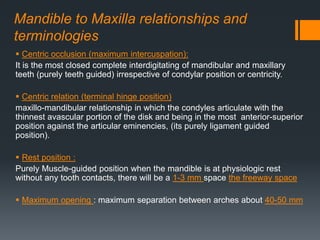
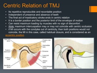
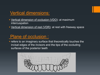
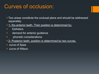
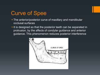
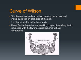
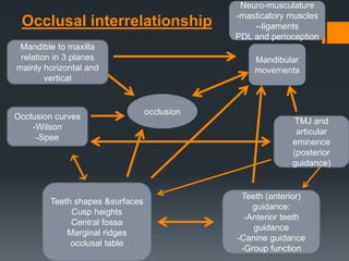
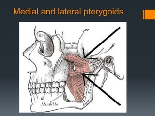
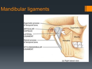
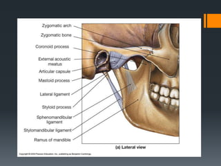
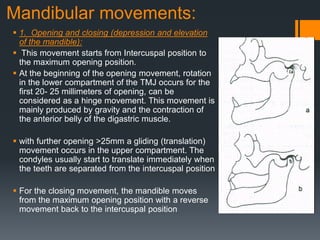
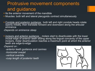
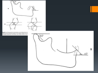
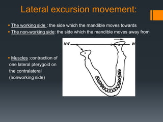
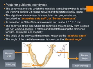
Occlusion in prosthodontics 
Dr. Razan Al Mghawich
Mandible to Maxilla relationships and 
terminologies 
 Centric occlusion (maximum intercuspation): 
It is the most closed complete interdigitating of mandibular and maxillary 
teeth (purely teeth guided) irrespective of condylar position or centricity. 
 Centric relation (terminal hinge position) 
maxillo-mandibular relationship in which the condyles articulate with the 
thinnest avascular portion of the disk and being in the most anterior-superior 
position against the articular eminencies, (its purely ligament guided 
position). 
 Rest position : 
Purely Muscle-guided position when the mandible is at physiologic rest 
without any tooth contacts, there will be a 1-3 mm space the freeway space 
 Maximum opening : maximum separation between arches about 40-50 mm
Centric Relation of TMJ 
• Its repetitive reproducible and recordable position 
• Independent of presence and absence of teeth 
• The final act of masticatory stroke ends in centric relation 
• It is a border position and the posterior limit of the envelope of motion 
• Can resist maximum loading by muscles with no sign of discomfort 
• Note: maximum intercuspation may or may not coincide with centric occlusion 
• if MI occurs with the condyles out of centricity, then both positions would not 
coincide, the MI in this case, called habitual closure, and is considered as an 
eccentric position
Vertical dimensions: 
 Vertical dimension of occlusion (VDO): at maximum 
intercuspation 
 Vertical dimension of rest (VDR): at rest with freeway space 
Plane of occlusion : 
 refers to an imaginary surface that theoretically touches the 
incisal edges of the incisors and the tips of the occluding 
surfaces of the posterior teeth
Curves of occlusion: 
 Two areas constitute the occlusal plane and should be addressed 
separately. 
 1- the anterior teeth, Their position is determined by: 
 Esthetics 
 demand for anterior guidance 
 phonetic considerations. 
 2- Posterior teeth, position is determined by two curves. 
 curve of Spee 
 curve of Wilson.
Curve of Spee 
 The anterior/posterior curve of maxillary and mandibular 
occlusal surfaces 
 It is designed so that the posterior teeth can be separated in 
protrusion, by the affects of condylar guidance and anterior 
guidance. This phenomenon reduces posterior interference
Curve of Wilson 
 “It is the mediolateral curve that contacts the buccal and 
lingual cusp tips on each side of the arch 
 it is always related to the lower arch 
 Allows for the lingual cusps (working cusps) of maxillary teeth 
to function with the lower occlusal scheme without 
interference.
Mandibular 
movements 
TMJ and 
articular 
eminence 
(posterior 
guidance) 
Occlusion curves 
Teeth shapes &surfaces 
Cusp heights 
Central fossa 
Marginal ridges 
occlusal table 
-Wilson 
-Spee 
Neuro-musculature 
-masticatory muscles 
--ligaments 
PDL and perioception 
occlusion 
Teeth (anterior) 
guidance: 
-Anterior teeth 
guidance 
-Canine guidance 
-Group function 
Occlusal interrelationship 
Mandible to maxilla 
relation in 3 planes 
mainly horizontal and 
vertical
Mandibular movement : 
 Mandibular movements are complex in nature. 
 The mandible moves in relation to maxilla to perform many different 
functional and parafunctional movements, they can be classified as 
masticatory and non masticatory. 
 The masticatory movements are necessary for introduction, grasping, 
crushing, grinding of food, and swallowing. 
 The non masticatory movements include the movements used in 
speech, wetting the lips, mouth breathing as well as, habitual or 
abnormal movements such as bruxing, clenching or tapping of teeth 
together. The time spent each day for the non masticatory movements 
exceeds the time used in masticatory movements 
 the dentist must understand the factors that regulate motion of the jaws. 
 These include contact of the opposing teeth, the anatomy and 
physiology of the TMJ, the axes around which the mandible rotates the 
action of the muscles, ligaments, and the neuromuscular integration of 
all these factors. 
 The dentist must relate an understanding of mandibular movements to 
their clinical application in the treatment of patients, particularly the 
edentulous. 
 Any prosthodontic work should aim to restore this functions probably 
with maximum imitation of the natural one
Mandibular movements 
 Mandibular movements are usually classified 
according to the main direction of movement. The 
starting position is the maximum intercuspal position. 
From this position the mandible can perform: 
 Opening and closing 
 Protrusive (forward )and retrusive ( backward) 
movements 
 Lateral movement (sideways-left & right) 
 The mandible during its function moves in all the 
three planes of space (horizontal, frontal, and 
sagittal). 
 Mandibular movement is controlled by the 
Neuromuscular system with perioceptive receptors 
 Influenced by two hard tissues that guide this 
movement, they are referred to as guidance systems: 
 Temporomandibular joints (the posterior guidance 
system) 
 Occlusal surfaces of teeth (the anterior guidance 
system)
The guidance systems 
 There are two systems that provide hard guidance during 
mandibular movements; they are interrelated and part of one 
system. 
 Posterior guidance 
 The temporomandibular joint ; made up of the head of the 
condyle, articular eminence, the intra-articular disc and the 
glenoid fossa 
 The anterior guidance system 
 is provided by which ever teeth naturally touch during 
function to guide the mandible movements; happens during 
dynamic occlusion or functional occlusion, and any other 
contact that happens prematurely, or inhibits harmony is 
referred to as Occlusal interference
Tempromandibular joint complex 
 It is a synovial joint but differs from 
most synovial joints in that the 
articular surfaces of the bony 
components are covered with 
dense fibrous connective tissue, 
instead of hyaline cartilage. In 
addition, the TMJ has a fibrous 
articular disc, to which muscle 
fibers are attached. This disc 
divides the joint into two 
compartments, upper and lower, 
thereby giving the mandible a wide 
range of movement.
Muscles of Mastication 
(Receive motor innervation for cranial nerve V-trigeminal SVE) 
Derived from components of 1st branchial arch 
MUSCLE ORIGIN INSERTION FUNCTION Excursive 
movement 
Masseter Zygomatic arch 
Lateral surface of 
ascending ramus 
Elevates mandible 
+ retrusion 
ipsilateral 
Temporalis Temporal bone 
Coronoid process & 
anterior border of 
ascending ramus 
Anterior fibers.-elevates 
mandible 
Posterior fibers-retrudes 
mandible 
ipsilateral 
Medial Pterygoid 
(Internal) 
Medial surface of 
lateral pterygoid 
plate 
Medial surface of 
ascending ramus 
Elevates mandible 
Protrusion 
contralateral 
Lateral Pterygoid 
(external) 
Superior head 
Greater wing of 
sphenoid bone 
Articular disc of TMJ 
and neck of 
mandibular condyle 
Stabilizes mandible contralateral 
Lateral Pterygoid 
(external) 
Inferior head 
Lateral surface of 
lateral pterygoid 
plate 
Anterior surface of 
condylar neck 
Depresses and protrudes 
mandible 
contralateral
Medial and lateral pterygoids
Temporalis & 
Masseter
Mandibular ligaments
Mandibular movements: 
 1. Opening and closing (depression and elevation 
of the mandible): 
 This movement starts from Intercuspal position to 
the maximum opening position. 
 At the beginning of the opening movement, rotation 
in the lower compartment of the TMJ occurs for the 
first 20- 25 millimeters of opening, can be 
considered as a hinge movement. This movement is 
mainly produced by gravity and the contraction of 
the anterior belly of the digastric muscle. 
 with further opening >25mm a gliding (translation) 
movement occurs in the upper compartment. The 
condyles usually start to translate immediately when 
the teeth are separated from the intercuspal position 
 For the closing movement, the mandible moves 
from the maximum opening position with a reverse 
movement back to the intercuspal position
Protrusive movement components 
and guidance 
 Its the anterior movement of the mandible 
 Muscles: both left and lateral pterygoids contract simultaneously 
 Condyle and posterior guidance : both left and right condyle heads rotate 
openly initially, then translate forward and downward along the articular 
eminence 
-Depends on eminence steep 
 incisors and anterior guidance : molars start to disarticulate with the lower 
incisal edge of anterior teeth sliding along the lingual concavity of the upper 
incisors, molar disarticulation reaches a maximum point at which the anterior 
teeth are edge-to-edge 
-Depend on : 
-anterior teeth guidance and canines 
-horizontal overjet 
-vertical overbite 
-cusp length of posterior teeth
Occlusal interrelationship 
 Steeper eminence -- taller cusps 
 Increased overbite--- taller cusps 
 Increased overjet– shorter cusps 
 Curve of Spee relation to this 
 more curved shorter cusps Its curvature can be described in 
terms of the length of the radius 
 short radius the curve is more acute that with a longer radius
Lateral excursion movement: 
 The working side : the side which the mandible moves towards 
 The non-working side: the side which the mandible moves away from 
 Muscles :contraction of 
one lateral pterygoid on 
the contralateral 
(nonworking side)
Posterior guidance (condyles): 
 The condyle at the side which the mandible is moving towards is called 
the working condyle , it rotates forward and translates slightly lateral 
 The slight lateral movement is immediate, non progressive and 
described as ‘immediate side shift’, or ‘Bennet movement’. 
 Its described in 86% of lateral movement and is about 0.5 to 3 mm 
 The condyles at the side which the condyle is moving away from is called 
the non working condyle, it rotates and translates along the eminence 
forward, downward and medially 
 The angle of the downward movement, known as the ‘condylar angle’ 
 The angle of the medial movement is known as the ‘Bennet angle’. 
Bennet 
angle 
Bennet 
shift 
Direction of 
movement 
Working 
Non-working
 Anterior guidance (teeth)for the lateral movement: 
 The lateral movement has 2 types of guidance : 
 Canine guidance : the sliding action contact between cusps of the 
opposing canines on the working side disarticulate the teeth on the 
non-working side 
 If any other premature contact between any other teeth happens on 
working and/or non-working side , is an interference 
 Or Group function : 
 The sliding contact between cusps of the opposing canines, 
premolars, and molars on the working side; disarticulate all teeth on 
the non-working side 
 If any other contact on the non-working side only happens is an 
interference
Intercondylar distance: 
 Intercondylar distance or length of the back axis. 
 It is the factor which has less significance in the lateral 
movements 
 because the variation in the width of the face among the 
different patients is small (Dawson, 1991) and the difference 
in the route of the cusps is not really important.
Relationship between anterior and 
posterior controlling factors
Relationship between anterior and 
posterior controlling factors
Posselt’s envelop of motion 
 It is a diagram illustrates a combination of border movements of the 
mandible in all 3 planes: Sagittal, Horizontal and Frontal 
 Following a movement of a point on the condyle or a tooth 
sagittal 
horizontal 
frontal
Chewing stroke 
Anterior opening 
border 
Sagittal view
Horizontal view 
 A: retruded contact 
 B: maximum intercuspation 
 C: edge-to-edge position 
 D: maximum protrusion 
 E: right lateral contact position 
 F: left lateral contact position 
F
Mutually Protected Occlusion 
An occlusal scheme in which the anterior teeth protect the posterior teeth, and 
vice versa. 
 The anterior teeth protect the posterior teeth by providing guidance during 
excursions 
 thus allowing the cusps of the posterior teeth to disoclude rather than strike 
one another during lateral or protrusive movements from centric relation; 
 Because posterior crowns are so much wider and possess cuspal projections 
in various configurations, they have an opportunity to bang into each other 
during chewing, speech or simply meeting together when one bites down. 
 To prevent this from happening, the anterior teeth of each arch will, ideally, be 
situated so as to come into contact before the cusps of the posterior teeth do, 
thus preventing wear on the posterior teeth. This requires less force because 
the anterior teeth are further from the joint (analogous to stopping a door 
further from its hinge). 
 The posterior teeth protect the anterior teeth by providing a stable vertical 
dimension of occlusion. While anterior teeth may retain their natural position 
even after loss of posterior teeth, the masticatory forces will eventually cause 
the single-rooted anterior to splay, thus leading to a collapsed bite
Balanced occlusion 
 It’s a type of occlusion used in dentures and artificial teeth 
 For lateral movement of the mandible 
 When the mandible moves laterally all posterior teeth with 
the canine on both sides ;the working and non-working 
;contact, unlike functional guidance where contact on the 
non-working side doesn’t happen 
 Compensating curves: 
 When artificial teeth have balanced occlusion the curvetures 
of the teeth occlusal surfaces said to have compensation 
curves; instead of curve of Spee and curve of Wilson
Reflexes and neural pathways 
 The mandible is controlled not only as a result of voluntary movement, but also 
by reflexes 
 The jaw-closing reflex: protects the mandible and associated structures during 
violent whole body movement; it can result in damage to the teeth, especially if 
the occlusal contacts are not in the long axis of the root. 
 The jaw opening reflex is to protect the teeth during sudden mastication of a hard 
object and to protect the lips, cheeks and tongue during mastication. 
 These voluntary and involuntary movements are controlled by the central and 
autonomic nervous systems via sensory and motor nerves. 
 specific proprioceptors are situated in deep muscle, and in the PDL 
 As a consequence there is the potential for any change in patient’s occlusion, by 
routine dentistry, to ‘be sensed’ by the patient’s nervous system. It is because of 
this consideration that dentists cannot ignore the effect of changing the occlusion 
when providing routine care.
Centric relation interocclusal 
record 
 Bimanual manipulation technique by dawson 2007
Anterior deprogramming devices 
 Devices that can manipulate the mandible to seat the condyles in the 
centric relation by deprogramming the perioceptive reflexes of the 
muscles 
 -leaf gauge 
 -Lucia jig :acrylic resin jig with acu-flow material on it that sets on 
45s
Materials used for inetrocclusal records 
 Thermoplastic impression waxes and natural resins, ex: Aluwax®: 
contains powdered aluminum; to increase the integrity of the 
compound and provide the heat retention properties needed for 
efficient modeling 
 Fast setting elastomeric impression materials (polyether, polyvinyl 
siloxane): 
-Elastomer: is a polymer with viscoelasticity ,having both viscosity and 
elasticity:( the tendency of solid materials to return to their original 
shape after being deformed)

Occlusion for prosthodontics

  • 1.
    Occlusion in prosthodontics Dr. Razan Al Mghawich
  • 2.
    Mandible to Maxillarelationships and terminologies  Centric occlusion (maximum intercuspation): It is the most closed complete interdigitating of mandibular and maxillary teeth (purely teeth guided) irrespective of condylar position or centricity.  Centric relation (terminal hinge position) maxillo-mandibular relationship in which the condyles articulate with the thinnest avascular portion of the disk and being in the most anterior-superior position against the articular eminencies, (its purely ligament guided position).  Rest position : Purely Muscle-guided position when the mandible is at physiologic rest without any tooth contacts, there will be a 1-3 mm space the freeway space  Maximum opening : maximum separation between arches about 40-50 mm
  • 3.
    Centric Relation ofTMJ • Its repetitive reproducible and recordable position • Independent of presence and absence of teeth • The final act of masticatory stroke ends in centric relation • It is a border position and the posterior limit of the envelope of motion • Can resist maximum loading by muscles with no sign of discomfort • Note: maximum intercuspation may or may not coincide with centric occlusion • if MI occurs with the condyles out of centricity, then both positions would not coincide, the MI in this case, called habitual closure, and is considered as an eccentric position
  • 4.
    Vertical dimensions: Vertical dimension of occlusion (VDO): at maximum intercuspation  Vertical dimension of rest (VDR): at rest with freeway space Plane of occlusion :  refers to an imaginary surface that theoretically touches the incisal edges of the incisors and the tips of the occluding surfaces of the posterior teeth
  • 5.
    Curves of occlusion:  Two areas constitute the occlusal plane and should be addressed separately.  1- the anterior teeth, Their position is determined by:  Esthetics  demand for anterior guidance  phonetic considerations.  2- Posterior teeth, position is determined by two curves.  curve of Spee  curve of Wilson.
  • 6.
    Curve of Spee  The anterior/posterior curve of maxillary and mandibular occlusal surfaces  It is designed so that the posterior teeth can be separated in protrusion, by the affects of condylar guidance and anterior guidance. This phenomenon reduces posterior interference
  • 7.
    Curve of Wilson  “It is the mediolateral curve that contacts the buccal and lingual cusp tips on each side of the arch  it is always related to the lower arch  Allows for the lingual cusps (working cusps) of maxillary teeth to function with the lower occlusal scheme without interference.
  • 8.
    Mandibular movements TMJand articular eminence (posterior guidance) Occlusion curves Teeth shapes &surfaces Cusp heights Central fossa Marginal ridges occlusal table -Wilson -Spee Neuro-musculature -masticatory muscles --ligaments PDL and perioception occlusion Teeth (anterior) guidance: -Anterior teeth guidance -Canine guidance -Group function Occlusal interrelationship Mandible to maxilla relation in 3 planes mainly horizontal and vertical
  • 9.
    Mandibular movement :  Mandibular movements are complex in nature.  The mandible moves in relation to maxilla to perform many different functional and parafunctional movements, they can be classified as masticatory and non masticatory.  The masticatory movements are necessary for introduction, grasping, crushing, grinding of food, and swallowing.  The non masticatory movements include the movements used in speech, wetting the lips, mouth breathing as well as, habitual or abnormal movements such as bruxing, clenching or tapping of teeth together. The time spent each day for the non masticatory movements exceeds the time used in masticatory movements  the dentist must understand the factors that regulate motion of the jaws.  These include contact of the opposing teeth, the anatomy and physiology of the TMJ, the axes around which the mandible rotates the action of the muscles, ligaments, and the neuromuscular integration of all these factors.  The dentist must relate an understanding of mandibular movements to their clinical application in the treatment of patients, particularly the edentulous.  Any prosthodontic work should aim to restore this functions probably with maximum imitation of the natural one
  • 10.
    Mandibular movements Mandibular movements are usually classified according to the main direction of movement. The starting position is the maximum intercuspal position. From this position the mandible can perform:  Opening and closing  Protrusive (forward )and retrusive ( backward) movements  Lateral movement (sideways-left & right)  The mandible during its function moves in all the three planes of space (horizontal, frontal, and sagittal).  Mandibular movement is controlled by the Neuromuscular system with perioceptive receptors  Influenced by two hard tissues that guide this movement, they are referred to as guidance systems:  Temporomandibular joints (the posterior guidance system)  Occlusal surfaces of teeth (the anterior guidance system)
  • 12.
    The guidance systems  There are two systems that provide hard guidance during mandibular movements; they are interrelated and part of one system.  Posterior guidance  The temporomandibular joint ; made up of the head of the condyle, articular eminence, the intra-articular disc and the glenoid fossa  The anterior guidance system  is provided by which ever teeth naturally touch during function to guide the mandible movements; happens during dynamic occlusion or functional occlusion, and any other contact that happens prematurely, or inhibits harmony is referred to as Occlusal interference
  • 13.
    Tempromandibular joint complex  It is a synovial joint but differs from most synovial joints in that the articular surfaces of the bony components are covered with dense fibrous connective tissue, instead of hyaline cartilage. In addition, the TMJ has a fibrous articular disc, to which muscle fibers are attached. This disc divides the joint into two compartments, upper and lower, thereby giving the mandible a wide range of movement.
  • 14.
    Muscles of Mastication (Receive motor innervation for cranial nerve V-trigeminal SVE) Derived from components of 1st branchial arch MUSCLE ORIGIN INSERTION FUNCTION Excursive movement Masseter Zygomatic arch Lateral surface of ascending ramus Elevates mandible + retrusion ipsilateral Temporalis Temporal bone Coronoid process & anterior border of ascending ramus Anterior fibers.-elevates mandible Posterior fibers-retrudes mandible ipsilateral Medial Pterygoid (Internal) Medial surface of lateral pterygoid plate Medial surface of ascending ramus Elevates mandible Protrusion contralateral Lateral Pterygoid (external) Superior head Greater wing of sphenoid bone Articular disc of TMJ and neck of mandibular condyle Stabilizes mandible contralateral Lateral Pterygoid (external) Inferior head Lateral surface of lateral pterygoid plate Anterior surface of condylar neck Depresses and protrudes mandible contralateral
  • 15.
  • 16.
  • 17.
  • 19.
    Mandibular movements: 1. Opening and closing (depression and elevation of the mandible):  This movement starts from Intercuspal position to the maximum opening position.  At the beginning of the opening movement, rotation in the lower compartment of the TMJ occurs for the first 20- 25 millimeters of opening, can be considered as a hinge movement. This movement is mainly produced by gravity and the contraction of the anterior belly of the digastric muscle.  with further opening >25mm a gliding (translation) movement occurs in the upper compartment. The condyles usually start to translate immediately when the teeth are separated from the intercuspal position  For the closing movement, the mandible moves from the maximum opening position with a reverse movement back to the intercuspal position
  • 20.
    Protrusive movement components and guidance  Its the anterior movement of the mandible  Muscles: both left and lateral pterygoids contract simultaneously  Condyle and posterior guidance : both left and right condyle heads rotate openly initially, then translate forward and downward along the articular eminence -Depends on eminence steep  incisors and anterior guidance : molars start to disarticulate with the lower incisal edge of anterior teeth sliding along the lingual concavity of the upper incisors, molar disarticulation reaches a maximum point at which the anterior teeth are edge-to-edge -Depend on : -anterior teeth guidance and canines -horizontal overjet -vertical overbite -cusp length of posterior teeth
  • 23.
    Occlusal interrelationship Steeper eminence -- taller cusps  Increased overbite--- taller cusps  Increased overjet– shorter cusps  Curve of Spee relation to this  more curved shorter cusps Its curvature can be described in terms of the length of the radius  short radius the curve is more acute that with a longer radius
  • 24.
    Lateral excursion movement:  The working side : the side which the mandible moves towards  The non-working side: the side which the mandible moves away from  Muscles :contraction of one lateral pterygoid on the contralateral (nonworking side)
  • 25.
    Posterior guidance (condyles):  The condyle at the side which the mandible is moving towards is called the working condyle , it rotates forward and translates slightly lateral  The slight lateral movement is immediate, non progressive and described as ‘immediate side shift’, or ‘Bennet movement’.  Its described in 86% of lateral movement and is about 0.5 to 3 mm  The condyles at the side which the condyle is moving away from is called the non working condyle, it rotates and translates along the eminence forward, downward and medially  The angle of the downward movement, known as the ‘condylar angle’  The angle of the medial movement is known as the ‘Bennet angle’. Bennet angle Bennet shift Direction of movement Working Non-working
  • 26.
     Anterior guidance(teeth)for the lateral movement:  The lateral movement has 2 types of guidance :  Canine guidance : the sliding action contact between cusps of the opposing canines on the working side disarticulate the teeth on the non-working side  If any other premature contact between any other teeth happens on working and/or non-working side , is an interference  Or Group function :  The sliding contact between cusps of the opposing canines, premolars, and molars on the working side; disarticulate all teeth on the non-working side  If any other contact on the non-working side only happens is an interference
  • 27.
    Intercondylar distance: Intercondylar distance or length of the back axis.  It is the factor which has less significance in the lateral movements  because the variation in the width of the face among the different patients is small (Dawson, 1991) and the difference in the route of the cusps is not really important.
  • 28.
    Relationship between anteriorand posterior controlling factors
  • 29.
    Relationship between anteriorand posterior controlling factors
  • 30.
    Posselt’s envelop ofmotion  It is a diagram illustrates a combination of border movements of the mandible in all 3 planes: Sagittal, Horizontal and Frontal  Following a movement of a point on the condyle or a tooth sagittal horizontal frontal
  • 31.
    Chewing stroke Anterioropening border Sagittal view
  • 32.
    Horizontal view A: retruded contact  B: maximum intercuspation  C: edge-to-edge position  D: maximum protrusion  E: right lateral contact position  F: left lateral contact position F
  • 33.
    Mutually Protected Occlusion An occlusal scheme in which the anterior teeth protect the posterior teeth, and vice versa.  The anterior teeth protect the posterior teeth by providing guidance during excursions  thus allowing the cusps of the posterior teeth to disoclude rather than strike one another during lateral or protrusive movements from centric relation;  Because posterior crowns are so much wider and possess cuspal projections in various configurations, they have an opportunity to bang into each other during chewing, speech or simply meeting together when one bites down.  To prevent this from happening, the anterior teeth of each arch will, ideally, be situated so as to come into contact before the cusps of the posterior teeth do, thus preventing wear on the posterior teeth. This requires less force because the anterior teeth are further from the joint (analogous to stopping a door further from its hinge).  The posterior teeth protect the anterior teeth by providing a stable vertical dimension of occlusion. While anterior teeth may retain their natural position even after loss of posterior teeth, the masticatory forces will eventually cause the single-rooted anterior to splay, thus leading to a collapsed bite
  • 34.
    Balanced occlusion It’s a type of occlusion used in dentures and artificial teeth  For lateral movement of the mandible  When the mandible moves laterally all posterior teeth with the canine on both sides ;the working and non-working ;contact, unlike functional guidance where contact on the non-working side doesn’t happen  Compensating curves:  When artificial teeth have balanced occlusion the curvetures of the teeth occlusal surfaces said to have compensation curves; instead of curve of Spee and curve of Wilson
  • 35.
    Reflexes and neuralpathways  The mandible is controlled not only as a result of voluntary movement, but also by reflexes  The jaw-closing reflex: protects the mandible and associated structures during violent whole body movement; it can result in damage to the teeth, especially if the occlusal contacts are not in the long axis of the root.  The jaw opening reflex is to protect the teeth during sudden mastication of a hard object and to protect the lips, cheeks and tongue during mastication.  These voluntary and involuntary movements are controlled by the central and autonomic nervous systems via sensory and motor nerves.  specific proprioceptors are situated in deep muscle, and in the PDL  As a consequence there is the potential for any change in patient’s occlusion, by routine dentistry, to ‘be sensed’ by the patient’s nervous system. It is because of this consideration that dentists cannot ignore the effect of changing the occlusion when providing routine care.
  • 36.
    Centric relation interocclusal record  Bimanual manipulation technique by dawson 2007
  • 37.
    Anterior deprogramming devices  Devices that can manipulate the mandible to seat the condyles in the centric relation by deprogramming the perioceptive reflexes of the muscles  -leaf gauge  -Lucia jig :acrylic resin jig with acu-flow material on it that sets on 45s
  • 38.
    Materials used forinetrocclusal records  Thermoplastic impression waxes and natural resins, ex: Aluwax®: contains powdered aluminum; to increase the integrity of the compound and provide the heat retention properties needed for efficient modeling  Fast setting elastomeric impression materials (polyether, polyvinyl siloxane): -Elastomer: is a polymer with viscoelasticity ,having both viscosity and elasticity:( the tendency of solid materials to return to their original shape after being deformed)