Temporomandibular joint/ dental courses
•
18 likes•3,901 views

The Indian Dental Academy is the Leader in continuing dental education , training dentists in all aspects of dentistry and offering a wide range of dental certified courses in different formats.for more details please visit www.indiandentalacademy.com

Recommended
More Related Content
What's hot
What's hot (20)
Viewers also liked
Viewers also liked (20)
Similar to Temporomandibular joint/ dental courses
Similar to Temporomandibular joint/ dental courses (20)
More from Indian dental academy
More from Indian dental academy (20)
Recently uploaded
Recently uploaded (20)
Temporomandibular joint/ dental courses
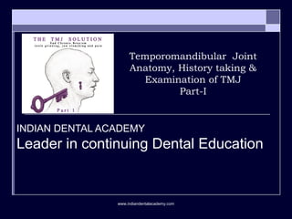
- 1. Temporomandibular Joint Anatomy, History taking & Examination of TMJ Part-I INDIAN DENTAL ACADEMY Leader in continuing Dental Education www.indiandentalacademy.com
- 2. Contents-I Functional Anatomy Biomechanics of the Masticatory System Etiology and Identification of functional disturbances in the masticatory system. Causes Signs and symptoms History and examination Investigations www.indiandentalacademy.com
- 3. Contents-II General considerations in treatment. Treatment of Masticatory muscle disorder Treatment of TMJ disorders Treatment of Chronic Mandibular hypomobility and growth disorders Treatment sequencing www.indiandentalacademy.com
- 4. Temporomandibular Joint Anatomy, History taking & Examination of TMJ Part-I Shweta Ujaoney Guided by: Dr Mukta Motwani Dr Shirish Degwekar (Prof & Guide) (Prof & HOD) www.indiandentalacademy.com
- 5. Functional Anatomy & Biomechanics of the Masticatory System Dentition and supporting structures The skeletal components of the TMJ The ligaments The muscles www.indiandentalacademy.com
- 6. Dentition and supporting structures www.indiandentalacademy.com
- 7. The skeletal components the TMJ www.indiandentalacademy.com
- 9. TMJ Ginglymoid joint Arthrodial joint Therefore, ginglymoarthroidal joint Compound joint Synovial joint www.indiandentalacademy.com
- 10. The ligaments Collateral ligament Capsular ligament Temporomandibular ligament Sphenomandibular ligamant Stylomandibular ligament www.indiandentalacademy.com
- 13. The muscles Masseter Temporalis Pterygoideus medialis Pterygoideus lateralis Inferior lateral pterygoid Superior lateral pterygoid Digastricus www.indiandentalacademy.com
- 17. Nerve supply: Dense plexus of unmyelinated fibres that weaves throughout the fibrous capsule and related fibrofatty tissue The small diameter afferent pain fibres enter the regionally related articular branches of Auricotemporal Masseteric Deep temporal Sometimes lateral pterygoid nerves– pass into the sensory root and spinal tract of the trigeminal nerve. No nerve endings of any type are present in the fibrous articular surface, the fibrocartlage, disc or synovia--- pain is probably only produced by mechanical or chemical irritation of the sensitive capsular tissues surrounding the joint. www.indiandentalacademy.com
- 21. Function of the neuromuscular system Muscle- Motor Unit Major function of the masticatory system www.indiandentalacademy.com
- 22. Muscle- Motor Unit The number of muscle fibres innervated by one motor neuron varies greatly according to the function of the motor unit. The fewer the muscle fibres per motor neuron, the more precise the movement. The inferior lateral pterygoid muscle has a relative low muscle fibre/motor neuron ratio; therefore it is capable of the fine adjustments in length needed to adapt to horizontal changes in the mandibular position. By contrast the masseter has a greater number of motor fibres per motor neuron, which corresponds to the more gross function of providing the force necessary during mastication.www.indiandentalacademy.com
- 24. Local event: Trauma- 1. post injection response to local anesthesia 2. mouth opened too widely (strain). Bruxism. Constant deep pain input (pain felt in the masticatory or associated structures often alter normal muscle function by way of the central excitatory effects) Systemic events: Increased levels of emotional stress. Acute and chronic disease Normal function +event > physiologic tolerance = TMD symptoms Etiology and identification of functional disturbances in the masticatory system www.indiandentalacademy.com
- 25. Normal function +event > physiologic tolerance = TMD symptoms Physiologic tolerance: Each patient has a ability to tolerate certain events without any adverse effect. Physiologic tolerance can be influenced by both local and systemic factors. www.indiandentalacademy.com
- 26. Etiology Five factors that are related to TMDs are Occlusal condition: 1. Presence of skeletal open bite 2. Retruded contact position & ICP slides of greater than 4mm. 3. Overjets greater than 4mm 4. Five or missing and unreplaced posterior teeth. Trauma Emotional stress Deep pain input Parafunctional activities www.indiandentalacademy.com
- 27. Trauma Trauma seems to have a greater influence on the intracapsular disorder than muscular disorders. Trauma can be divided into: Macrotrauma Microtrauma Macrotrauma is considered any sudden force that results in structural alterations, such as direct blow to the face. Microtrauma refers to any small force that is repeatedly applied to the structures over a long period of time. Activities like bruxism or clenching can produce microtrauma to the teeth, joints and muscles that are being loaded.www.indiandentalacademy.com
- 28. Emotional stress Increased levels of emotional stress – increases the tonicity of head and neck muscles increase levels of non-functional muscle activity, such as bruxism or tooth clenching. Another systemic factor that can influence an individual’s physiologic tolerance to certain events - his/her sympathetic activity or tone. It is closely related to the fight or flight reflex activated by the stressor. In the presence of stress, the capillary blood flow in the outer tissues is constricted, permitting increased blood flow to the more important musculoskeletal structures and internal organs. It has been suggested that increased sympathetic activity or tone can influence TMD symptoms and plays an important role in chronic pain. Deep pain input Parafunctional activities www.indiandentalacademy.com
- 29. History The purpose of history and examination is to identify any area or structure of the masticatory system that shows breakdown or pathologic damage. Pain and dysfunction both signify breakdown in the masticatory system. History can be obtained by Direct conversation Written questionnaire www.indiandentalacademy.com
- 30. Features to be included in a thororugh orofacial pain history I. Chief complaint A. Location of pain B. Onset of pain Associated with other factors Progression C. Characteristics of pain 1. Quality of pain 2. Behaviour of pain a.Temporal b. Duration c. Localization 3. Intensity of pain 4. Concomitant symptoms 5. Flow of the pain www.indiandentalacademy.com
- 31. D. Aggravating and alleviating factors 1. Function and parafunction 2. Physical modalities 3. Medication 4. Emotional stress 5. Sleep disturbances 6. Litigation E. Past consultations and /or treatments H. Relationship to other pain complaints II. Medical history III. Review of systems IV. Psychological assessment www.indiandentalacademy.com
- 33. Symptoms If the weakest structures in the system are the muscles, the individual commonly experiences muscle tenderness and pain during mandibular movements-limited jaw movements with related pain. If TMJ’s are the weakest link, joint tenderness and pain will often be reported. The joints then produce sounds such as clicking or grating. Sometimes the muscles and joints can tolerate the changes, but because of increased activity of the muscles(e.g bruxism) the weakest link is either the supportive structures of the teeth or the teeth themselves. The teeth then show mobility or wear. www.indiandentalacademy.com
- 34. To summarize them some of the most common symptoms are: 1. Tooth wear 2. Pulpitis 3. Tooth mobility 4. Masticatory muscle pain 5. TMJ pain 6. Ear pain 7. Headache www.indiandentalacademy.com
- 35. The clinical signs and symptoms are grouped into three categories according to structures that are affected: Muscles TMJ’s Dentition www.indiandentalacademy.com
- 36. Functional disorder of muscle Two major symptoms that can be observed are: Pain Dysfunction Muscle pain may range from slight tenderness to extreme discomfort. Pain felt in muscle tissue is called myalgia. Myalgia can arise from increased levels of muscular use. The symptoms are often associated with a feeling of muscle fatigue and tightness. Some authors suggest it is related to vasoconstriction of relevant nutrient arteries and the accumulation of metabolic waste products in the muscle tissues. within the ischemic area of the muscle, certain algogenic substances like bradykinins, prostaglandins are released causing muscle pain. www.indiandentalacademy.com
- 37. e.g when a pt reports of pain during chewing or speaking, these functional activities are not usually the cause of the disorder, instead they heighten the patients awareness of it. More likely some type of activity or central nervous system effect has lead to the muscle pain. Thus the need to be directed at diminishing the hyperactivity of the muscle or the CNS effect. Also the myogenous pain is a deep type of pain and if it becomes constant it can produce central excitatory effects, thus muscle pain can reinitiate more muscle pain called the cyclic muscle pain. Another very common symptom commonly associated with muscle pain is headache. www.indiandentalacademy.com
- 38. Dysfunction It is a common clinical symptom associated with myalgia. It is usually seen as a decrease in the range of the mandibular movement. When the muscle tissues have been compromised by overuse, any contraction or stretching increases the pain. Thus to maintain comfort the pt restricts the range of movement. Acute malocclusion is another type of dysfunction. It refers to sudden change in the occlusal condition that has been created by a disorder. It may result from a sudden change in the resting length of a muscle that controls jaw position. www.indiandentalacademy.com
- 39. E.g slight shortening of inferior lateral pterygoid will cause disocclusion of the posterior teeth on the ipsilateral side and premature contact of the ant teeth esp canines on the contralateral side. Thus acute malocclusion is the result of rather than the cause of muscle disorder and thus the treatment should be aimed at eliminating the muscle disorder. Some intracapsular disorders can also lead to acute malocclusions. www.indiandentalacademy.com
- 40. Five types of muscle disorders Protective co-contraction Local muscle soreness Myofascial pain Myospasm Chronic centrally mediated myalgia Fibromyalgia www.indiandentalacademy.com
- 41. TMJ signs n symptoms Pain Dysfunction Pain in any joint structure is called arthralgia. It would seem that arthralgic pain would originate from the articular surfaces when the muscles load the joint, however this is impossible in a healthy join because there are no innervations of the articular surfaces. Arthralgia therefore originates only from the nociceptors located in the soft tissue surrounding a joint. Three pre-articular tissues contain such nociceptors The discal ligaments The capsular ligaments The retrodiscal tissues www.indiandentalacademy.com
- 42. When these ligaments are elongated or retrodiscal tissue compressed the nociceptors send out signals and pain is perceived. Stimulation of the nociceptors creates inhibitory action in the muscles that move the mandible. Therefore when pain is suddenly and unexpectedly felt mandibular movement immediately ceases (nociceptive reflex). When chronic pain is felt, movement becomes limited and very deliberate (protective co-contraction). Arthalgia from normally healthy structures of the joint is a sharp, sudden, and intense pain that is closely associated with joint movement. When the joint is rested, the pain resolves quickly. If the joint structures breakdown, inflammation can produce a constant pain that is accentuated by joint movement. www.indiandentalacademy.com
- 43. Dysfunction: It usually presents as a disruption of the normal condyle-disc movement with the production of joint sounds may be a single event of short duration known as a click. If the sound is loud, it may be referred to as a pop. Crepitation is a multiple rough gravel-like sound described as grating and complicated. Dysfunction of the TMJ may also present as catching sensations when the patient opens the mouth. Sometimes jaw can actually lock. Dysfunction of the jaw is always directly related to jaw movement. www.indiandentalacademy.com
- 44. TMJ disorders are Derrangement of the condyle-disc complex Structural incompatibility of the articular surfaces… Both these are also called Disc-interference disorders by Welden Bell. Inflammatory joint disorders www.indiandentalacademy.com
- 45. Cranial nerve examination CN V: Trigeminal Setup Patient sitting over edge of bed. Motor: pt opens mouth, clenches teeth (pterygoids). • Palpate temporal, masseter muscles as they clench. Test jaw jerk: Dr's finger should be placed on tip of jaw. Grip patellar hammer halfway up shaft and tap Dr's finger lightly. Usually nothing happens, or just a slight closure. www.indiandentalacademy.com
- 47. Cervical examination Cervico spinal pain and dysfunction can be referred to the masticatory apparatus . The mobility of the neck is examined for range and symptoms. The pt is asked to look first to the right and then to the left. There should be atleast 70 degree of rotation in each direction. Next the patient is asked to look upwards as far as possible. The head should normally extend backwards some 60 degree and flex downwards 45 degree. Finally the pt is asked to bend the neck to the right and to the left. This should be possible approximately 40 degrees each way. Any pain recorded and any limitation of movement carefully investigated to determine whether its source is a muscular or a vertebral problem.www.indiandentalacademy.com
- 48. When pts with limited range of movement can be passively stretched to a greater range the source is usually muscular. Pts with vertebral problems cannot be stretched to a greater range. If the clinician suspects that the patient has a cranio cervical disorder, proper referral for a more complete evaluation is indicated. The masticatory apparatus examination consists of evaluating three major structures Muscles Joints Teeth. www.indiandentalacademy.com
- 49. Examination of TMJ Inspection: Asymmetry Presence of any swelling/growth Depression Discharge Colour change of the skin over the TMJ area Surface of the overlying skin in the preauricular area. Mouth opening- normal/ restricted. Deviation/deflection www.indiandentalacademy.com
- 50. Palpation Tenderness over the TMJ during movement. Palpation of the TMJ- joint sounds Hypermobility/ Hypomobility Palpation of the muscles www.indiandentalacademy.com
- 60. Medial pterygoid Contraction: It is an elevator muscle and hence contracts when the teeth are coming together. If it is a source of pain then clenching the teeth together will increase the pain. When the tongue blades is placed between the posterior teeth and the patient clenches against it, the pain is still increased because the elevators are still contracting. Stretching: it stretches when the mouth is opened widely. Therefore if it is the source of pain, opening mouth wide will increase the pain. www.indiandentalacademy.com
- 67. Joint sounds can be perceived by placing the fingertips over the lateral surfaces of the joint and having the patient open and close the mouth. For more careful examination, the clinician can place a stethoscope over the joint area. If a stethoscope is used, the clinician must appreciate that this instrument will detect many more sounds than mere palpation, and the significance of these sounds needs to be assessed. Not all joint sounds should be considered a problem worthy of treatment. www.indiandentalacademy.com
- 72. Diagnostic tests OPG Transcranial Transpharyngeal Transorbital Other extraoral technique-SMV, Reverse Towne’s view Arthrography CT MRI Arthroscopy Mounted Cast Electromyography Mandibular tracking device Sonography Vibration analysis Thermography www.indiandentalacademy.com
- 77. Conventional tomography Advantages: Tomograms are superior to conventional radiography because of their ability to depict a greater portion of the joint. By providing a series of sectional radiographs, tomography can reproduce changes in the central portion of the TMJ and therefore decrease false-negative interpretations. Limitations: Tomography however is limited in its ability to detect early lesions. Although tomograms may supply superior diagnostic information, patients undergoing tomography are exposed to substantially more radiation, and the difference in cost is considerable. www.indiandentalacademy.com
- 78. Arthrography Arthrography is defined as the injection of contrast material, radiolucent and / or radio opaque, into a synovial space followed by radiography of the joint. The following information can be obtained from an arthrogram 1. Position of the disc relative to the condyle and articular eminence a. With mandible closed b. At various positions of mandibular movements 2. Morphology of the disc 3. Presence of tears/perforations in the disc or its attachments. 4. Presence of adhesions in the joint spaces 5. Presence of “loose bodies” in the joint spaces 6. Irregularities in the posterior attachments of the disc. 7. Possibly synovial proliferations. www.indiandentalacademy.com
- 79. Indications: To confirm the diagnosis before operating for a TMD. To obtain an arthrogram to have information about soft tissues. To obtain specific information about disc position and morphology to plan a surgical procedure. To some extent for documentary proof to avoid litigation. Contraindication: Allergic reaction to iodinated contrast medium. Bleeding disorders and patients on anticoagulant medications. In presence of localized skin infection. Disadvantages: Invasiveness Pain (intra operative or postoperative) Risk of infection Potential damage to disc, capsule, and fibro cartilage Allergy to contrast material (or local anesthetic)www.indiandentalacademy.com
- 81. CT Advantages: It provides images without superimposition inherent in conventional tomography and permits a section of optimal views through multiple plane reconstructions CT may yield information concerning the position of the soft tissue disc in addition to depicting the osseous structures. www.indiandentalacademy.com
- 82. Limitations: The expense and radiation involved is significantly greater for CT than for conventional tomography. Some studies have shown that conventional tomography is superior to CT in the diagnosis of single structural bone changes and comparable for the comprehensive diagnosis of TMJ disorders. www.indiandentalacademy.com
- 83. MRI MRI has overtaken arthrography and CT as the imaging modality of choice for th diagnosis of joint abnormalities. Using a strong magnetic field, MRI can depict soft-tissue anatomy with details through its effect on tissues with high water content. www.indiandentalacademy.com
- 84. Advantages: MRI does not use ionizing radiation and is non- invasive. It has been shown to be superior to arthrography in demonstrating medial and lateral displacements of the disc. www.indiandentalacademy.com
- 85. The ability of MRI to depict osseous changes in the TMJ may prove to be better than originally anticipated. The use of coronal MRI in addition to the sagittal view, may provide increased information concerning bony changes. Limitation: It does not detect perforation as consistently. www.indiandentalacademy.com
- 86. Arthroscopy Arthroscopy is the fundamental procedure in the diagnosis and traetment of various orthopedic disorders for many years. Arthroscopy of the upper compartment permits direct inspection of the articular surfaces of the temporal bone and superior aspect of the disc. Advantages: As a diagnostic tool it can help confirm the impression derived from preceding clinical, radiographic and imaging findings. Most studies that have evaluated the diagnostic capabilities of TMJ arthroscopy and have found arthroscopic diagnosis of Arthrosis, remodeling, adhesions, or perforation to be reliable (high specificity) An additional diagnostic use for TMJ arthroscopy is the ability to perform synovial biopsies to ascertain the presence of histologic changes, such as inflammation (i.e synovitis) or proliferation.www.indiandentalacademy.com
- 87. Limitations: The risk of under-diagnosis of pathologic changes is significant (low sensitivity). Under-diagnosis of perforations, especially when they occur on the lateral aspect of the disc where good visualization is difficult, appears to be a particular problem in arthroscopic diagnosis. The size of the instrumentation prevents its use in the smaller lower compartment where pathologic changes frequently occur. www.indiandentalacademy.com
- 88. Mounted Cast If during the examination the clinician finds significant orthopedic instability, accurately mounted study casts may be helpful to further assess the occlusal condition. Mounted casts are not indicated for all patients being examined for TMds. Dental study casts can be of value not only as a baseline record for tooth and jaw relations but also for evaluating the effects of bruxism over time. www.indiandentalacademy.com
- 89. Electromyography The use of EMG is based on the assumption that certain pathologic or dysfunctional conditions can be identified by abnormal activity of the masticatory muscles. An analysis of literature conducted by Mohl and colleagues have uncovered several major deficiencies as : The lack of adequate control groups, the lack of studies showing reliability and validity of the methods, the inadequacy or non existence of statistical comparisons, and the declaration of conclusions that were not supported by the study results. The most significant problem, however, was the large inter-individual variability in normal and patient groups, resulting in considerable overlap between them. www.indiandentalacademy.com
- 90. Advantages: EMG has been proven to provide excellent information on muscle function under research conditions. It is also useful with various biofeedback techniques to enable the patient to monitor muscle tension during relaxation training. www.indiandentalacademy.com
- 91. Mandibular tracking device Computerised mandibular scanning It is a device to track the movement of the entire mandible relative to the maxilla. It has been suggested that these device can be used to diagnose and monitor the treatment of TMDs www.indiandentalacademy.com
- 92. Several investigators have examined jaw movement parameters that are considered to be of potential diagnostic value are: Amplitude of jaw movement Reproducibility or consistency of jaw movements Velocity and smoothness of jaw trajectories No evidence suggests that the sensitivity and specificity of jaw tracking devices are reliable enough to be used for diagnosis or treatment. www.indiandentalacademy.com
- 93. Sonography Sonography is a technique of recording and graphically demonstrating joint sounds. Some techniques use audio-amplifying devices, whereas others rely on ultrasound echo recordings (Doppler ultrasonography) The joint sounds are often related to specific disc derrangements; therefore there presence may have meaning. Anterior disc displacement with reduction- reciprocal click or popping sound Anterior disc displacement without reduction- less discrete soft tissue sounds Degenerative joint disease- crepitation or grating noises www.indiandentalacademy.com
- 94. On the other hand, the presence of joint sounds does not , in itself, denote a problem. If sonography is to have a meaning, it must be able to separate sounds that have significance to treatment from those that do not. Presently sonography does not provide the clinician with any additional diagnostic information over manual palpation or stethoscopic evaluation www.indiandentalacademy.com
- 95. Vibration analysis To help in diagnosing intracapsular TMD, internal derrangements in particular. It measures the minute vibrations made by the condyle as it translates and has been shown to be reliable.www.indiandentalacademy.com
- 96. Although some studies do report encouraging accuracy in detection of joint vibrations, little data demonstrates that vibration analysis is a useful adjunct in the selection of appropriate patient therapy. www.indiandentalacademy.com
- 97. Thermography Thermography is a technique that records and graphically illustrates surface skin temperatures. Recorded– different colours producing a map that depicts the surface being studied. www.indiandentalacademy.com
- 98. Normal- bilaterally symmetrical thermograms Otherwise it reveals a problem such as TMD Studies show variable results. The sensitivity and specificity of identifying myofascial trigger points with thermography has not been demonstrated to be reliable. At this time– thermography is not a useful technique for the diagnosis and management of TMDs. www.indiandentalacademy.com
- 99. References: www.clinicalexam.com/pda/n_cranial_nerves_exam.htm Technological devices in the diagnosis of TMD; Gonzales etal oral Maxillofacial Surg Clin N Am 20(2008) 211-220. Management of temporomandibular disorder and occlusion, Jeffrey P. Okeson 5th edition Temporomandibular disorders, classification, diagnosis, management, 3rd edition, Welden Bell Colour Atlas Temporomandibular joint surgery, Peter D Quinn. Common disorders of TMJ, H.D Ogus, PA Toller, dental practitioner handbook, 2nd edition. Management of Temporomandibular Disorder in General Dental Practice, Gunnar E. Carlsson, Tomas Magnusson. Burket’s Oral medicine diagnosis and treatment, ninth edition. Textbook of oral medicine, Ghom, first edition 2005 B.D Chourasia’s Human anatomy fourth edition, HNF. www.indiandentalacademy.com
- 100. Reliability and validity of diagnostic modalities for temporomandibular disorders Mohl ND;Adv Dent Res. 1993 Aug;7(2):113-9 www.indiandentalacademy.com