SlideShare a Scribd company logo
1 of 79
Download to read offline
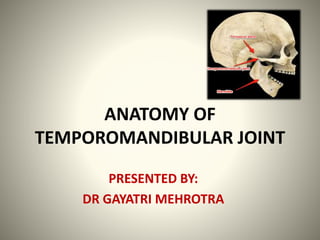
ANATOMY OF
TEMPOROMANDIBULAR JOINT
PRESENTED BY:
DR GAYATRI MEHROTRA
INTRODUCTION
 TMJ is the articulation between the head of
the mandible and the mandibular fossa and
articular tubercle of the temporal bone.
 Also known as:
 Bicondylar joint
 Ginglymodiarthroidal joint
Provides hinging
movement in one
plane, thus
considered as
ginglymoid joint
Provides for
gliding
movements,thus
considered as
arthroidal joint
Ginglymodiarthroidal
joint
 It is considered as a synovial joint of the
condylar variety.
 It is classified as a compound joint because it
is made up of 3 bones:
 Anterior part of mandibular fossa and articular
tubercle of temporal bone.
 Head of the mandible.
 Articular disc ( serves as a nonossified bone ).
DEVELOPMENT OF TMJ
 At approximately 10 weeks the components of
future joint become evident in the mesenchyme
between the condylar cartilage of mandible and
temporal bone
 Two slitlike joint cavities and an intervening
disk make their appearance in this region by 12
weeks.
The mesenchyme around the joint begins to
form the fibrous joint capsule.
 Mandibular fossa is flat at birth and there is
no articular eminence, this becomes
prominent only following the eruption of the
deciduous dentition.
 The developing disc is highly cellular and
vascular.
 All components of the joint mature by 14th
week of gestation.
The fetal disc contains nerve fibres and blood
vessels in its periphery.
 They disappear from the disc proper but
remain at the disc attachment after birth.
PARTS OF TMJ
1) Articular
Surfaces
2) Glenoid Fossa
3) Articular
Eminence
4) Articular Disc
5) Capsule
6) Condyle
7) Ligaments
ARTICULAR SURFACES
 Upper articular surface is formed by :
 Articular eminence
 Anterior part of mandibular fossa
 The inferior articular surface is formed by the
head of mandible.
GLENOID FOSSA
 The glenoid or mandibular fossa is the concave
depression on the inferior surface of the petrous
part of the temporal bone.
 It is bounded posteriorly by the petrotympanic
fissure and anteriorly by the articular eminence.
ARTICULAR EMINENCE
 It is present anterior to the glenoid fossa.
 It is covered by dense,compact,fibrous tissue
 The fibrous covering is thickest at the
descending slope of the eminence.
 Underlying the fibrous
tissue covering is
chondroid bone
and then compact bone.
CONDYLE
 The adult condyle is elliptical in shape with
mediolateral width more than anteroposterior
width.
 mediolateral width= 15-20mm
 anteroposterior width= 8-10mm
 The components of the condylar covering vary
with age and with the region of the condyle.
 In the centre of the condyle, cartilage develops
and works as secondary cartilage.
 The cartilage contributes to enlargement of
condyle in adulthood as part of adaptive
changes in response to overloading.
 The condyle is vascular at birth and vessels
anastomose over the articular surface, but
these disappear by the age of 3 yrs.
CAPSULE
 The joint is surrounded by a capsule which is
attached beyond the limits of the articular
surface.
 The capsule attaches inferiorly to the priosteum
of the neck of the condyle.
 Lateral aspect attaches to the zygomatic
tubercle, lateral rim of glenoid fossa and
postglenoid tubercle.
 Medially, the capsule attaches to the medial
rim of the glenoid fossa.
 The articular surface of condyle is covered by
fibrocartilaginous tissue and not hyaline
cartilage.
Spine of sphenoid
Spenomandibular
ligament
Middle meningeal
artery
Closely related
to the medial
capsule
ARTICULAR DISC
 The articular disc is composed of dense fibrous
connective tissue , devoid of blood veesels or
nerve fibers.
 In sagittal plane,it can be divided into three
regions:
1) Intermediate zone
2) Anterior band
3) Posterior band
 The disc is thinnest in its center and thickens to
form anterior and posterior bands.
 During movements the disc is somewhat flexible
and can adapt to the functional demands of the
articular surfaces.
 The disc maintains its morphology unless
destructive forces or structural changes occur in
the joint .
 If these changes occur, the morphology of disc
can be irreversibly altered, producing
biomechanical changes during function.
 The articular disc is attached posteriorly to a
region of loose connective tissue that is highly
vascularized and innervated known as retrodiscal
tissue or posterior attachment.
Superior
Retrodiscal
Lamina
Inferior
Retrodiscal
Lamina
Articular disc
Retrodiscal
lamina
Posterior
attachment
Superior lamina Inferior lamina
Origin Arises from the posterior
band of the disc
Arises from the
posterior band of the
disc
Insertion It inserts to the
squamotympanic fissure
and tympanic part of the
temporal bone
Inserts into the
inferior margin of the
posterior articular
slope of the condyle
Composition Consists primarily of
elastic fibers
Consist mainly of
collagen fibers
 The disc and its attachment divide the joint into
upper and lower compartment.
Upper
Compartment
Passive Volume
Of Synovial
Fluid=1.2ml
Roof Is The
Mandibular Fossa
Floor Is The
Superior Surface
Of The Disc
Lower
Compartment
Passive Volume
Of Synovial Fluid
= 0.9ml
Roof Is Inferior
Surface Of Disc
Floor Is The
Articulating
Surface Of
Mandibular
Condyle
SYNOVIAL FLUID
 Joint cavity is filled with synovial fluid, thus TMJ
is referred to as synovial joint.
 Synovial fluid is a filtrate of plasma with added
mucins and proteins.
 Its main constituent is hyaluronic acid.
 Functions Of Synovial Fluid:
 Act as a medium for providing metabolic
requirement since the articular surfaces are
nonvascular.
 Also serves as a lubricant between articular
surfaces during function, thus helps to minimize
friction.
 Joint lubrication is achieved by 2 mechanisms:
1) Boundary Lubrication
2) Weeping Lubrication
Moving joint
Synovial fluid forced from one area to
other of cavity
Boundary lubrication comes into action
Prevents friction in moving joint
During funtion of a joint
Forces are created between articular
forces
Synovial fluid is released
Weeping lubrication comes into action
Helps eliminate friction in
compressed joint
HISTOLOGY OF ARTICULAR SURFACES
 The articular surfaces of the mandibular
condyle and fossa are composed of 4 distinct
layers or zones:
1) Articular
2) Proliferative
3) Fibrocartilaginous
4) Calcified cartilage
LIGAMENTS
The ligaments of the joint are made up of
collagenous connective tissues.
 If extensive forces are applied the ligament
can be elongated altering the joint function.
 3 functional ligaments:
1) The collateral ligament
2) The capsular ligament
3) The temporomandibular ligament(lateral
ligament)
 There are also two accessory ligaments:
1) The sphenomandibular ligament
2) The stylomandibular ligament
 The Collateral Ligament:
 These ligaments attach the medial and lateral
borders of the articular disc to the poles of the
condyle
 Commonly called discal ligaments
1) medial discal ligament
2) lateral discal ligament
 Collateral ligaments are true ligaments and do
not stretch.
 They are responsible for the hinging
movement of the tmj.
 The Capsular Ligament:
 The entire TMJ is surrounded and
encompassed by the capsular ligament.
 Superiorly, it is attached to margins of
Articular disc and Articular fossa.
 Inferiorly, it is attached to the neck of the
condyle.
 A significant function of the capsular ligament
is to encompass the joint, thus retaining the
synovial fluid.
 The Temporomandibular Ligament Or Lateral
Ligament:
 The lateral aspect of the capsular ligament is
reinforced by strong,tight fibers that make up
TM ligament.
IHP = Inner Horizontal
Portion
OOP= Outer Oblique
Portion
 Attached above to the tubercle of the root of
the zygoma.
 Below to the lateral surface and posterior
border of the neck of mandible.
Force is applied to mandible
Inner horizontal portion of ligament becomes tight
Prevents the condyle from moving into posterior region
of mandibular fossa
Thus, protects the retrodiscal tissues from trauma
Also, protects lateral pterygoid muscle from over-
lengthening or extension
 The Sphenomandibular Ligament:
 It is one of the accessory ligament.
 Attached above to the spine of sphenoid.
 Below to the lingula of the mandible.
 The Stylomandibular Ligament:
 It is the thickened part of the deep part of the
capsule of the parotid gland derived from deep
cervical fascia.
 Attached above to the styloid process of
temporal bone.
 Below to the angle of mandible and adjacent
part of the posterior part of the ramus of
mandible.
 It limits the excessive protrusive movements
of the mandible.
RELATIONS OF TMJ
BLOOD SUPPLY
 TMJ is richly supplied by a variety of vessels
that surround it.
Includes following:
1) Superficial Temporal Artery from posterior
2) Middle Meningeal Artery from anterior
3) Internal Maxillary Artery from inferior
 Other important arteries are:
1) Deep auricular artery
2) Anterior tympanic artery
3) Ascending pharyngeal artery
 Condyle receives its vascular supply through
its marrow spaces by way of inferior alveolar
artery and “feeder vessels” that enter directly
into condylar head.
NERVE SUPPLY
 TMJ is innervated by the Trigeminal Nerve that
provides sensory and motor innervation to
muscles that control it.
 Branches of mandibular nerve
 Auriculotemporal nerve
 Deep temporal nerve
 Masseteric nerves
MOVEMENTS OF TMJ
 Protraction of mandible:
 The articular disc glides forward over the upper
articular surface, the head of the mandible
moving with it.
 Retraction of mandible:
 Reversal of protraction.
 Depression of mandible:
 The head of mandible moves on the
undersurface of the disc like a hinge.
 In wide opening, this hinge like movement is
followed by gliding of the disc and the head of
mandible.
 Elevation of mandible:
 The movements are reversed in this as they
were in depression of mandible.
 Side to side movements:
 Head of one side glides forward along with
the disc as in protraction, but the head of
other side rotates on a vertical axis.
MUSCLES ACTING ON TMJ
1) Depression:
 Lateral pterygoid
 Platysma
 Mylohyoid
 Geniohyoid
 Anterior belly of digastric
2) Elevation:
 Temporalis
 Masseter
 Medial pterygoid
3) Protraction:
 Medial pterygoid
 Lateral pterygoid
 Superficial fibres of the
masseter
4) Retraction:
 Temporalis posterior horizontal fibres
 Masseter deep fibres
5) Side to side:
 Medial pterygoid
 Lateral pterygoid
CLINICAL ANATOMY
 Dislocation Of Mandible:
 During excessive opening of mouth or during a
convulsion, the head of the mandible of one or
more sides may slip anteriorly into the
infratemporal fossa, as a result of which there is
inability to close the mouth.
 Reduction is done by depressing the jaw with
the thumbs placed on the last molar, and at the
same time elevating the chin.
 Deflection And Deviation Of Mandible:
The difference between deviation and
deflection is represented by the deviated joint
returns to center and the deflected TMJ stays to
one side.
 Derangement Of The Articular Disc:
 It may result from any injury like overclosure or
malocclusion.
This give rise to clicking and pain during
movements of the jaw.
 Trismus:
 It is defined as a prolonged , tetanic spasm of
the jaw muscles by which normal opening of
the mouth is restricted (locked jaw).
 Causes :
 Infection
 Trauma
 Tetany
 Tetanus
 Neurological disorders
 Drug induced
 Ankylosis:
 Ankylosis means abnormal immobility of a
joint.
 Causes:
 Trauma
 Infection
 Autoimmunedisease
 Long standing
immobilization
 Jaw Jerk Reflex And Jaw Opening Reflex:
 In Operations On The Joint, the seventh cranial
nerve that is facial nerve should be preserved
with care.
REFERENCES
 Management Of Temporomandibular
Disorders And Occlusion- 5 Edition- By Jeffrey
P. Okeson
 B D Chaurasia Human Anatomy- 4 Edition-
Vol3
 Principles Of Anatomy And Oral Anatomy- By
M.E.Alkinson And F.H.White
 Human Anatomy For Dental Students-2
Edition By M V Ramasay
 Burkets Oral Medicine-11 Edition-by
Greenberg And Glick And Ship
 Textbook Of Oral And Maxillofacial Surgery-by
S M Balaji
 Textbook Of Oral And Maxillofacial Surgery- 2
Edition- Neelima Anil Malik
QUESTIONS
 Ligaments of tmj?
 Movements of tmj?
 Tmj capsule is formed by?
Muscle acting on Tmj?
 Various projections for imaging tmj?
 Other names for tmj?
 Nerve supply for tmj?
Anatomy of the Temporomandibular Joint

More Related Content

What's hot

Presentation temporomandibular joint anatomy
Presentation temporomandibular joint anatomyPresentation temporomandibular joint anatomy
Presentation temporomandibular joint anatomyLeena Gangil
 
Temporomandibular joint /disorders /management / treatment
Temporomandibular joint /disorders /management / treatmentTemporomandibular joint /disorders /management / treatment
Temporomandibular joint /disorders /management / treatmentCairo University
 
TMJ PPT By Dr.Nasser
TMJ PPT  By Dr.NasserTMJ PPT  By Dr.Nasser
TMJ PPT By Dr.NasserGamal Nasser
 
Temparo mandibular joint disorders /certified fixed orthodontic courses by In...
Temparo mandibular joint disorders /certified fixed orthodontic courses by In...Temparo mandibular joint disorders /certified fixed orthodontic courses by In...
Temparo mandibular joint disorders /certified fixed orthodontic courses by In...Indian dental academy
 
Surgical anatomy of temporomandibular joint
Surgical anatomy of temporomandibular jointSurgical anatomy of temporomandibular joint
Surgical anatomy of temporomandibular jointAaisha Ansari
 
Mandibular movements
Mandibular movementsMandibular movements
Mandibular movementsAmal Kaddah
 
Muscles of mastication powerpoint
Muscles of mastication powerpointMuscles of mastication powerpoint
Muscles of mastication powerpointAvisha Agrawal
 
Anatomy of temporomandibular joint(tmj)
Anatomy of temporomandibular joint(tmj)Anatomy of temporomandibular joint(tmj)
Anatomy of temporomandibular joint(tmj)oorvi
 
TMJ Disorder And its Management
TMJ Disorder And its ManagementTMJ Disorder And its Management
TMJ Disorder And its ManagementPriyanka Parihar
 
Temporomandibular Joint
Temporomandibular JointTemporomandibular Joint
Temporomandibular Jointckeat
 
Temporomandibular joint development and applied aspects
Temporomandibular joint development and applied aspectsTemporomandibular joint development and applied aspects
Temporomandibular joint development and applied aspectsRavi banavathu
 
Temporomandibular joint anatomy and its prosthodontic implications
Temporomandibular joint anatomy and its prosthodontic implicationsTemporomandibular joint anatomy and its prosthodontic implications
Temporomandibular joint anatomy and its prosthodontic implicationsFALAKNAZ121
 

What's hot (20)

Tmj
TmjTmj
Tmj
 
Presentation temporomandibular joint anatomy
Presentation temporomandibular joint anatomyPresentation temporomandibular joint anatomy
Presentation temporomandibular joint anatomy
 
Tmj
TmjTmj
Tmj
 
TMJ
TMJTMJ
TMJ
 
Temporomandibular joint /disorders /management / treatment
Temporomandibular joint /disorders /management / treatmentTemporomandibular joint /disorders /management / treatment
Temporomandibular joint /disorders /management / treatment
 
TMJ PPT By Dr.Nasser
TMJ PPT  By Dr.NasserTMJ PPT  By Dr.Nasser
TMJ PPT By Dr.Nasser
 
Temparo mandibular joint disorders /certified fixed orthodontic courses by In...
Temparo mandibular joint disorders /certified fixed orthodontic courses by In...Temparo mandibular joint disorders /certified fixed orthodontic courses by In...
Temparo mandibular joint disorders /certified fixed orthodontic courses by In...
 
anatomy of TMJ
anatomy of TMJanatomy of TMJ
anatomy of TMJ
 
Surgical anatomy of temporomandibular joint
Surgical anatomy of temporomandibular jointSurgical anatomy of temporomandibular joint
Surgical anatomy of temporomandibular joint
 
Mandibular movements
Mandibular movementsMandibular movements
Mandibular movements
 
Muscles of mastication powerpoint
Muscles of mastication powerpointMuscles of mastication powerpoint
Muscles of mastication powerpoint
 
Muscles of mastication
Muscles of masticationMuscles of mastication
Muscles of mastication
 
Anatomy of temporomandibular joint(tmj)
Anatomy of temporomandibular joint(tmj)Anatomy of temporomandibular joint(tmj)
Anatomy of temporomandibular joint(tmj)
 
TMJ Disorder And its Management
TMJ Disorder And its ManagementTMJ Disorder And its Management
TMJ Disorder And its Management
 
Temporomandibular Joint
Temporomandibular JointTemporomandibular Joint
Temporomandibular Joint
 
Tmj anatomy
Tmj anatomyTmj anatomy
Tmj anatomy
 
Temporomandibular joint development and applied aspects
Temporomandibular joint development and applied aspectsTemporomandibular joint development and applied aspects
Temporomandibular joint development and applied aspects
 
anatomy of tmj.pptx
anatomy of tmj.pptxanatomy of tmj.pptx
anatomy of tmj.pptx
 
Temporomandibular joint
Temporomandibular jointTemporomandibular joint
Temporomandibular joint
 
Temporomandibular joint anatomy and its prosthodontic implications
Temporomandibular joint anatomy and its prosthodontic implicationsTemporomandibular joint anatomy and its prosthodontic implications
Temporomandibular joint anatomy and its prosthodontic implications
 

Similar to Anatomy of the Temporomandibular Joint

ANATOMY TEMPOROMANDIBULAR JUNCTION OF HUMAN
ANATOMY TEMPOROMANDIBULAR JUNCTION OF HUMANANATOMY TEMPOROMANDIBULAR JUNCTION OF HUMAN
ANATOMY TEMPOROMANDIBULAR JUNCTION OF HUMANDesiFitriani85
 
Anatomy of temporomandibular joint
Anatomy of temporomandibular jointAnatomy of temporomandibular joint
Anatomy of temporomandibular jointDrGayatriMehrotra
 
DR SHAKIR New anatomy of tmj
DR SHAKIR New anatomy of tmjDR SHAKIR New anatomy of tmj
DR SHAKIR New anatomy of tmjdoctorshakir
 
Anatomy of TMJ & its applied anatomy.pptx
Anatomy of TMJ & its applied anatomy.pptxAnatomy of TMJ & its applied anatomy.pptx
Anatomy of TMJ & its applied anatomy.pptxVishaltrivedi62
 
Temporomandibular joint by dr.vibhuti amin
Temporomandibular joint by dr.vibhuti aminTemporomandibular joint by dr.vibhuti amin
Temporomandibular joint by dr.vibhuti aminDr.Vibhuti Amin
 
temporomandibular joint.pptx
temporomandibular joint.pptxtemporomandibular joint.pptx
temporomandibular joint.pptxSumedhaThosar
 
Temporo mandibular joint
Temporo mandibular jointTemporo mandibular joint
Temporo mandibular jointMartin Bush
 
histology of tempromandibular joint
histology of tempromandibular jointhistology of tempromandibular joint
histology of tempromandibular jointNeppoliyan S
 
Temporomandibular Joint (TMJ )
Temporomandibular Joint  (TMJ )Temporomandibular Joint  (TMJ )
Temporomandibular Joint (TMJ )Dr Monika Negi
 
Temporomandibular joint 1
Temporomandibular joint 1Temporomandibular joint 1
Temporomandibular joint 1ANIL KUMAR
 
Surgical anatomy of the temporomandibular joint and surgical (nx power lite) ...
Surgical anatomy of the temporomandibular joint and surgical (nx power lite) ...Surgical anatomy of the temporomandibular joint and surgical (nx power lite) ...
Surgical anatomy of the temporomandibular joint and surgical (nx power lite) ...Indian dental academy
 
1 anatomy & physiology of tmj
1 anatomy & physiology of tmj1 anatomy & physiology of tmj
1 anatomy & physiology of tmjDrKamini Dadsena
 
TMJ and its relation to periodontics
TMJ and its relation to periodonticsTMJ and its relation to periodontics
TMJ and its relation to periodonticsChittoor Deals
 
Tmj surgical anatomy and approaches
Tmj surgical anatomy and approachesTmj surgical anatomy and approaches
Tmj surgical anatomy and approachesJoel D'silva
 
Anatomy and physiology of TMJ /orthodontic courses by Indian dental academy
Anatomy and physiology of TMJ   /orthodontic courses by Indian dental academy Anatomy and physiology of TMJ   /orthodontic courses by Indian dental academy
Anatomy and physiology of TMJ /orthodontic courses by Indian dental academy Indian dental academy
 

Similar to Anatomy of the Temporomandibular Joint (20)

ANATOMY TEMPOROMANDIBULAR JUNCTION OF HUMAN
ANATOMY TEMPOROMANDIBULAR JUNCTION OF HUMANANATOMY TEMPOROMANDIBULAR JUNCTION OF HUMAN
ANATOMY TEMPOROMANDIBULAR JUNCTION OF HUMAN
 
Anatomy of temporomandibular joint
Anatomy of temporomandibular jointAnatomy of temporomandibular joint
Anatomy of temporomandibular joint
 
DR SHAKIR New anatomy of tmj
DR SHAKIR New anatomy of tmjDR SHAKIR New anatomy of tmj
DR SHAKIR New anatomy of tmj
 
Anatomy of TMJ & its applied anatomy.pptx
Anatomy of TMJ & its applied anatomy.pptxAnatomy of TMJ & its applied anatomy.pptx
Anatomy of TMJ & its applied anatomy.pptx
 
Temporomandibular joint by dr.vibhuti amin
Temporomandibular joint by dr.vibhuti aminTemporomandibular joint by dr.vibhuti amin
Temporomandibular joint by dr.vibhuti amin
 
temporomandibular joint.pptx
temporomandibular joint.pptxtemporomandibular joint.pptx
temporomandibular joint.pptx
 
Temporo mandibular joint
Temporo mandibular jointTemporo mandibular joint
Temporo mandibular joint
 
histology of tempromandibular joint
histology of tempromandibular jointhistology of tempromandibular joint
histology of tempromandibular joint
 
Temporomandibular Joint (TMJ )
Temporomandibular Joint  (TMJ )Temporomandibular Joint  (TMJ )
Temporomandibular Joint (TMJ )
 
Temporomandibular joint 1
Temporomandibular joint 1Temporomandibular joint 1
Temporomandibular joint 1
 
Surgical anatomy of the temporomandibular joint and surgical (nx power lite) ...
Surgical anatomy of the temporomandibular joint and surgical (nx power lite) ...Surgical anatomy of the temporomandibular joint and surgical (nx power lite) ...
Surgical anatomy of the temporomandibular joint and surgical (nx power lite) ...
 
Tmj
TmjTmj
Tmj
 
TMJ written report
TMJ written reportTMJ written report
TMJ written report
 
Occlusion ppt
Occlusion pptOcclusion ppt
Occlusion ppt
 
1 anatomy & physiology of tmj
1 anatomy & physiology of tmj1 anatomy & physiology of tmj
1 anatomy & physiology of tmj
 
TMJ and its relation to periodontics
TMJ and its relation to periodonticsTMJ and its relation to periodontics
TMJ and its relation to periodontics
 
Tmj surgical anatomy and approaches
Tmj surgical anatomy and approachesTmj surgical anatomy and approaches
Tmj surgical anatomy and approaches
 
Temperomandibular joint
Temperomandibular jointTemperomandibular joint
Temperomandibular joint
 
Tmj
TmjTmj
Tmj
 
Anatomy and physiology of TMJ /orthodontic courses by Indian dental academy
Anatomy and physiology of TMJ   /orthodontic courses by Indian dental academy Anatomy and physiology of TMJ   /orthodontic courses by Indian dental academy
Anatomy and physiology of TMJ /orthodontic courses by Indian dental academy
 

More from snithiyuvarajayuvara (17)

SUBMANDIBULAR-REGION.pdf
SUBMANDIBULAR-REGION.pdfSUBMANDIBULAR-REGION.pdf
SUBMANDIBULAR-REGION.pdf
 
5-ANATOMY OF SMALL INTESTINE.ppt
5-ANATOMY OF SMALL INTESTINE.ppt5-ANATOMY OF SMALL INTESTINE.ppt
5-ANATOMY OF SMALL INTESTINE.ppt
 
Lecture 2- Anatomy of Ureter, Urinary Bladder & Urethra.ppt
Lecture 2- Anatomy of Ureter, Urinary Bladder & Urethra.pptLecture 2- Anatomy of Ureter, Urinary Bladder & Urethra.ppt
Lecture 2- Anatomy of Ureter, Urinary Bladder & Urethra.ppt
 
gastrointestinal_tract.pdf
gastrointestinal_tract.pdfgastrointestinal_tract.pdf
gastrointestinal_tract.pdf
 
Larynx-III.pdf
Larynx-III.pdfLarynx-III.pdf
Larynx-III.pdf
 
EAR-III.pdf
EAR-III.pdfEAR-III.pdf
EAR-III.pdf
 
Larynx-II.pdf
Larynx-II.pdfLarynx-II.pdf
Larynx-II.pdf
 
TONGUE-II.pdf
TONGUE-II.pdfTONGUE-II.pdf
TONGUE-II.pdf
 
CRANIAL CAVITY-II.pdf
CRANIAL CAVITY-II.pdfCRANIAL CAVITY-II.pdf
CRANIAL CAVITY-II.pdf
 
HISTORY OF ANATOMY.ppt
HISTORY OF ANATOMY.pptHISTORY OF ANATOMY.ppt
HISTORY OF ANATOMY.ppt
 
TONGUE-I.pdf
TONGUE-I.pdfTONGUE-I.pdf
TONGUE-I.pdf
 
sem1-tmj-150430104820-conversion-gate01.pdf
sem1-tmj-150430104820-conversion-gate01.pdfsem1-tmj-150430104820-conversion-gate01.pdf
sem1-tmj-150430104820-conversion-gate01.pdf
 
8temporomandibularjoint-141024112046-conversion-gate02.pdf
8temporomandibularjoint-141024112046-conversion-gate02.pdf8temporomandibularjoint-141024112046-conversion-gate02.pdf
8temporomandibularjoint-141024112046-conversion-gate02.pdf
 
3-180318193312.pdf
3-180318193312.pdf3-180318193312.pdf
3-180318193312.pdf
 
3- Anatomy of Adrenal Glands (Updated).pdf
3- Anatomy of Adrenal Glands (Updated).pdf3- Anatomy of Adrenal Glands (Updated).pdf
3- Anatomy of Adrenal Glands (Updated).pdf
 
The_Heart_Pericardium2.pdf
The_Heart_Pericardium2.pdfThe_Heart_Pericardium2.pdf
The_Heart_Pericardium2.pdf
 
arches_of_foot.ppt
arches_of_foot.pptarches_of_foot.ppt
arches_of_foot.ppt
 

Recently uploaded

Presentation by Andreas Schleicher Tackling the School Absenteeism Crisis 30 ...
Presentation by Andreas Schleicher Tackling the School Absenteeism Crisis 30 ...Presentation by Andreas Schleicher Tackling the School Absenteeism Crisis 30 ...
Presentation by Andreas Schleicher Tackling the School Absenteeism Crisis 30 ...EduSkills OECD
 
Accessible design: Minimum effort, maximum impact
Accessible design: Minimum effort, maximum impactAccessible design: Minimum effort, maximum impact
Accessible design: Minimum effort, maximum impactdawncurless
 
Kisan Call Centre - To harness potential of ICT in Agriculture by answer farm...
Kisan Call Centre - To harness potential of ICT in Agriculture by answer farm...Kisan Call Centre - To harness potential of ICT in Agriculture by answer farm...
Kisan Call Centre - To harness potential of ICT in Agriculture by answer farm...Krashi Coaching
 
Hybridoma Technology ( Production , Purification , and Application )
Hybridoma Technology  ( Production , Purification , and Application  ) Hybridoma Technology  ( Production , Purification , and Application  )
Hybridoma Technology ( Production , Purification , and Application ) Sakshi Ghasle
 
POINT- BIOCHEMISTRY SEM 2 ENZYMES UNIT 5.pptx
POINT- BIOCHEMISTRY SEM 2 ENZYMES UNIT 5.pptxPOINT- BIOCHEMISTRY SEM 2 ENZYMES UNIT 5.pptx
POINT- BIOCHEMISTRY SEM 2 ENZYMES UNIT 5.pptxSayali Powar
 
Mastering the Unannounced Regulatory Inspection
Mastering the Unannounced Regulatory InspectionMastering the Unannounced Regulatory Inspection
Mastering the Unannounced Regulatory InspectionSafetyChain Software
 
18-04-UA_REPORT_MEDIALITERAСY_INDEX-DM_23-1-final-eng.pdf
18-04-UA_REPORT_MEDIALITERAСY_INDEX-DM_23-1-final-eng.pdf18-04-UA_REPORT_MEDIALITERAСY_INDEX-DM_23-1-final-eng.pdf
18-04-UA_REPORT_MEDIALITERAСY_INDEX-DM_23-1-final-eng.pdfssuser54595a
 
_Math 4-Q4 Week 5.pptx Steps in Collecting Data
_Math 4-Q4 Week 5.pptx Steps in Collecting Data_Math 4-Q4 Week 5.pptx Steps in Collecting Data
_Math 4-Q4 Week 5.pptx Steps in Collecting DataJhengPantaleon
 
Introduction to ArtificiaI Intelligence in Higher Education
Introduction to ArtificiaI Intelligence in Higher EducationIntroduction to ArtificiaI Intelligence in Higher Education
Introduction to ArtificiaI Intelligence in Higher Educationpboyjonauth
 
Introduction to AI in Higher Education_draft.pptx
Introduction to AI in Higher Education_draft.pptxIntroduction to AI in Higher Education_draft.pptx
Introduction to AI in Higher Education_draft.pptxpboyjonauth
 
Concept of Vouching. B.Com(Hons) /B.Compdf
Concept of Vouching. B.Com(Hons) /B.CompdfConcept of Vouching. B.Com(Hons) /B.Compdf
Concept of Vouching. B.Com(Hons) /B.CompdfUmakantAnnand
 
CARE OF CHILD IN INCUBATOR..........pptx
CARE OF CHILD IN INCUBATOR..........pptxCARE OF CHILD IN INCUBATOR..........pptx
CARE OF CHILD IN INCUBATOR..........pptxGaneshChakor2
 
Organic Name Reactions for the students and aspirants of Chemistry12th.pptx
Organic Name Reactions  for the students and aspirants of Chemistry12th.pptxOrganic Name Reactions  for the students and aspirants of Chemistry12th.pptx
Organic Name Reactions for the students and aspirants of Chemistry12th.pptxVS Mahajan Coaching Centre
 
Science 7 - LAND and SEA BREEZE and its Characteristics
Science 7 - LAND and SEA BREEZE and its CharacteristicsScience 7 - LAND and SEA BREEZE and its Characteristics
Science 7 - LAND and SEA BREEZE and its CharacteristicsKarinaGenton
 
Enzyme, Pharmaceutical Aids, Miscellaneous Last Part of Chapter no 5th.pdf
Enzyme, Pharmaceutical Aids, Miscellaneous Last Part of Chapter no 5th.pdfEnzyme, Pharmaceutical Aids, Miscellaneous Last Part of Chapter no 5th.pdf
Enzyme, Pharmaceutical Aids, Miscellaneous Last Part of Chapter no 5th.pdfSumit Tiwari
 
A Critique of the Proposed National Education Policy Reform
A Critique of the Proposed National Education Policy ReformA Critique of the Proposed National Education Policy Reform
A Critique of the Proposed National Education Policy ReformChameera Dedduwage
 
Crayon Activity Handout For the Crayon A
Crayon Activity Handout For the Crayon ACrayon Activity Handout For the Crayon A
Crayon Activity Handout For the Crayon AUnboundStockton
 
The Most Excellent Way | 1 Corinthians 13
The Most Excellent Way | 1 Corinthians 13The Most Excellent Way | 1 Corinthians 13
The Most Excellent Way | 1 Corinthians 13Steve Thomason
 

Recently uploaded (20)

Presentation by Andreas Schleicher Tackling the School Absenteeism Crisis 30 ...
Presentation by Andreas Schleicher Tackling the School Absenteeism Crisis 30 ...Presentation by Andreas Schleicher Tackling the School Absenteeism Crisis 30 ...
Presentation by Andreas Schleicher Tackling the School Absenteeism Crisis 30 ...
 
Accessible design: Minimum effort, maximum impact
Accessible design: Minimum effort, maximum impactAccessible design: Minimum effort, maximum impact
Accessible design: Minimum effort, maximum impact
 
Kisan Call Centre - To harness potential of ICT in Agriculture by answer farm...
Kisan Call Centre - To harness potential of ICT in Agriculture by answer farm...Kisan Call Centre - To harness potential of ICT in Agriculture by answer farm...
Kisan Call Centre - To harness potential of ICT in Agriculture by answer farm...
 
Hybridoma Technology ( Production , Purification , and Application )
Hybridoma Technology  ( Production , Purification , and Application  ) Hybridoma Technology  ( Production , Purification , and Application  )
Hybridoma Technology ( Production , Purification , and Application )
 
POINT- BIOCHEMISTRY SEM 2 ENZYMES UNIT 5.pptx
POINT- BIOCHEMISTRY SEM 2 ENZYMES UNIT 5.pptxPOINT- BIOCHEMISTRY SEM 2 ENZYMES UNIT 5.pptx
POINT- BIOCHEMISTRY SEM 2 ENZYMES UNIT 5.pptx
 
Mastering the Unannounced Regulatory Inspection
Mastering the Unannounced Regulatory InspectionMastering the Unannounced Regulatory Inspection
Mastering the Unannounced Regulatory Inspection
 
18-04-UA_REPORT_MEDIALITERAСY_INDEX-DM_23-1-final-eng.pdf
18-04-UA_REPORT_MEDIALITERAСY_INDEX-DM_23-1-final-eng.pdf18-04-UA_REPORT_MEDIALITERAСY_INDEX-DM_23-1-final-eng.pdf
18-04-UA_REPORT_MEDIALITERAСY_INDEX-DM_23-1-final-eng.pdf
 
_Math 4-Q4 Week 5.pptx Steps in Collecting Data
_Math 4-Q4 Week 5.pptx Steps in Collecting Data_Math 4-Q4 Week 5.pptx Steps in Collecting Data
_Math 4-Q4 Week 5.pptx Steps in Collecting Data
 
Introduction to ArtificiaI Intelligence in Higher Education
Introduction to ArtificiaI Intelligence in Higher EducationIntroduction to ArtificiaI Intelligence in Higher Education
Introduction to ArtificiaI Intelligence in Higher Education
 
Introduction to AI in Higher Education_draft.pptx
Introduction to AI in Higher Education_draft.pptxIntroduction to AI in Higher Education_draft.pptx
Introduction to AI in Higher Education_draft.pptx
 
Concept of Vouching. B.Com(Hons) /B.Compdf
Concept of Vouching. B.Com(Hons) /B.CompdfConcept of Vouching. B.Com(Hons) /B.Compdf
Concept of Vouching. B.Com(Hons) /B.Compdf
 
CARE OF CHILD IN INCUBATOR..........pptx
CARE OF CHILD IN INCUBATOR..........pptxCARE OF CHILD IN INCUBATOR..........pptx
CARE OF CHILD IN INCUBATOR..........pptx
 
Código Creativo y Arte de Software | Unidad 1
Código Creativo y Arte de Software | Unidad 1Código Creativo y Arte de Software | Unidad 1
Código Creativo y Arte de Software | Unidad 1
 
Organic Name Reactions for the students and aspirants of Chemistry12th.pptx
Organic Name Reactions  for the students and aspirants of Chemistry12th.pptxOrganic Name Reactions  for the students and aspirants of Chemistry12th.pptx
Organic Name Reactions for the students and aspirants of Chemistry12th.pptx
 
Science 7 - LAND and SEA BREEZE and its Characteristics
Science 7 - LAND and SEA BREEZE and its CharacteristicsScience 7 - LAND and SEA BREEZE and its Characteristics
Science 7 - LAND and SEA BREEZE and its Characteristics
 
Enzyme, Pharmaceutical Aids, Miscellaneous Last Part of Chapter no 5th.pdf
Enzyme, Pharmaceutical Aids, Miscellaneous Last Part of Chapter no 5th.pdfEnzyme, Pharmaceutical Aids, Miscellaneous Last Part of Chapter no 5th.pdf
Enzyme, Pharmaceutical Aids, Miscellaneous Last Part of Chapter no 5th.pdf
 
A Critique of the Proposed National Education Policy Reform
A Critique of the Proposed National Education Policy ReformA Critique of the Proposed National Education Policy Reform
A Critique of the Proposed National Education Policy Reform
 
Crayon Activity Handout For the Crayon A
Crayon Activity Handout For the Crayon ACrayon Activity Handout For the Crayon A
Crayon Activity Handout For the Crayon A
 
Staff of Color (SOC) Retention Efforts DDSD
Staff of Color (SOC) Retention Efforts DDSDStaff of Color (SOC) Retention Efforts DDSD
Staff of Color (SOC) Retention Efforts DDSD
 
The Most Excellent Way | 1 Corinthians 13
The Most Excellent Way | 1 Corinthians 13The Most Excellent Way | 1 Corinthians 13
The Most Excellent Way | 1 Corinthians 13
 

Anatomy of the Temporomandibular Joint

  • 2. INTRODUCTION  TMJ is the articulation between the head of the mandible and the mandibular fossa and articular tubercle of the temporal bone.
  • 3.  Also known as:  Bicondylar joint  Ginglymodiarthroidal joint Provides hinging movement in one plane, thus considered as ginglymoid joint Provides for gliding movements,thus considered as arthroidal joint Ginglymodiarthroidal joint
  • 4.  It is considered as a synovial joint of the condylar variety.  It is classified as a compound joint because it is made up of 3 bones:  Anterior part of mandibular fossa and articular tubercle of temporal bone.  Head of the mandible.  Articular disc ( serves as a nonossified bone ).
  • 5.
  • 6.
  • 7. DEVELOPMENT OF TMJ  At approximately 10 weeks the components of future joint become evident in the mesenchyme between the condylar cartilage of mandible and temporal bone  Two slitlike joint cavities and an intervening disk make their appearance in this region by 12 weeks. The mesenchyme around the joint begins to form the fibrous joint capsule.
  • 8.  Mandibular fossa is flat at birth and there is no articular eminence, this becomes prominent only following the eruption of the deciduous dentition.
  • 9.  The developing disc is highly cellular and vascular.  All components of the joint mature by 14th week of gestation. The fetal disc contains nerve fibres and blood vessels in its periphery.  They disappear from the disc proper but remain at the disc attachment after birth.
  • 10. PARTS OF TMJ 1) Articular Surfaces 2) Glenoid Fossa 3) Articular Eminence 4) Articular Disc 5) Capsule 6) Condyle 7) Ligaments
  • 11. ARTICULAR SURFACES  Upper articular surface is formed by :  Articular eminence  Anterior part of mandibular fossa  The inferior articular surface is formed by the head of mandible.
  • 12.
  • 13. GLENOID FOSSA  The glenoid or mandibular fossa is the concave depression on the inferior surface of the petrous part of the temporal bone.  It is bounded posteriorly by the petrotympanic fissure and anteriorly by the articular eminence.
  • 14. ARTICULAR EMINENCE  It is present anterior to the glenoid fossa.  It is covered by dense,compact,fibrous tissue  The fibrous covering is thickest at the descending slope of the eminence.  Underlying the fibrous tissue covering is chondroid bone and then compact bone.
  • 15. CONDYLE  The adult condyle is elliptical in shape with mediolateral width more than anteroposterior width.  mediolateral width= 15-20mm  anteroposterior width= 8-10mm  The components of the condylar covering vary with age and with the region of the condyle.  In the centre of the condyle, cartilage develops and works as secondary cartilage.
  • 16.  The cartilage contributes to enlargement of condyle in adulthood as part of adaptive changes in response to overloading.  The condyle is vascular at birth and vessels anastomose over the articular surface, but these disappear by the age of 3 yrs.
  • 17.
  • 18. CAPSULE  The joint is surrounded by a capsule which is attached beyond the limits of the articular surface.  The capsule attaches inferiorly to the priosteum of the neck of the condyle.  Lateral aspect attaches to the zygomatic tubercle, lateral rim of glenoid fossa and postglenoid tubercle.
  • 19.  Medially, the capsule attaches to the medial rim of the glenoid fossa.  The articular surface of condyle is covered by fibrocartilaginous tissue and not hyaline cartilage. Spine of sphenoid Spenomandibular ligament Middle meningeal artery Closely related to the medial capsule
  • 20.
  • 21. ARTICULAR DISC  The articular disc is composed of dense fibrous connective tissue , devoid of blood veesels or nerve fibers.  In sagittal plane,it can be divided into three regions: 1) Intermediate zone 2) Anterior band 3) Posterior band
  • 22.
  • 23.  The disc is thinnest in its center and thickens to form anterior and posterior bands.
  • 24.  During movements the disc is somewhat flexible and can adapt to the functional demands of the articular surfaces.  The disc maintains its morphology unless destructive forces or structural changes occur in the joint .  If these changes occur, the morphology of disc can be irreversibly altered, producing biomechanical changes during function.
  • 25.  The articular disc is attached posteriorly to a region of loose connective tissue that is highly vascularized and innervated known as retrodiscal tissue or posterior attachment. Superior Retrodiscal Lamina Inferior Retrodiscal Lamina
  • 27. Superior lamina Inferior lamina Origin Arises from the posterior band of the disc Arises from the posterior band of the disc Insertion It inserts to the squamotympanic fissure and tympanic part of the temporal bone Inserts into the inferior margin of the posterior articular slope of the condyle Composition Consists primarily of elastic fibers Consist mainly of collagen fibers
  • 28.
  • 29.  The disc and its attachment divide the joint into upper and lower compartment. Upper Compartment Passive Volume Of Synovial Fluid=1.2ml Roof Is The Mandibular Fossa Floor Is The Superior Surface Of The Disc
  • 30. Lower Compartment Passive Volume Of Synovial Fluid = 0.9ml Roof Is Inferior Surface Of Disc Floor Is The Articulating Surface Of Mandibular Condyle
  • 31.
  • 32.
  • 33. SYNOVIAL FLUID  Joint cavity is filled with synovial fluid, thus TMJ is referred to as synovial joint.  Synovial fluid is a filtrate of plasma with added mucins and proteins.  Its main constituent is hyaluronic acid.
  • 34.  Functions Of Synovial Fluid:  Act as a medium for providing metabolic requirement since the articular surfaces are nonvascular.  Also serves as a lubricant between articular surfaces during function, thus helps to minimize friction.
  • 35.  Joint lubrication is achieved by 2 mechanisms: 1) Boundary Lubrication 2) Weeping Lubrication Moving joint Synovial fluid forced from one area to other of cavity Boundary lubrication comes into action Prevents friction in moving joint
  • 36. During funtion of a joint Forces are created between articular forces Synovial fluid is released Weeping lubrication comes into action Helps eliminate friction in compressed joint
  • 37. HISTOLOGY OF ARTICULAR SURFACES  The articular surfaces of the mandibular condyle and fossa are composed of 4 distinct layers or zones: 1) Articular 2) Proliferative 3) Fibrocartilaginous 4) Calcified cartilage
  • 38. LIGAMENTS The ligaments of the joint are made up of collagenous connective tissues.  If extensive forces are applied the ligament can be elongated altering the joint function.  3 functional ligaments: 1) The collateral ligament 2) The capsular ligament 3) The temporomandibular ligament(lateral ligament)
  • 39.  There are also two accessory ligaments: 1) The sphenomandibular ligament 2) The stylomandibular ligament
  • 40.  The Collateral Ligament:  These ligaments attach the medial and lateral borders of the articular disc to the poles of the condyle  Commonly called discal ligaments 1) medial discal ligament 2) lateral discal ligament
  • 41.  Collateral ligaments are true ligaments and do not stretch.  They are responsible for the hinging movement of the tmj.  The Capsular Ligament:  The entire TMJ is surrounded and encompassed by the capsular ligament.  Superiorly, it is attached to margins of Articular disc and Articular fossa.
  • 42.  Inferiorly, it is attached to the neck of the condyle.  A significant function of the capsular ligament is to encompass the joint, thus retaining the synovial fluid.
  • 43.  The Temporomandibular Ligament Or Lateral Ligament:  The lateral aspect of the capsular ligament is reinforced by strong,tight fibers that make up TM ligament. IHP = Inner Horizontal Portion OOP= Outer Oblique Portion
  • 44.  Attached above to the tubercle of the root of the zygoma.  Below to the lateral surface and posterior border of the neck of mandible.
  • 45.
  • 46. Force is applied to mandible Inner horizontal portion of ligament becomes tight Prevents the condyle from moving into posterior region of mandibular fossa Thus, protects the retrodiscal tissues from trauma Also, protects lateral pterygoid muscle from over- lengthening or extension
  • 47.  The Sphenomandibular Ligament:  It is one of the accessory ligament.  Attached above to the spine of sphenoid.  Below to the lingula of the mandible.
  • 48.  The Stylomandibular Ligament:  It is the thickened part of the deep part of the capsule of the parotid gland derived from deep cervical fascia.  Attached above to the styloid process of temporal bone.  Below to the angle of mandible and adjacent part of the posterior part of the ramus of mandible.
  • 49.  It limits the excessive protrusive movements of the mandible.
  • 51. BLOOD SUPPLY  TMJ is richly supplied by a variety of vessels that surround it. Includes following: 1) Superficial Temporal Artery from posterior 2) Middle Meningeal Artery from anterior 3) Internal Maxillary Artery from inferior
  • 52.
  • 53.  Other important arteries are: 1) Deep auricular artery 2) Anterior tympanic artery 3) Ascending pharyngeal artery  Condyle receives its vascular supply through its marrow spaces by way of inferior alveolar artery and “feeder vessels” that enter directly into condylar head.
  • 54.
  • 55. NERVE SUPPLY  TMJ is innervated by the Trigeminal Nerve that provides sensory and motor innervation to muscles that control it.  Branches of mandibular nerve  Auriculotemporal nerve  Deep temporal nerve  Masseteric nerves
  • 56.
  • 57. MOVEMENTS OF TMJ  Protraction of mandible:  The articular disc glides forward over the upper articular surface, the head of the mandible moving with it.  Retraction of mandible:  Reversal of protraction.
  • 58.  Depression of mandible:  The head of mandible moves on the undersurface of the disc like a hinge.  In wide opening, this hinge like movement is followed by gliding of the disc and the head of mandible.  Elevation of mandible:  The movements are reversed in this as they were in depression of mandible.
  • 59.  Side to side movements:  Head of one side glides forward along with the disc as in protraction, but the head of other side rotates on a vertical axis.
  • 60.
  • 61. MUSCLES ACTING ON TMJ 1) Depression:  Lateral pterygoid  Platysma  Mylohyoid  Geniohyoid  Anterior belly of digastric
  • 62. 2) Elevation:  Temporalis  Masseter  Medial pterygoid 3) Protraction:  Medial pterygoid  Lateral pterygoid  Superficial fibres of the masseter
  • 63. 4) Retraction:  Temporalis posterior horizontal fibres  Masseter deep fibres 5) Side to side:  Medial pterygoid  Lateral pterygoid
  • 64.
  • 65. CLINICAL ANATOMY  Dislocation Of Mandible:  During excessive opening of mouth or during a convulsion, the head of the mandible of one or more sides may slip anteriorly into the infratemporal fossa, as a result of which there is inability to close the mouth.  Reduction is done by depressing the jaw with the thumbs placed on the last molar, and at the same time elevating the chin.
  • 66.
  • 67.  Deflection And Deviation Of Mandible: The difference between deviation and deflection is represented by the deviated joint returns to center and the deflected TMJ stays to one side.
  • 68.  Derangement Of The Articular Disc:  It may result from any injury like overclosure or malocclusion. This give rise to clicking and pain during movements of the jaw.
  • 69.
  • 70.
  • 71.  Trismus:  It is defined as a prolonged , tetanic spasm of the jaw muscles by which normal opening of the mouth is restricted (locked jaw).  Causes :  Infection  Trauma  Tetany  Tetanus  Neurological disorders  Drug induced
  • 72.  Ankylosis:  Ankylosis means abnormal immobility of a joint.  Causes:  Trauma  Infection  Autoimmunedisease  Long standing immobilization
  • 73.  Jaw Jerk Reflex And Jaw Opening Reflex:
  • 74.  In Operations On The Joint, the seventh cranial nerve that is facial nerve should be preserved with care.
  • 75. REFERENCES  Management Of Temporomandibular Disorders And Occlusion- 5 Edition- By Jeffrey P. Okeson  B D Chaurasia Human Anatomy- 4 Edition- Vol3  Principles Of Anatomy And Oral Anatomy- By M.E.Alkinson And F.H.White  Human Anatomy For Dental Students-2 Edition By M V Ramasay  Burkets Oral Medicine-11 Edition-by Greenberg And Glick And Ship
  • 76.  Textbook Of Oral And Maxillofacial Surgery-by S M Balaji  Textbook Of Oral And Maxillofacial Surgery- 2 Edition- Neelima Anil Malik
  • 77. QUESTIONS  Ligaments of tmj?  Movements of tmj?  Tmj capsule is formed by? Muscle acting on Tmj?
  • 78.  Various projections for imaging tmj?  Other names for tmj?  Nerve supply for tmj?