SlideShare a Scribd company logo
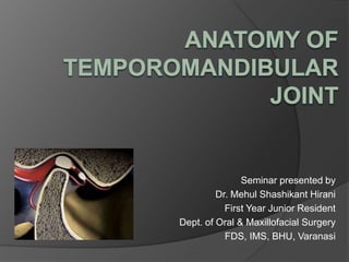
Seminar presented by
Dr. Mehul Shashikant Hirani
First Year Junior Resident
Dept. of Oral & Maxillofacial Surgery
FDS, IMS, BHU, Varanasi
CONTENTS
 INTRODUCTION
 CLASSIFICATION OF JOINT
 DEVELOPMENT OF TMJ
 MANDIBULAR FOSSA
 CONDYLE
 ARTTICULAR DISC
 INNERVATION
 VASCULAR SUPPLY
 LIGAMENTS
 BIOMECHANICS
 SURGICAL APPROACHES
INTRODUCTION
 The Temporomandibular Joint is the one
which connects the mandible to the skull
and regulates mandibular movements
 It is a bicondyler joint in which the
condyles ,located at the ends of
mandible, function at the same time.
 The right & left TMJ form Bicondylar
Articulation and Ellipsoid variety of
synovial joints similar to Knee joint.
 TMJ is GINGLIMOARTHRODIAL joint
Ginglymus - Hinge, allowing motion only
forward and backward in one plane.
Arthrodia - Joint which permits gliding
movements of surfaces.
 The common features of synovial Joint
exhibited by this joint includes a disk, bone,
fibrous capsule, fluid, synovial membrane
and ligaments. However, TMJ is unique in
a way that the articular surface is made of
Fibrocartilage instead of Hyaline cartilage
as seen in other synovial joints.
PECULIARITY OF TMJ
1. Bilateral diarthrosis – right & left
function together
2. Articular surface covered by
Fibrocartilage – instead of Hyaline
cartilage
3. Only joint in the body to have a Rigid
endpoint of closure ( that of teeth
making occlusal contact )
4. Has 4 articular surfaces.
5. In contrast to other diarthrodial joints TMJ is
the last joint to start developing –in about 7th
week in utero
6. Develops from two distict blastema
i) temporal
ii) condylar
7. TMJ acts like Class III Lever
Development Of TMJ
 The TMJ develops from
mesenchyme lying between
developing mandibular
condyle below and the bone
above, which develops
intramembranously.
 During the 12th week of IU
life, 2 clefts appear in the
mesenchyme – producing
upper and lower joint
cavities.
 The remaining intervening mesenchyme
becomes the Intra articular disc.
 The joint capsule develops from a
condensation of mesenchyme
surrounding the developing joint.
 Mandibular Fossa is flat at birth and
there is no articular eminence, this
becomes prominent only following the
eruption of the deciduous dentition.
Mandibular/ Glenoid Fossa
Bounderies-
 Anterior aspect of articular
eminence
 Posterior non articular fossa is
part of squamous temporal bone
& formed by tympanic plate (
Forms anterior bony wall of
external Acoustic meatus )
ARTICULAR EMINENCE
 This is the entire transverse bony bar that
forms the anterior root of zygoma.
 This articular surface is most heavily traveled
by the condyle and disk as they ride forward
and backward in normal jaw function.
ARTICULAR TUBERCLE
 This is a small, raised, rough, bony knob on
the outer end of the articular eminence.
 It projects below the level of the articular
surface and serves to attach the lateral
collateral ligament of the joint.
PREGLENOID PLANE
 This is the slightly hollowed, almost
horizontal, articular surface continuing
anteriorly from the height of the articular
eminence
E: Articular eminence;
enp: entogolenoid
process;
t:articular tubercle;
lb: lateral border of the
mandibular fossa;
pep: preglenoid
plane;
Gf: glenoid fossa
MANDIBULAR CONDYLE
 An ovoid process seated atop a narrow
mandibular neck.
 It’s the articulating surface of the mandible.
 It is convex in all directions but
wider medio-laterally (15 to 20mm)
than antero-posteriorly (8 to10mm).
 It has a medial and lateral pole.The medial
pole is directed more posteriorly.
Mainly 4 forms are
seen-
1. Convex-58%
2. Flat- 25%
3. Pointed-12%
4. Round- 3%
( mainly in children)
If the long axes of two
condyles are extended
medially, they meet at
approximately the basion
on the anterior limit of the
foramen magnum, forming
an angle that opens
toward the front ranging
from 145° to160°
 The lateral pole of the condyle is
rough, bluntly pointed, and
projects only moderately from the
plane of ramus, while the medial
pole extends sharply inward from
this plane.
 The articular surface lies on its
anterosuperior aspect, thus facing
the posterior slope of the articular
eminence of the temporal bone.
ARTICULAR DISC
 Biconcave fibro cartilaginous structure
located between the mandibular condyle and
the temporal bone component of the joint.
 Functions to accommodate a hinging action
as well as the gliding actions between the
temporal and mandibular articular bone
 Is avascular and aneural in its central part but is
vascular and innervated in the peripheral areas,
where load-bearing is minimal
 The main load-bearing areas are located on the
lateral aspect; this is an area of potential
perforation
 Merges around its periphery, attaching to the
capsule
 The articular disc is a roughly oval, firm, fibrous plate.
1. anterior band = 2 mm in thickness,
2. posterior band = 3 mm thick,
3. thin in the center intermediate band of 1 mm
thickness.
 More posteriorly there is a bilaminar or retrodiscal
region.
Anterior Band
Posterior Band
Retrodiscal Tissue
Located posterior to the articular disc
Highly distortable, especially on opening the mouth
Composed of:
● Superior lamina—contains elastic fibers and anchors
the superior aspect of the posterior portion of the disc
to the capsule and bone at the postglenoid tubercle
and tympanic plate
● Retrodiscal pad—the highly vascular and neural
portion of the TMJ, made of collagen, elastic fibers,
fat, nerves, and blood vessels (a large venous plexus
fills with blood when the condyle moves anteriorly)
● Inferior lamina—contains mainly collagen fibers and
anchors the inferior aspect of the posterior portion of
the disc to the condyle
Bilaminar zone (posterior attachment complex)
TMJ compartments
 The articular disc divides the TMJ into superior
and inferior compartments
 The internal surface of both compartments
contain specialized endothelial cells that form
a synovial lining that produces synovial fluid,
making the TMJ a synovial joint
 Synovial fluid acts as:
A lubricant
An instrument for providing the metabolic
requirements to the articular surfaces of the
TMJ
Superior Compartment
 Between the squamous
portion of the temporal
bone and the articular disc
 Volume = 1.2mL
 Provides for the
translational movement
of the TMJ
Inferior Compartment
 Between the articular disc
and the condyle
 Volume = 0.9mL
 Provides for the rotational
movement of the TMJ
Open mouth,
Sagittal section of TMJ
CAPSULE
 Completely encloses the articular surface of the temporal
bone and the condyle
 Composed of fibrous connective tissue
 Toughened along the medial and lateral aspects by
ligaments
 Lined by a highly vascular synovial membrane
 Has various sensory receptors including nociceptors
Attachments:
● Superior—along the rim of the temporal articular surfaces
● Inferior—along the condylar neck
● Medial—blends along the medial collateral ligament
● Lateral—blends along the lateral collateral ligament
● Anterior—blends with the superior head of the lateral
pterygoid muscle
● Posterior—along the retrodiscal pad
LIGAMENTS
Collateral Ligaments
 Composed of 2 ligaments:
Medial collateral ligament—connects the medial
aspect of the articular disc to the medial pole
of the condyle
Lateral collateral ligament—connects the lateral
aspect of the articular disc to the lateral pole
of the condyle
● Frequently called the discal ligaments
● Composed of collagenous connective tissue;
thus, they do not stretch
LATERAL VIEW
JOINT CAPSULE
LATERAL LIGAMENT
SPHENOMANDIBULAR LIGAMENT
STYLOID PROCESS
STYLOMANDIBULAR LIGAMENT
Temporomandibular (Lateral) Ligament
● The thickened ligament on the lateral aspect of
the capsule
● Prevents lateral and posterior displacement of the
condyle
● Composed of 2 separate bands:
 Outer oblique part—largest portion; attached to
the articular tubercle; travels posteroinferiorly to
attach immediately inferior to the condyle; this
limits the opening of the mandible
 Inner horizontal part—smaller band attached to
the articular tubercle running horizontally to
attach to the lateral part of the condyle and disc;
this limits posterior movement of the articular disc
and the condyle
Stylomandibular Ligament
● Composed of a thickening of deep
cervical fascia
● Extends from the styloid process to the
posterior margin of the angle and the
ramus of the mandible
● Helps limit anterior protrusion of the
mandible
Sphenomandibular Ligament
● Remnant of Meckel’s cartilage
● Extends from the spine of the sphenoid
to the lingula of the mandible
● May help act as a pivot on the mandible
by maintaining the same amount of
tension during both opening and closing
of the mouth
ARTERIAL SUPPLY
ANTERIOR TYMPANIC
ARTERY
MAXILLARY ARTERY
DEEP AURICULAR
ARTERY
POSTERIOR
AURICULAR
ARTERY
EXTERNAL
CAROTID ARTERY
SUPERFICIAL
TEMPORAL ARTERY
Artery Source Course
SUPERFICIAL
TEMPORAL
Terminal branch of
EXTERNAL CAROTID
ARTERY
Begins in the parotid gland and
initially is located posterior to the
mandible, where it provides small
branches to the TMJ
DEEP AURICULAR MAXILLARY ARTERY Arising in the same area as that
of the anterior tympanic artery
Lies in the parotid gland,
posterior to the TMJ, where it
gives branches to the TMJ
ANTERIOR
TYMPANIC
Arising in the same area as that
of the deep auricular artery.
Passes superiorly behind the
TMJ to enter the tympanic
cavity through the petrotympanic
fissure, where it gives
branches to the TMJ
VENOUS DRAINAGE
VEIN COURSE
SUPERFICIAL TEMPORAL Receives some branches from the TMJ
Then joins the maxillary vein to form the
retromandibular vein
MAXILLARY Receives some branches from the TMJ
Joins the superficial temporal vein to
form the retromandibular vein
SENSORY
INNERVATION
MANDIBULAR NERVE
& OTIC GANGLIONAURICULOTEM
PORAL NERVE
INFERIOR
ALVEOLAR
NERVE
LINGUAL NERVE
MAXILLARY
ARTERY
NERVE SOURCE COMMENT
AURICULOTEM
PORAL
MANDIBULAR DIVISION
OF TRIGEMINAL NERVE
From the posterior division of the mandibular
division of the trigeminal nerve.
Splits around the middle meningeal artery
and passes between the sphenomandibular
ligament and the condylar neck.
Supplies sensory branches all along the
capsule.
Sensory but carries autonomic function to the
parotid Gland.
MASSETERIC ANTERIOR DIVISION OF
MANDIBULAR DIVISION
OF TRIGEMINAL NERVE
Lies anterior to the TMJ and provides
branches to the joint before passing over the
masseteric notch to reach the masseter
muscle.
Sensory branches aid the auriculotemporal
nerve.
POSTERIOR
DEEP
TEMPORAL
Lies anterior to the TMJ and provides
branches to the joint before innervating the
temporalis muscle.
Sensory branches aid the auriculotemporal
nerve in supplying the anterior part of the
TMJ.
Mainly motor, but carries additional sensory
function to the TMJ
JOINT MOVEMENTS
 Rotational / hinge movement in first 20-
25mm of mouth opening
 Translational movement after that when
the mouth is excessively opened.
 Translatory movement – in the superior
part of the joint as the disc and the
condyle traverse anteriorly along the
inclines of the anterior tubercle to
provide an anterior and inferior
movement of the mandible
 Hinge movement – the inferior portion of
the joint between the head of the
condyle and the lower surface of the
disc to permit opening of the mandible.
WIDE OPEN
HINGE + GLIDING
SLIGHT OPEN
HINGE PREDOMINATES
SURGICAL APPROACHES
 Preauricular :
Dingman’s, Blair’s, Thoma’s, Al-kayat and
Bramley’s, Popowitch’s
 Postauricular .
 Endaural approach
 Post ramal/ Hind’s approach
 Submandibular/Risdon’s approach
 Hemicoronal
 Bicoronal
 Rhytidectomy approach
PREAURICULAR
APPROACH
 Basic incision given
by Dingman(1951)
 Most basic &
standard approach
to TMJ.
MODIFICATIONS OF
PREAURICULAR APPROACH
 Blair & Ivy (1936) –
“Inverted hockey stick “
incision.
 Facilities exposure of
arch along with
condylar area.
 Thoma in 1958
 Angulated vertical
incision.
 Carried out across
zygomatic arch
infront of ear to
avoid main trunk of
facial nerve
AL-KAYAT & BRAMLEY
APPROCH
 1979.
 Modified preauricular
approach.
 Facial nerve divides in
front of auditory canal as
near as 0.8cm & as far
as 3.5cm
 Protection achieved by
making incision through
temporal fascia &
periosteum down to arch
not more than 0.8 cm.
POST – AURICULAR
APPROACH
 Hoops et al (1970),
 Alexander and
James (1975)
 Highly cosmetic
incision
 Disadvantage- poor
access & visibility, the
risk of external
auditory meatus
stenosis, infection &
deformity of the
auricle.
 Lempart (1938)
 Short facial skin
incision extending in to
external Auditory
meatus
 Excellent cosmetics
 Disadvantage-Meatal
stenosis or chondritis,
injury to the branches
of the facial nerve
END AURAL Approach
Post Ramal / Hind’s Approach
 Indication – surgeries of
condylar neck & ramus
area.
 Incision- 1cm behind
ramus of mandible and
extends 1cm below the
lobe of ear.
 Highly cosmetic, excellent
visibility and accessibility.
 Injury may occur to
posterior facial vein and
main trunk of facial nerve.
Submandibular Risdon
Approach
 Risdon (1934)
 Mainly used for
neck of condyle &
ramus region.
 Supplement to
different TMJ
approaches for
tunneling through
the soft tissues to
place a graft
Coronal Approach
 Hemicoronal (unilateral) or bicoronal
(bilateral) approach is used.
 More extensive but versatile approach
for upper & middle regions of facial
skeleton, zygomatic arch & TMJ.
 Advantage- scar is hidden in the
hairline.
Rhytidectomy Approach
 Incision made in pre
auricular area and in
the neck hairline
 Skin and
subcutaneous
tissues are incised,
and dissection
carried out above the
level of SMAS
Anatomy of Temporomandibular Joint

More Related Content

What's hot

Anatomy and physiology of temporomandibular joint
Anatomy and physiology of temporomandibular joint Anatomy and physiology of temporomandibular joint
Anatomy and physiology of temporomandibular joint
Akshay Karve
 
Applied anatomy of tmj
Applied anatomy of tmjApplied anatomy of tmj
Applied anatomy of tmj
Dr. Samarth Johari
 
Temporomandibular joint 1
Temporomandibular joint 1Temporomandibular joint 1
Temporomandibular joint 1
ANIL KUMAR
 
Tmj anatomy
Tmj anatomyTmj anatomy
Tmj anatomy
Tony Pious
 
anatomy of tmj.pptx
anatomy of tmj.pptxanatomy of tmj.pptx
anatomy of tmj.pptx
praveena943549
 
TMJ surgical anatomy and applied aspects
TMJ surgical anatomy and applied aspectsTMJ surgical anatomy and applied aspects
TMJ surgical anatomy and applied aspectsJoel D'silva
 
TEMPROMANDIBULAR JOINT
TEMPROMANDIBULAR JOINTTEMPROMANDIBULAR JOINT
TEMPROMANDIBULAR JOINT
Menatalla Elhindawy
 
TMJ and its relation to periodontics
TMJ and its relation to periodonticsTMJ and its relation to periodontics
TMJ and its relation to periodontics
Chittoor Deals
 
Temporomandibular Joint
Temporomandibular JointTemporomandibular Joint
Temporomandibular Joint
Divya Gaur
 
anatomy of TMJ
anatomy of TMJanatomy of TMJ
anatomy of TMJ
Dhaval Trivedi
 
Presentation temporomandibular joint anatomy
Presentation temporomandibular joint anatomyPresentation temporomandibular joint anatomy
Presentation temporomandibular joint anatomy
Leena Gangil
 
Temporo mandibular joint
Temporo mandibular jointTemporo mandibular joint
Temporo mandibular joint
nitya Krishna
 
mandible
mandiblemandible
mandible
KevinMungasia
 
Muscles of mastication/prosthodontic courses
Muscles of mastication/prosthodontic coursesMuscles of mastication/prosthodontic courses
Muscles of mastication/prosthodontic courses
Indian dental academy
 
temporomandibular joint-development and anatomy
temporomandibular joint-development and anatomytemporomandibular joint-development and anatomy
temporomandibular joint-development and anatomy
spsangeetaporiya
 
Surgical Anatomy of Temporomandibular Joint
Surgical Anatomy of Temporomandibular JointSurgical Anatomy of Temporomandibular Joint
Surgical Anatomy of Temporomandibular Joint
Dibya Falgoon Sarkar
 
TEMPOROMANDIBULAR JOINT
TEMPOROMANDIBULAR JOINT TEMPOROMANDIBULAR JOINT
TEMPOROMANDIBULAR JOINT
Siddharth Tevatia
 
Tmj
TmjTmj
Enamel
EnamelEnamel
Development of Mandible
Development of MandibleDevelopment of Mandible
Development of Mandible
Menatalla Elhindawy
 

What's hot (20)

Anatomy and physiology of temporomandibular joint
Anatomy and physiology of temporomandibular joint Anatomy and physiology of temporomandibular joint
Anatomy and physiology of temporomandibular joint
 
Applied anatomy of tmj
Applied anatomy of tmjApplied anatomy of tmj
Applied anatomy of tmj
 
Temporomandibular joint 1
Temporomandibular joint 1Temporomandibular joint 1
Temporomandibular joint 1
 
Tmj anatomy
Tmj anatomyTmj anatomy
Tmj anatomy
 
anatomy of tmj.pptx
anatomy of tmj.pptxanatomy of tmj.pptx
anatomy of tmj.pptx
 
TMJ surgical anatomy and applied aspects
TMJ surgical anatomy and applied aspectsTMJ surgical anatomy and applied aspects
TMJ surgical anatomy and applied aspects
 
TEMPROMANDIBULAR JOINT
TEMPROMANDIBULAR JOINTTEMPROMANDIBULAR JOINT
TEMPROMANDIBULAR JOINT
 
TMJ and its relation to periodontics
TMJ and its relation to periodonticsTMJ and its relation to periodontics
TMJ and its relation to periodontics
 
Temporomandibular Joint
Temporomandibular JointTemporomandibular Joint
Temporomandibular Joint
 
anatomy of TMJ
anatomy of TMJanatomy of TMJ
anatomy of TMJ
 
Presentation temporomandibular joint anatomy
Presentation temporomandibular joint anatomyPresentation temporomandibular joint anatomy
Presentation temporomandibular joint anatomy
 
Temporo mandibular joint
Temporo mandibular jointTemporo mandibular joint
Temporo mandibular joint
 
mandible
mandiblemandible
mandible
 
Muscles of mastication/prosthodontic courses
Muscles of mastication/prosthodontic coursesMuscles of mastication/prosthodontic courses
Muscles of mastication/prosthodontic courses
 
temporomandibular joint-development and anatomy
temporomandibular joint-development and anatomytemporomandibular joint-development and anatomy
temporomandibular joint-development and anatomy
 
Surgical Anatomy of Temporomandibular Joint
Surgical Anatomy of Temporomandibular JointSurgical Anatomy of Temporomandibular Joint
Surgical Anatomy of Temporomandibular Joint
 
TEMPOROMANDIBULAR JOINT
TEMPOROMANDIBULAR JOINT TEMPOROMANDIBULAR JOINT
TEMPOROMANDIBULAR JOINT
 
Tmj
TmjTmj
Tmj
 
Enamel
EnamelEnamel
Enamel
 
Development of Mandible
Development of MandibleDevelopment of Mandible
Development of Mandible
 

Similar to Anatomy of Temporomandibular Joint

Tmj surgical anatomy and approaches
Tmj surgical anatomy and approachesTmj surgical anatomy and approaches
Tmj surgical anatomy and approaches
Joel D'silva
 
Tmj
TmjTmj
DR SHAKIR New anatomy of tmj
DR SHAKIR New anatomy of tmjDR SHAKIR New anatomy of tmj
DR SHAKIR New anatomy of tmj
doctorshakir
 
Tmj
TmjTmj
1 anatomy & physiology of tmj
1 anatomy & physiology of tmj1 anatomy & physiology of tmj
1 anatomy & physiology of tmj
DrKamini Dadsena
 
Examination of tmj &muscles of mastication (2)
Examination of tmj &muscles of mastication (2)Examination of tmj &muscles of mastication (2)
Examination of tmj &muscles of mastication (2)
rachitajainr
 
temporomandibular joint.pptx
temporomandibular joint.pptxtemporomandibular joint.pptx
temporomandibular joint.pptx
SumedhaThosar
 
Temporomandibular joint
Temporomandibular jointTemporomandibular joint
Temporomandibular joint
Chitransha03
 
ANATOMY TEMPOROMANDIBULAR JUNCTION OF HUMAN
ANATOMY TEMPOROMANDIBULAR JUNCTION OF HUMANANATOMY TEMPOROMANDIBULAR JUNCTION OF HUMAN
ANATOMY TEMPOROMANDIBULAR JUNCTION OF HUMAN
DesiFitriani85
 
TMJ, Muscles of Mastication and their Applied Anatomy .pptx
TMJ, Muscles of Mastication and their Applied Anatomy .pptxTMJ, Muscles of Mastication and their Applied Anatomy .pptx
TMJ, Muscles of Mastication and their Applied Anatomy .pptx
Dr. Mukesh Kumar Dey
 
Temporomandibular joint
Temporomandibular jointTemporomandibular joint
Temporomandibular joint
Parikshit Kadam
 
Temporomandibular joint [Autosaved].pptx
Temporomandibular joint [Autosaved].pptxTemporomandibular joint [Autosaved].pptx
Temporomandibular joint [Autosaved].pptx
Abhidha Tripathi
 
TMJ AND ITS IMPLICATIONS IN PROSTHODONTICS
TMJ AND ITS IMPLICATIONS IN PROSTHODONTICSTMJ AND ITS IMPLICATIONS IN PROSTHODONTICS
TMJ AND ITS IMPLICATIONS IN PROSTHODONTICS
Dr Jibi Sara Varghese
 
anatomyoftemporomandibularjoint-210112114624.pdf
anatomyoftemporomandibularjoint-210112114624.pdfanatomyoftemporomandibularjoint-210112114624.pdf
anatomyoftemporomandibularjoint-210112114624.pdf
snithiyuvarajayuvara
 
Temporomandibular joint anatomy and function
Temporomandibular joint anatomy and functionTemporomandibular joint anatomy and function
Temporomandibular joint anatomy and function
DR POOJA
 
Surgical anatomy of temporomandibular joint
Surgical anatomy of temporomandibular jointSurgical anatomy of temporomandibular joint
Surgical anatomy of temporomandibular joint
Aaisha Ansari
 
Temperomandibular joint Anatomy
Temperomandibular joint AnatomyTemperomandibular joint Anatomy
Temperomandibular joint Anatomy
Ashish Ranghani
 
Surgical anatomy of tmj
Surgical anatomy of tmjSurgical anatomy of tmj
Surgical anatomy of tmj
MinalSonare2
 

Similar to Anatomy of Temporomandibular Joint (20)

Tmj surgical anatomy and approaches
Tmj surgical anatomy and approachesTmj surgical anatomy and approaches
Tmj surgical anatomy and approaches
 
Tmj
TmjTmj
Tmj
 
DR SHAKIR New anatomy of tmj
DR SHAKIR New anatomy of tmjDR SHAKIR New anatomy of tmj
DR SHAKIR New anatomy of tmj
 
Tmj
TmjTmj
Tmj
 
1 anatomy & physiology of tmj
1 anatomy & physiology of tmj1 anatomy & physiology of tmj
1 anatomy & physiology of tmj
 
Examination of tmj &muscles of mastication (2)
Examination of tmj &muscles of mastication (2)Examination of tmj &muscles of mastication (2)
Examination of tmj &muscles of mastication (2)
 
temporomandibular joint.pptx
temporomandibular joint.pptxtemporomandibular joint.pptx
temporomandibular joint.pptx
 
Occlusion ppt
Occlusion pptOcclusion ppt
Occlusion ppt
 
Temporomandibular joint
Temporomandibular jointTemporomandibular joint
Temporomandibular joint
 
ANATOMY TEMPOROMANDIBULAR JUNCTION OF HUMAN
ANATOMY TEMPOROMANDIBULAR JUNCTION OF HUMANANATOMY TEMPOROMANDIBULAR JUNCTION OF HUMAN
ANATOMY TEMPOROMANDIBULAR JUNCTION OF HUMAN
 
TMJ, Muscles of Mastication and their Applied Anatomy .pptx
TMJ, Muscles of Mastication and their Applied Anatomy .pptxTMJ, Muscles of Mastication and their Applied Anatomy .pptx
TMJ, Muscles of Mastication and their Applied Anatomy .pptx
 
Temporomandibular joint
Temporomandibular jointTemporomandibular joint
Temporomandibular joint
 
Temporomandibular joint [Autosaved].pptx
Temporomandibular joint [Autosaved].pptxTemporomandibular joint [Autosaved].pptx
Temporomandibular joint [Autosaved].pptx
 
TMJ AND ITS IMPLICATIONS IN PROSTHODONTICS
TMJ AND ITS IMPLICATIONS IN PROSTHODONTICSTMJ AND ITS IMPLICATIONS IN PROSTHODONTICS
TMJ AND ITS IMPLICATIONS IN PROSTHODONTICS
 
anatomyoftemporomandibularjoint-210112114624.pdf
anatomyoftemporomandibularjoint-210112114624.pdfanatomyoftemporomandibularjoint-210112114624.pdf
anatomyoftemporomandibularjoint-210112114624.pdf
 
Temporomandibular joint anatomy and function
Temporomandibular joint anatomy and functionTemporomandibular joint anatomy and function
Temporomandibular joint anatomy and function
 
Surgical anatomy of temporomandibular joint
Surgical anatomy of temporomandibular jointSurgical anatomy of temporomandibular joint
Surgical anatomy of temporomandibular joint
 
Temperomandibular joint Anatomy
Temperomandibular joint AnatomyTemperomandibular joint Anatomy
Temperomandibular joint Anatomy
 
Surgical anatomy of tmj
Surgical anatomy of tmjSurgical anatomy of tmj
Surgical anatomy of tmj
 
TMJ written report
TMJ written reportTMJ written report
TMJ written report
 

Recently uploaded

Novas diretrizes da OMS para os cuidados perinatais de mais qualidade
Novas diretrizes da OMS para os cuidados perinatais de mais qualidadeNovas diretrizes da OMS para os cuidados perinatais de mais qualidade
Novas diretrizes da OMS para os cuidados perinatais de mais qualidade
Prof. Marcus Renato de Carvalho
 
New Drug Discovery and Development .....
New Drug Discovery and Development .....New Drug Discovery and Development .....
New Drug Discovery and Development .....
NEHA GUPTA
 
NVBDCP.pptx Nation vector borne disease control program
NVBDCP.pptx Nation vector borne disease control programNVBDCP.pptx Nation vector borne disease control program
NVBDCP.pptx Nation vector borne disease control program
Sapna Thakur
 
BENIGN PROSTATIC HYPERPLASIA.BPH. BPHpdf
BENIGN PROSTATIC HYPERPLASIA.BPH. BPHpdfBENIGN PROSTATIC HYPERPLASIA.BPH. BPHpdf
BENIGN PROSTATIC HYPERPLASIA.BPH. BPHpdf
DR SETH JOTHAM
 
Knee anatomy and clinical tests 2024.pdf
Knee anatomy and clinical tests 2024.pdfKnee anatomy and clinical tests 2024.pdf
Knee anatomy and clinical tests 2024.pdf
vimalpl1234
 
basicmodesofventilation2022-220313203758.pdf
basicmodesofventilation2022-220313203758.pdfbasicmodesofventilation2022-220313203758.pdf
basicmodesofventilation2022-220313203758.pdf
aljamhori teaching hospital
 
Couples presenting to the infertility clinic- Do they really have infertility...
Couples presenting to the infertility clinic- Do they really have infertility...Couples presenting to the infertility clinic- Do they really have infertility...
Couples presenting to the infertility clinic- Do they really have infertility...
Sujoy Dasgupta
 
Ocular injury ppt Upendra pal optometrist upums saifai etawah
Ocular injury  ppt  Upendra pal  optometrist upums saifai etawahOcular injury  ppt  Upendra pal  optometrist upums saifai etawah
Ocular injury ppt Upendra pal optometrist upums saifai etawah
pal078100
 
Flu Vaccine Alert in Bangalore Karnataka
Flu Vaccine Alert in Bangalore KarnatakaFlu Vaccine Alert in Bangalore Karnataka
Flu Vaccine Alert in Bangalore Karnataka
addon Scans
 
Charaka Samhita Sutra sthana Chapter 15 Upakalpaniyaadhyaya
Charaka Samhita Sutra sthana Chapter 15 UpakalpaniyaadhyayaCharaka Samhita Sutra sthana Chapter 15 Upakalpaniyaadhyaya
Charaka Samhita Sutra sthana Chapter 15 Upakalpaniyaadhyaya
Dr KHALID B.M
 
How to Give Better Lectures: Some Tips for Doctors
How to Give Better Lectures: Some Tips for DoctorsHow to Give Better Lectures: Some Tips for Doctors
How to Give Better Lectures: Some Tips for Doctors
LanceCatedral
 
Physiology of Special Chemical Sensation of Taste
Physiology of Special Chemical Sensation of TastePhysiology of Special Chemical Sensation of Taste
Physiology of Special Chemical Sensation of Taste
MedicoseAcademics
 
Hemodialysis: Chapter 3, Dialysis Water Unit - Dr.Gawad
Hemodialysis: Chapter 3, Dialysis Water Unit - Dr.GawadHemodialysis: Chapter 3, Dialysis Water Unit - Dr.Gawad
Hemodialysis: Chapter 3, Dialysis Water Unit - Dr.Gawad
NephroTube - Dr.Gawad
 
Phone Us ❤85270-49040❤ #ℂall #gIRLS In Surat By Surat @ℂall @Girls Hotel With...
Phone Us ❤85270-49040❤ #ℂall #gIRLS In Surat By Surat @ℂall @Girls Hotel With...Phone Us ❤85270-49040❤ #ℂall #gIRLS In Surat By Surat @ℂall @Girls Hotel With...
Phone Us ❤85270-49040❤ #ℂall #gIRLS In Surat By Surat @ℂall @Girls Hotel With...
Savita Shen $i11
 
Pulmonary Thromboembolism - etilogy, types, medical- Surgical and nursing man...
Pulmonary Thromboembolism - etilogy, types, medical- Surgical and nursing man...Pulmonary Thromboembolism - etilogy, types, medical- Surgical and nursing man...
Pulmonary Thromboembolism - etilogy, types, medical- Surgical and nursing man...
VarunMahajani
 
Triangles of Neck and Clinical Correlation by Dr. RIG.pptx
Triangles of Neck and Clinical Correlation by Dr. RIG.pptxTriangles of Neck and Clinical Correlation by Dr. RIG.pptx
Triangles of Neck and Clinical Correlation by Dr. RIG.pptx
Dr. Rabia Inam Gandapore
 
Prix Galien International 2024 Forum Program
Prix Galien International 2024 Forum ProgramPrix Galien International 2024 Forum Program
Prix Galien International 2024 Forum Program
Levi Shapiro
 
Pharynx and Clinical Correlations BY Dr.Rabia Inam Gandapore.pptx
Pharynx and Clinical Correlations BY Dr.Rabia Inam Gandapore.pptxPharynx and Clinical Correlations BY Dr.Rabia Inam Gandapore.pptx
Pharynx and Clinical Correlations BY Dr.Rabia Inam Gandapore.pptx
Dr. Rabia Inam Gandapore
 
Evaluation of antidepressant activity of clitoris ternatea in animals
Evaluation of antidepressant activity of clitoris ternatea in animalsEvaluation of antidepressant activity of clitoris ternatea in animals
Evaluation of antidepressant activity of clitoris ternatea in animals
Shweta
 
Physiology of Chemical Sensation of smell.pdf
Physiology of Chemical Sensation of smell.pdfPhysiology of Chemical Sensation of smell.pdf
Physiology of Chemical Sensation of smell.pdf
MedicoseAcademics
 

Recently uploaded (20)

Novas diretrizes da OMS para os cuidados perinatais de mais qualidade
Novas diretrizes da OMS para os cuidados perinatais de mais qualidadeNovas diretrizes da OMS para os cuidados perinatais de mais qualidade
Novas diretrizes da OMS para os cuidados perinatais de mais qualidade
 
New Drug Discovery and Development .....
New Drug Discovery and Development .....New Drug Discovery and Development .....
New Drug Discovery and Development .....
 
NVBDCP.pptx Nation vector borne disease control program
NVBDCP.pptx Nation vector borne disease control programNVBDCP.pptx Nation vector borne disease control program
NVBDCP.pptx Nation vector borne disease control program
 
BENIGN PROSTATIC HYPERPLASIA.BPH. BPHpdf
BENIGN PROSTATIC HYPERPLASIA.BPH. BPHpdfBENIGN PROSTATIC HYPERPLASIA.BPH. BPHpdf
BENIGN PROSTATIC HYPERPLASIA.BPH. BPHpdf
 
Knee anatomy and clinical tests 2024.pdf
Knee anatomy and clinical tests 2024.pdfKnee anatomy and clinical tests 2024.pdf
Knee anatomy and clinical tests 2024.pdf
 
basicmodesofventilation2022-220313203758.pdf
basicmodesofventilation2022-220313203758.pdfbasicmodesofventilation2022-220313203758.pdf
basicmodesofventilation2022-220313203758.pdf
 
Couples presenting to the infertility clinic- Do they really have infertility...
Couples presenting to the infertility clinic- Do they really have infertility...Couples presenting to the infertility clinic- Do they really have infertility...
Couples presenting to the infertility clinic- Do they really have infertility...
 
Ocular injury ppt Upendra pal optometrist upums saifai etawah
Ocular injury  ppt  Upendra pal  optometrist upums saifai etawahOcular injury  ppt  Upendra pal  optometrist upums saifai etawah
Ocular injury ppt Upendra pal optometrist upums saifai etawah
 
Flu Vaccine Alert in Bangalore Karnataka
Flu Vaccine Alert in Bangalore KarnatakaFlu Vaccine Alert in Bangalore Karnataka
Flu Vaccine Alert in Bangalore Karnataka
 
Charaka Samhita Sutra sthana Chapter 15 Upakalpaniyaadhyaya
Charaka Samhita Sutra sthana Chapter 15 UpakalpaniyaadhyayaCharaka Samhita Sutra sthana Chapter 15 Upakalpaniyaadhyaya
Charaka Samhita Sutra sthana Chapter 15 Upakalpaniyaadhyaya
 
How to Give Better Lectures: Some Tips for Doctors
How to Give Better Lectures: Some Tips for DoctorsHow to Give Better Lectures: Some Tips for Doctors
How to Give Better Lectures: Some Tips for Doctors
 
Physiology of Special Chemical Sensation of Taste
Physiology of Special Chemical Sensation of TastePhysiology of Special Chemical Sensation of Taste
Physiology of Special Chemical Sensation of Taste
 
Hemodialysis: Chapter 3, Dialysis Water Unit - Dr.Gawad
Hemodialysis: Chapter 3, Dialysis Water Unit - Dr.GawadHemodialysis: Chapter 3, Dialysis Water Unit - Dr.Gawad
Hemodialysis: Chapter 3, Dialysis Water Unit - Dr.Gawad
 
Phone Us ❤85270-49040❤ #ℂall #gIRLS In Surat By Surat @ℂall @Girls Hotel With...
Phone Us ❤85270-49040❤ #ℂall #gIRLS In Surat By Surat @ℂall @Girls Hotel With...Phone Us ❤85270-49040❤ #ℂall #gIRLS In Surat By Surat @ℂall @Girls Hotel With...
Phone Us ❤85270-49040❤ #ℂall #gIRLS In Surat By Surat @ℂall @Girls Hotel With...
 
Pulmonary Thromboembolism - etilogy, types, medical- Surgical and nursing man...
Pulmonary Thromboembolism - etilogy, types, medical- Surgical and nursing man...Pulmonary Thromboembolism - etilogy, types, medical- Surgical and nursing man...
Pulmonary Thromboembolism - etilogy, types, medical- Surgical and nursing man...
 
Triangles of Neck and Clinical Correlation by Dr. RIG.pptx
Triangles of Neck and Clinical Correlation by Dr. RIG.pptxTriangles of Neck and Clinical Correlation by Dr. RIG.pptx
Triangles of Neck and Clinical Correlation by Dr. RIG.pptx
 
Prix Galien International 2024 Forum Program
Prix Galien International 2024 Forum ProgramPrix Galien International 2024 Forum Program
Prix Galien International 2024 Forum Program
 
Pharynx and Clinical Correlations BY Dr.Rabia Inam Gandapore.pptx
Pharynx and Clinical Correlations BY Dr.Rabia Inam Gandapore.pptxPharynx and Clinical Correlations BY Dr.Rabia Inam Gandapore.pptx
Pharynx and Clinical Correlations BY Dr.Rabia Inam Gandapore.pptx
 
Evaluation of antidepressant activity of clitoris ternatea in animals
Evaluation of antidepressant activity of clitoris ternatea in animalsEvaluation of antidepressant activity of clitoris ternatea in animals
Evaluation of antidepressant activity of clitoris ternatea in animals
 
Physiology of Chemical Sensation of smell.pdf
Physiology of Chemical Sensation of smell.pdfPhysiology of Chemical Sensation of smell.pdf
Physiology of Chemical Sensation of smell.pdf
 

Anatomy of Temporomandibular Joint

  • 1. Seminar presented by Dr. Mehul Shashikant Hirani First Year Junior Resident Dept. of Oral & Maxillofacial Surgery FDS, IMS, BHU, Varanasi
  • 2. CONTENTS  INTRODUCTION  CLASSIFICATION OF JOINT  DEVELOPMENT OF TMJ  MANDIBULAR FOSSA  CONDYLE  ARTTICULAR DISC  INNERVATION  VASCULAR SUPPLY  LIGAMENTS  BIOMECHANICS  SURGICAL APPROACHES
  • 3. INTRODUCTION  The Temporomandibular Joint is the one which connects the mandible to the skull and regulates mandibular movements  It is a bicondyler joint in which the condyles ,located at the ends of mandible, function at the same time.  The right & left TMJ form Bicondylar Articulation and Ellipsoid variety of synovial joints similar to Knee joint.
  • 4.  TMJ is GINGLIMOARTHRODIAL joint Ginglymus - Hinge, allowing motion only forward and backward in one plane. Arthrodia - Joint which permits gliding movements of surfaces.  The common features of synovial Joint exhibited by this joint includes a disk, bone, fibrous capsule, fluid, synovial membrane and ligaments. However, TMJ is unique in a way that the articular surface is made of Fibrocartilage instead of Hyaline cartilage as seen in other synovial joints.
  • 5. PECULIARITY OF TMJ 1. Bilateral diarthrosis – right & left function together 2. Articular surface covered by Fibrocartilage – instead of Hyaline cartilage 3. Only joint in the body to have a Rigid endpoint of closure ( that of teeth making occlusal contact ) 4. Has 4 articular surfaces.
  • 6. 5. In contrast to other diarthrodial joints TMJ is the last joint to start developing –in about 7th week in utero 6. Develops from two distict blastema i) temporal ii) condylar 7. TMJ acts like Class III Lever
  • 7. Development Of TMJ  The TMJ develops from mesenchyme lying between developing mandibular condyle below and the bone above, which develops intramembranously.  During the 12th week of IU life, 2 clefts appear in the mesenchyme – producing upper and lower joint cavities.
  • 8.  The remaining intervening mesenchyme becomes the Intra articular disc.  The joint capsule develops from a condensation of mesenchyme surrounding the developing joint.  Mandibular Fossa is flat at birth and there is no articular eminence, this becomes prominent only following the eruption of the deciduous dentition.
  • 9. Mandibular/ Glenoid Fossa Bounderies-  Anterior aspect of articular eminence  Posterior non articular fossa is part of squamous temporal bone & formed by tympanic plate ( Forms anterior bony wall of external Acoustic meatus )
  • 10. ARTICULAR EMINENCE  This is the entire transverse bony bar that forms the anterior root of zygoma.  This articular surface is most heavily traveled by the condyle and disk as they ride forward and backward in normal jaw function. ARTICULAR TUBERCLE  This is a small, raised, rough, bony knob on the outer end of the articular eminence.  It projects below the level of the articular surface and serves to attach the lateral collateral ligament of the joint.
  • 11. PREGLENOID PLANE  This is the slightly hollowed, almost horizontal, articular surface continuing anteriorly from the height of the articular eminence
  • 12. E: Articular eminence; enp: entogolenoid process; t:articular tubercle; lb: lateral border of the mandibular fossa; pep: preglenoid plane; Gf: glenoid fossa
  • 13. MANDIBULAR CONDYLE  An ovoid process seated atop a narrow mandibular neck.  It’s the articulating surface of the mandible.  It is convex in all directions but wider medio-laterally (15 to 20mm) than antero-posteriorly (8 to10mm).  It has a medial and lateral pole.The medial pole is directed more posteriorly.
  • 14. Mainly 4 forms are seen- 1. Convex-58% 2. Flat- 25% 3. Pointed-12% 4. Round- 3% ( mainly in children)
  • 15. If the long axes of two condyles are extended medially, they meet at approximately the basion on the anterior limit of the foramen magnum, forming an angle that opens toward the front ranging from 145° to160°
  • 16.  The lateral pole of the condyle is rough, bluntly pointed, and projects only moderately from the plane of ramus, while the medial pole extends sharply inward from this plane.  The articular surface lies on its anterosuperior aspect, thus facing the posterior slope of the articular eminence of the temporal bone.
  • 18.  Biconcave fibro cartilaginous structure located between the mandibular condyle and the temporal bone component of the joint.  Functions to accommodate a hinging action as well as the gliding actions between the temporal and mandibular articular bone  Is avascular and aneural in its central part but is vascular and innervated in the peripheral areas, where load-bearing is minimal  The main load-bearing areas are located on the lateral aspect; this is an area of potential perforation  Merges around its periphery, attaching to the capsule
  • 19.  The articular disc is a roughly oval, firm, fibrous plate. 1. anterior band = 2 mm in thickness, 2. posterior band = 3 mm thick, 3. thin in the center intermediate band of 1 mm thickness.  More posteriorly there is a bilaminar or retrodiscal region. Anterior Band Posterior Band Retrodiscal Tissue
  • 20. Located posterior to the articular disc Highly distortable, especially on opening the mouth Composed of: ● Superior lamina—contains elastic fibers and anchors the superior aspect of the posterior portion of the disc to the capsule and bone at the postglenoid tubercle and tympanic plate ● Retrodiscal pad—the highly vascular and neural portion of the TMJ, made of collagen, elastic fibers, fat, nerves, and blood vessels (a large venous plexus fills with blood when the condyle moves anteriorly) ● Inferior lamina—contains mainly collagen fibers and anchors the inferior aspect of the posterior portion of the disc to the condyle Bilaminar zone (posterior attachment complex)
  • 21. TMJ compartments  The articular disc divides the TMJ into superior and inferior compartments  The internal surface of both compartments contain specialized endothelial cells that form a synovial lining that produces synovial fluid, making the TMJ a synovial joint  Synovial fluid acts as: A lubricant An instrument for providing the metabolic requirements to the articular surfaces of the TMJ
  • 22. Superior Compartment  Between the squamous portion of the temporal bone and the articular disc  Volume = 1.2mL  Provides for the translational movement of the TMJ Inferior Compartment  Between the articular disc and the condyle  Volume = 0.9mL  Provides for the rotational movement of the TMJ Open mouth, Sagittal section of TMJ
  • 23. CAPSULE  Completely encloses the articular surface of the temporal bone and the condyle  Composed of fibrous connective tissue  Toughened along the medial and lateral aspects by ligaments  Lined by a highly vascular synovial membrane  Has various sensory receptors including nociceptors Attachments: ● Superior—along the rim of the temporal articular surfaces ● Inferior—along the condylar neck ● Medial—blends along the medial collateral ligament ● Lateral—blends along the lateral collateral ligament ● Anterior—blends with the superior head of the lateral pterygoid muscle ● Posterior—along the retrodiscal pad
  • 24. LIGAMENTS Collateral Ligaments  Composed of 2 ligaments: Medial collateral ligament—connects the medial aspect of the articular disc to the medial pole of the condyle Lateral collateral ligament—connects the lateral aspect of the articular disc to the lateral pole of the condyle ● Frequently called the discal ligaments ● Composed of collagenous connective tissue; thus, they do not stretch
  • 25. LATERAL VIEW JOINT CAPSULE LATERAL LIGAMENT SPHENOMANDIBULAR LIGAMENT STYLOID PROCESS STYLOMANDIBULAR LIGAMENT
  • 26. Temporomandibular (Lateral) Ligament ● The thickened ligament on the lateral aspect of the capsule ● Prevents lateral and posterior displacement of the condyle ● Composed of 2 separate bands:  Outer oblique part—largest portion; attached to the articular tubercle; travels posteroinferiorly to attach immediately inferior to the condyle; this limits the opening of the mandible  Inner horizontal part—smaller band attached to the articular tubercle running horizontally to attach to the lateral part of the condyle and disc; this limits posterior movement of the articular disc and the condyle
  • 27. Stylomandibular Ligament ● Composed of a thickening of deep cervical fascia ● Extends from the styloid process to the posterior margin of the angle and the ramus of the mandible ● Helps limit anterior protrusion of the mandible Sphenomandibular Ligament ● Remnant of Meckel’s cartilage ● Extends from the spine of the sphenoid to the lingula of the mandible ● May help act as a pivot on the mandible by maintaining the same amount of tension during both opening and closing of the mouth
  • 28. ARTERIAL SUPPLY ANTERIOR TYMPANIC ARTERY MAXILLARY ARTERY DEEP AURICULAR ARTERY POSTERIOR AURICULAR ARTERY EXTERNAL CAROTID ARTERY SUPERFICIAL TEMPORAL ARTERY
  • 29. Artery Source Course SUPERFICIAL TEMPORAL Terminal branch of EXTERNAL CAROTID ARTERY Begins in the parotid gland and initially is located posterior to the mandible, where it provides small branches to the TMJ DEEP AURICULAR MAXILLARY ARTERY Arising in the same area as that of the anterior tympanic artery Lies in the parotid gland, posterior to the TMJ, where it gives branches to the TMJ ANTERIOR TYMPANIC Arising in the same area as that of the deep auricular artery. Passes superiorly behind the TMJ to enter the tympanic cavity through the petrotympanic fissure, where it gives branches to the TMJ
  • 30. VENOUS DRAINAGE VEIN COURSE SUPERFICIAL TEMPORAL Receives some branches from the TMJ Then joins the maxillary vein to form the retromandibular vein MAXILLARY Receives some branches from the TMJ Joins the superficial temporal vein to form the retromandibular vein
  • 31. SENSORY INNERVATION MANDIBULAR NERVE & OTIC GANGLIONAURICULOTEM PORAL NERVE INFERIOR ALVEOLAR NERVE LINGUAL NERVE MAXILLARY ARTERY
  • 32. NERVE SOURCE COMMENT AURICULOTEM PORAL MANDIBULAR DIVISION OF TRIGEMINAL NERVE From the posterior division of the mandibular division of the trigeminal nerve. Splits around the middle meningeal artery and passes between the sphenomandibular ligament and the condylar neck. Supplies sensory branches all along the capsule. Sensory but carries autonomic function to the parotid Gland. MASSETERIC ANTERIOR DIVISION OF MANDIBULAR DIVISION OF TRIGEMINAL NERVE Lies anterior to the TMJ and provides branches to the joint before passing over the masseteric notch to reach the masseter muscle. Sensory branches aid the auriculotemporal nerve. POSTERIOR DEEP TEMPORAL Lies anterior to the TMJ and provides branches to the joint before innervating the temporalis muscle. Sensory branches aid the auriculotemporal nerve in supplying the anterior part of the TMJ. Mainly motor, but carries additional sensory function to the TMJ
  • 33. JOINT MOVEMENTS  Rotational / hinge movement in first 20- 25mm of mouth opening  Translational movement after that when the mouth is excessively opened.  Translatory movement – in the superior part of the joint as the disc and the condyle traverse anteriorly along the inclines of the anterior tubercle to provide an anterior and inferior movement of the mandible
  • 34.
  • 35.  Hinge movement – the inferior portion of the joint between the head of the condyle and the lower surface of the disc to permit opening of the mandible. WIDE OPEN HINGE + GLIDING SLIGHT OPEN HINGE PREDOMINATES
  • 37.  Preauricular : Dingman’s, Blair’s, Thoma’s, Al-kayat and Bramley’s, Popowitch’s  Postauricular .  Endaural approach  Post ramal/ Hind’s approach  Submandibular/Risdon’s approach  Hemicoronal  Bicoronal  Rhytidectomy approach
  • 38. PREAURICULAR APPROACH  Basic incision given by Dingman(1951)  Most basic & standard approach to TMJ.
  • 39. MODIFICATIONS OF PREAURICULAR APPROACH  Blair & Ivy (1936) – “Inverted hockey stick “ incision.  Facilities exposure of arch along with condylar area.
  • 40.  Thoma in 1958  Angulated vertical incision.  Carried out across zygomatic arch infront of ear to avoid main trunk of facial nerve
  • 41. AL-KAYAT & BRAMLEY APPROCH  1979.  Modified preauricular approach.  Facial nerve divides in front of auditory canal as near as 0.8cm & as far as 3.5cm  Protection achieved by making incision through temporal fascia & periosteum down to arch not more than 0.8 cm.
  • 42. POST – AURICULAR APPROACH  Hoops et al (1970),  Alexander and James (1975)  Highly cosmetic incision  Disadvantage- poor access & visibility, the risk of external auditory meatus stenosis, infection & deformity of the auricle.
  • 43.  Lempart (1938)  Short facial skin incision extending in to external Auditory meatus  Excellent cosmetics  Disadvantage-Meatal stenosis or chondritis, injury to the branches of the facial nerve END AURAL Approach
  • 44. Post Ramal / Hind’s Approach  Indication – surgeries of condylar neck & ramus area.  Incision- 1cm behind ramus of mandible and extends 1cm below the lobe of ear.  Highly cosmetic, excellent visibility and accessibility.  Injury may occur to posterior facial vein and main trunk of facial nerve.
  • 45. Submandibular Risdon Approach  Risdon (1934)  Mainly used for neck of condyle & ramus region.  Supplement to different TMJ approaches for tunneling through the soft tissues to place a graft
  • 46. Coronal Approach  Hemicoronal (unilateral) or bicoronal (bilateral) approach is used.  More extensive but versatile approach for upper & middle regions of facial skeleton, zygomatic arch & TMJ.  Advantage- scar is hidden in the hairline.
  • 47.
  • 48. Rhytidectomy Approach  Incision made in pre auricular area and in the neck hairline  Skin and subcutaneous tissues are incised, and dissection carried out above the level of SMAS