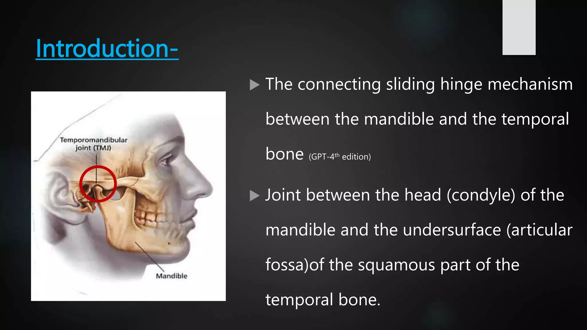
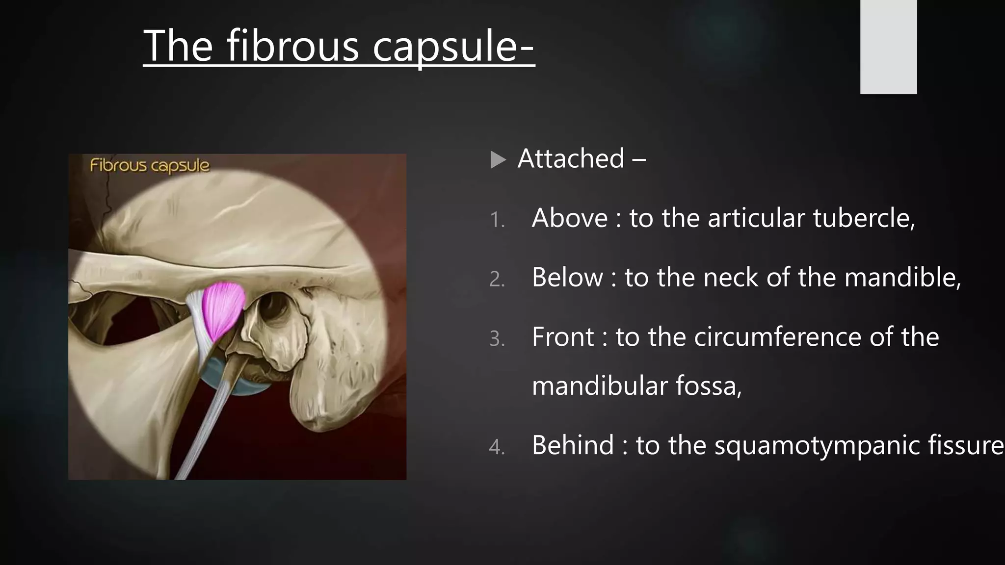
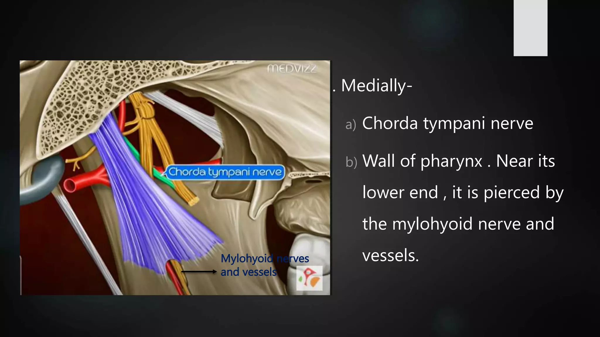
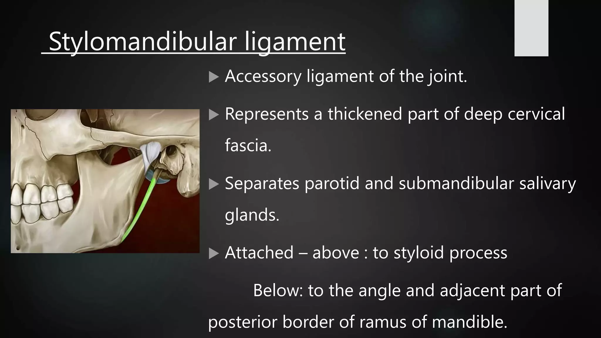
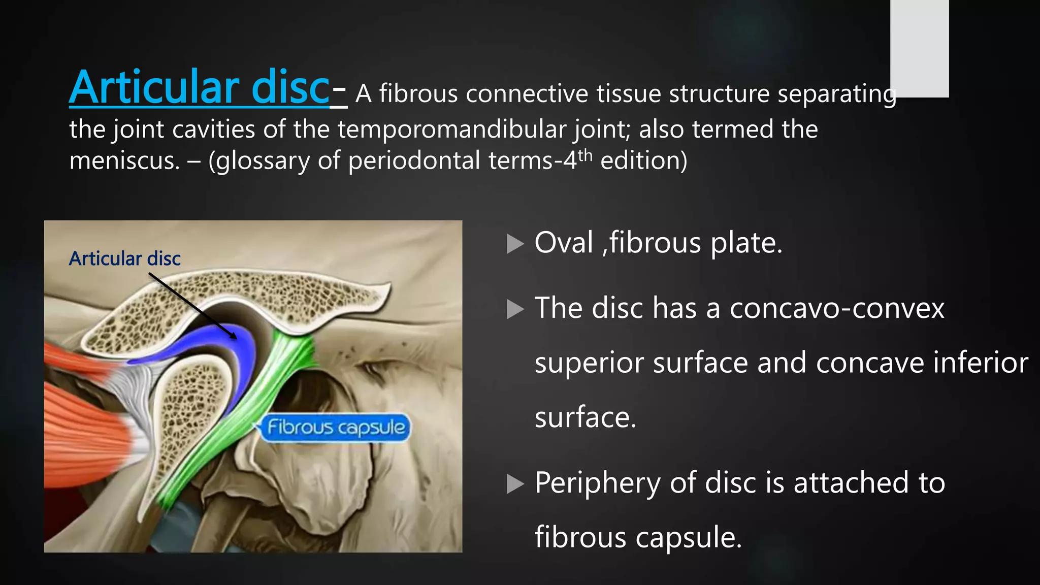
DEFINITION, ANATOMY, AND FUNCTIONS OF TEMPOROMANDIBULAR JOINT.
Joint between the head (condyle) of the mandible and the undersurface (articular fossa)of the squamous part of the temporal bone is the temporomandibular joint.
Type of joint : synovial joint (condylar variety).
Capable of providing-hinging (rotation) -gliding (translation) movement.
Sustains incredible forces of mastication.
articulating surfaces-articualar tubercle, mandibular fossa.
functions-Chewing
Sucking
Swallowing
Phonation
Facial expressions
Breathing Protrusion,
Retrusion,
Lateralization of the jaw
Opening the mouth
Maintain the correct pressure of the middle ear
Blood supply- Branches from superficial temporal and maxillary artery.
Veins follow the arteries.
Nerve supply-Auriculotemporal nerve (branch of mandibular nerve) and masseteric nerve (motar branch of anterior division of mandibular nerve).
movemnets of tmj- protraction, retraction, elevation, depression, side to side grinding.
examination of tmj- preauricular method and intraauricular method.
Jalandhar Female Call Girls Contact Number 9053900678 💚Jalandhar Female Call...
temporomandibular joint.pptx
1. PRESENTED BY –DR. SUMEDHA THOSAR
I-MDS
GUIDED BY- DR. SWAPNA MAHALE MA’AM
2. Table of contents-
Introduction
Development
Type of joint
Articular surfaces
Bony surfaces
Ligaments
Articular disc
Relations
Attachment
Blood supply
Nerve supply
Functions
Movements
Examination of tmj
Clinical aspect
References
3. Introduction-
The connecting sliding hinge mechanism
between the mandible and the temporal
bone (GPT-4th edition)
Joint between the head (condyle) of the
mandible and the undersurface (articular
fossa)of the squamous part of the
temporal bone.
4. The cranium, with which the
mandible articulates, is also
mechanically a single
component, with a mandibular
fossa on each side.
5. The temporomandibular joints are the
bilateral components of a
craniomandibular articulation.
Movement cannot take place at one
temporomandibular joint without a
concomitant movement occurring at the
joint on the opposite side.
6. Development-
There are 3 stages that define
the normal embryonic
development of TMJ-
blastemic stage ,
cavitation stage ,
maturation stage
7. At 3 months of gestation-1st evidence of TMJ - two distinct regions of
mesenchymal condensation : 1)temporal and 2)condylar blastema
{between developing ramus and developing squamous tympanic bone}.
Ossification : first in temporal blastema and ,condylar blastema is
condensed mesenchyme.
Cleft appears immediately above condylar blastema- becomes the
inferior joint cavity.
The condylar blastema differentiates into i)condylar cartilage
ii)second cleft appears in relation to temporal ossification that becomes
superior joint cavity
With the appearance of this cleft, the primitive articular disk is formed.
9. At 6th week- Articular disc first
appears.
At 7th week- Meckels cartilage
extends from chin to base of skull
acts as scaffolding for mandible
development.
Two ectomesenchymal
condensation appears.
At 12th week- Condylar growth
appears and condyle begins to
develop.
At 22nd week- Articular capsule
becomes recognisable and merges
peripherally with condensation.
At 26th week- All components of
joints appears expect articular
eminence.
At 31st week- Sphenomandibular
ligament appears.
At 39th week- Ossification of bony
components starts and resembles
the future joint.
10. Type of joint
Type of joint : synovial joint (condylar
variety).
Capable of providing
-hinging (rotation)
-gliding (translation) movement.
Sustains incredible forces of mastication.
15. The inferior articular surface is formed by the head of the
mandible
16. The articular surfaces are covered
by fibrocartilage
The joint cavity is divided into
upper and lower parts by an intra
articular disc- superior joint
cavity and inferior joint cavity.
Upper
joint
cavity
Lower
joint
cavity
19. Ligaments-
ligaments play an
important role in
protecting the
structures.
made up of collagenous
connective tissues fibres that have
particular lengths & do not
stretch.
Ligaments do not enter actively
into joint function but instead
act as passive restraining
devices to limit and restrict
border movements.
21. Other ligaments-"oto-mandibular ligaments“
connect the middle ear (malleus) with the temporomandibular
joint.
• discomallear (or disco-malleolar) ligament,
• malleomandibular (or malleolar-mandibular) ligament.
22. The fibrous capsule-
Attached –
1. Above : to the articular tubercle,
2. Below : to the neck of the mandible,
3. Front : to the circumference of the
mandibular fossa,
4. Behind : to the squamotympanic fissure
23.
24. The synovial membrane lines the fibrous capsule and the neck of
mandible
Capsule is loose above the intra articular disc and tight above it.
Function-
1.To resist medial, lateral, or inferior forces that tend to separate or
dislocate the articular surfaces.
2.To encompass the joint, to retaining the synovial fluid.
3.It is well innervated & provides proprioceptive feedback
regarding position and movement of the joint.
25. Lateral (temporomandibular) ligament
Attached- above to the articular tubercle,
and below to the posterolateral aspect of
the neck of the mandible.
The lateral temporomandibular ligament
strengthens the lateral part of the
capsular ligament.
Fibers are directed downwards and
backwards.
26. • The outer oblique portion of the TM ligament functions to resist
the impingement of vital submandibular and retromandibular structures of
the neck.
•The inner horizontal portion of the TM ligament limits posterior movement
of the condyle and disc. The TM ligament therefore protects the retrodiscal
tissues from trauma created by the posterior displacement of the condyle.
•The inner horizontal portion also protects the lateral pterygoid muscle
from overlengthening or extension.
The effectiveness of this ligament is demonstrated during cases of extreme
trauma to the mandible.
Functions of
temporo-
mandibular
ligament
27. Sphenonmandibular ligament
It is an accessory ligament, that lies on
a deep plane away from the fibrous
capsule.
Remanent of dorsal part of Meckel's
cartilage.
Attached superiorly to the spine of
sphenoid and inferiorly to the lingula
of the mandibular foramen.
28. Relations:
1. Laterally-
a) lateral pterygoid muscle
b) Auriculotemporal nerve
c) Maxillary artery
Maxillary artery
29. 2. Medially-
a) Chorda tympani nerve
b) Wall of pharynx . Near its
lower end , it is pierced by
the mylohyoid nerve and
vessels.
Mylohyoid nerves
and vessels
30. Stylomandibular ligament
Accessory ligament of the joint.
Represents a thickened part of deep cervical
fascia.
Separates parotid and submandibular salivary
glands.
Attached – above : to styloid process
Below: to the angle and adjacent part of
posterior border of ramus of mandible.
31. It becomes taut when the mandible is
protruded, but is most relaxed when
the mandible is opened.
The stylomandibular ligament
therefore limits excessive protrusive
movements of the mandible.
This ligament becomes tense only in
extreme protrusive movements.
Functions of stylo-
mandibular
ligament
32. Colateral ligament
Attach the medial and lateral borders of the
articular disc to the poles of the condyle.
The medial discal ligament attaches the medial
edge of the disc to the medial pole of the
condyle.
The lateral discal ligament attaches the lateral
edge of the disc to the lateral pole of the
condyle .
33. Divide the joint mediolaterally into the superior and
inferior joint cavities.
True ligaments, composed of collagenous
connective tissue fibers; therefore they do not
stretch.
34. • to restrict movement of the disc away from the condyle.
•allow the disc to move passively with the condyle as it
glides anteriorly and posteriorly.
• responsible for the hinging movement of the TMJ,
which occurs between the condyle and the articular disc.
have a vascular supply and are innervated. & provides
information regarding joint position and movement.
•Strain on them produces pain
Functions of
collateral ligament
35. Articular disc- A fibrous connective tissue structure separating
the joint cavities of the temporomandibular joint; also termed the
meniscus. – (glossary of periodontal terms-4th edition)
Oval ,fibrous plate.
The disc has a concavo-convex
superior surface and concave inferior
surface.
Periphery of disc is attached to
fibrous capsule.
Articular disc
36. Divides the joint into-
1. Upper compartment permits gliding movements
2. Lower compartment provides rotatory as well as gliding
movements.
Composed of – anterior extension , anterior thick band,
intermediate zone, posterior thick band, and bilaminar
region containing venous plexus.
The disc represents primitive insertion of lateral
pterygoid.
37. Functions-
1. Friction between the articulating surfaces.
2. Acts as cushion and helps in shock absorption.
3. Stabilises the condyle by filling up the space between articulating
surfaces.
4. Proprioceptive fibres present in the disc help to regulate
movements of the joint.
5. Helps in distribution of weight across the TMJ by increasing the
area of contact.
38. Relations of temporomandibular joint-
Lateral –
1. Skin and fascia
2. Parotid gland
3. Temporal branches of the facial nerve.
Medial –
1. The tympanic plate separates the joint from the internal carotid artery.
2. Spine of sphenoid , with upper end of the spheno-mandibular ligament attached
to it.
3. Auriculotemporal and chorda tympani nerves.
39. Anterior –
1. Lateral pterygoid
2. Masseteric nerve and artery
Posterior –
1. The parotid gland separates the joint from the external auditory
meatus
2. Superficial temporal vessels
3. Auriculotemporal nerve
40. Superior –
1. Middle cranial fossa
2. Middle meningeal vessels
Inferior-
1. Maxillary artery and vein
42. Blood supply-
Branches from superficial
temporal and maxillary
artery.
Veins follow the arteries.
43. Nerve supply-
Auriculotemporal nerve
(branch of mandibular
nerve) and masseteric
nerve (motar branch of
anterior division of
mandibular nerve).
44. Function of temporomandibular joint-
Chewing
Sucking
Swallowing
Phonation
Facial expressions
Breathing Protrusion,
Retrusion,
Lateralization of the jaw
Opening the mouth
Maintain the correct
pressure of the middle ear
50. Disorders of TMJ-Temporomandibular disorders (TMD) is a collective term embracing a
number of clinical problems that involve the masticatory musculature, the temporomandibular joint (TMJ)
and associated structures, or both.
Extrinsic disorders
MPDS
Myositis
Protective muscle splinting
Intrinsic disorders
Traumatic
arthritis
fracture
Internal disc
derangement
tendonitis
Myofibrotic contractures
51. Deviation in form
Disc displacement
1. Disc displacement with
reduction
2. Disc displacement without
reduction
Dislocation
1. Inflammatory conditions
2. Synovitis
Capsulitis
Arthritides
1. Osteoarthrosis
2. Osteoarthritis
3. Polyarthritides
Ankylosis
1. Fibrous
2. Bony
Recommended Diagnostic Classification for
Temporomandibular joint disorders
52. Trismus-
Trismus, or lockjaw, is a painful
condition in which the jaws do not
open fully.
It can lead to problems with eating,
speaking, and oral hygiene.
The defining symptom of trismus is
the jaw not opening fully or
opening to 35 mm or less.
53. Most cases of trismus are temporary,
typically lasting for less than 2 weeks, but
some may be permanent.
Can occur as a result of trauma to the jaw,
oral surgery, infection, cancer, or radiation
treatment for cancers of the head and
throat
54. Treatment-
1. warm compress (loosens the muscles).
2. Using cold packs for pain relief
3. Rectifying your posture to prevent the lockjaw condition from
worsening.
4. Practicing jaw exercises
5. Staying hydrated
6. Taking magnesium and calcium rich foods
55. MPDS- myofascial pain dysfunction
Myofascial pain dysfunction
syndrome (MPDS) is a stomatognathic system
disturbance, which consists of pain, jaw
movement irregularities, and muscle spasm.
Hyperexcitation of peripheral sensory neurons
causes a reaction of induction in the motor
neuron and then spasms of the masticatory
muscles follow.
56. Clinical features-
• Pain or discomfort.
• Limited motion of the jaw
• Tenderness to palpation of muscles
of mastication
• Joint noises i.e clicking ,snapping.
57.
58. Myositis-
Myositis consists of localized
transient muscle and facial
tissue swelling caused by
infection or injury.
mandibular movement and
localized tenderness have a
tendency to increase the pain
threshold.
Protective splinting
59. Traumatic arthritis-
A traumatic injury can result in
arthritis of the temporomandibular
joint causing limited motion, pain,
and tenderness.
Treatment typically involves anti-
inflammatory medications, heat
application, restricted jaw
movement, and softer diet.
Types -
60. Fracture of tmj-
A blow to the anterior mandibular body
is the most common reason for the
condylar fracture. (indirect).
The force is transmitted from the body
of the mandible to the condyle.
The condyle is trapped in the glenoid
fossa.
61. If the impact is in the midline of the
mandible ,fracture of the bilateral
condylar region is very common.
Commonly a blow to the ipsilateral
mandible causes contralateral
fracture in the condylar region.
62. Luxation / dislocation-
Dislocation can be defined
as long lasting ability to
close the mouth due to the
complete translation of the
condyle anterior to the
articular eminence.
Unilateral or bilateral.
63. The standard treatment for
acute TMJ dislocation
is reduction of the mandible, a
technique by which the
dislocated jaw is pushed
downward and backward into its
normal position.
Frequently takes place in
emergency rooms under
sedation or GA
66. Subluxation-
Subluxation is defined as a
self reducing partial
dislocation of tmj during
which the condyle passes
anterior to the articular
eminence.
67. Patient is himself able to
manipulate it back into
normal position.
This is recurrent ,
incomplete, self-reducing,
habitual dislocation
68. Ankylosis of TMJ
Temporomandibular joint (TMJ) ankylosis
is a pathologic condition where the
mandible is fused to the fossa by bony or
fibrotic tissues.
This interferes with mastication, speech,
oral hygiene, and normal life activities,
and can be potentially life threatening
when struggling to acquire an airway in an
emergency.
69.
70. Treatment-surgery: complete ankylotic block removal and
subsequent arthroplasty(), possibly with autologous graft
between articular surfaces (skin, rib cartilage, temporal muscle)
or heterologous graft (Silastic) to fully restore the anatomic
structure.
72. Hypoplasia or aplasia of
mandibular condyle of the
mandibular condyle
indicates underdevelopment or
nondevelopment associated
mainly with various craniofacial
abnormalities.
These may be either congenital or
acquired
73. Treacher Collins syndrome, mandibulofacial
dysostosis (MFD) -rare syndrome characterized
by underdeveloped facial bones and
micrognathia. The most obvious facial
differences are the underdeveloped cheekbones
and the drooping lower eyelids.
Treatment is focused on correcting facial
structure, and can involve plastic surgery,
orthodontics and dental care.
74. Hemifacial microsomia is a congenital
condition in which the tissues on one side
the face are underdeveloped.
Surgery for children- involve one or more
procedures to correct underdevelopment.
Common surgeries for children dealing with
HFM include: Lowering the upper jaw to
match the opposite side and lengthening the
lower jaw. Using a bone graft to lengthen the
jaw.
75. Goldenhar syndrome is a rare
congenital defect characterized by
incomplete development of the ear,
nose, soft palate, lip and
mandible on usually one side of the
body.
Hallermann-Streiff syndrome (HSS) is a
rare disorder characterized by
dyscephalia, with facial and dental
abnormalities.
76. Condylar hyperplasia, also known as
mandibular hyperplasia, is the presence of an
enlarged mandible bone within the skull.
First identified in 1836 by Robert Adams
(associated it with the overdevelopment of
mandible).
Cause unknown but several theories exists.
One theory states that an event of a trauma
leading to increase in number of repair
mechanism and hormones in that area may lead
to increase in growth of mandible on that side.
77. Bifid condyle is a rare
anatomic variation of
mandibular condyle. It can be
symptomatic or diagnosed
incidentally on routine
radiographic examination.
No definite etiologic factor,
could be a developmental
anomaly or secondary to
trauma.
78. Relation between periodontitis and TMJ
Study indicates that unilateral mastication due to chronic periodontitis could induce
not only pain but also structural TMJ changes if adequate treatment is not
administered and supported within a short time from the onset of the condition.
Therefore, immediate treatment of chronic periodontitis is recommended to prevent
not only the primary progress of periodontal disease, but also secondary TMJ-related
problems.
Furthermore, subjects who have suffered chronic long-term periodontitis without
treatment should be urged to undergo a TMJ examination.
79. References-
BD charasia -4th edition
Carranza 10th edition
Carranza 13th south asia edition
Neelima malik textbook of oral and maxillofacial studies -4thh edition.
Journal of maxillofcaila surgery Natl J Maxillofac Surg. 2015 Jan-Jun; 6(1): 16–20.
doi: 10.4103/0975-5950.168212 PMCID: PMC4668726 PMID: 26668447
J Periodontal Implant Sci. 2017 Aug; 47(4): 211–218.
Published online 2017 Aug 21. doi: 10.5051/jpis.2017.47.4.211 PMCID: PMC5577439
PMID: 28861285 Pattern analysis of patients with temporomandibular disorders
resulting from unilateral mastication due to chronic periodontitis