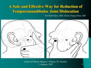
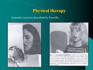
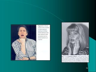
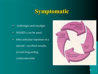
Presenter - Dr. Apurva Mehta
Postgraduate.
Moderator - Dr. Raghvendra
Assistant. Prof
1
• Introduction
• Definitions & terminologies
• Normal jaw biomechanics
• Hypermobility & its management
• Subluxation management
• Dislocation , clinical presentation & diagnosis
• Etiology & predisposing factors for dislocation
• Treatment options
• Nonsurgical
• Surgical
• Conclusion
2
• Dislocation of mandible is one of the earliest afflictions
of the jaws to be described in the literatures.
• Hippocrates in he 5th century described the condition
and its management.
• Mandibular dislocation uncommon compared to other
joint dislocation (3%)
• Higher incidence in females than in males.
3
Hypermobility
Subluxation
Dislocation.
4
5
Def – Excessive anterior movement of the condyle at
maximum mouth opening without strain /
symptoms.
-- Acquired
-- Systemic
6
An excessive range of movement occurring in one or more joints
as a result of a pathologic process.
1) Isolated,
2) Generalized.
7
Neuropathic arthropathy (Charcot’s joint).
Traumatic rupture of ligaments.
Rheumatoid arthritis and related disorders.
Late osteoarthrosis.
8
Acromegaly- lowering of the elastic modulus of joints
(Grahame et al) collagen defect due to GH disorders.
Pregnancy - Joint laxity increases ( Bird et al) hormonal
influence.
Hyperparathyroidism ↑ collagenase activity.
Chronic alcoholism.
9
• Familial hypermobility syndromes
• Ehlers – Danlos syndrome
• Marfan syndrome
• Familial joint laxity
• wide spectrum of dislocations and subluxations
10
Normal joint stability depends on:
i. Bony architecture of joint surfaces
ii. Integrity of joint ligaments
iii. Activity of muscles acting on the joint
11
Morphologic conditions of the condyle & eminence.
Previous capsule & ligament injury.
Laxity of ligaments (TMJ)
Degenerative joint disease.
Dyssynchronous muscle function.
TMJ overextensions + Dyssynchronous muscle function.
Internal derangement.
12
An incomplete joint dislocation wherein the articular surfaces
maintain partial contact and the condyle is
able to return to the glenoid fossa voluntarily or aided
by self-manipulation.
13
Anatomic variation of the fossa-
steep short posterior slope with longer flat anterior
slope of the articular eminence
Associated with disc derangement
14
Momentary sticking / catching open for a short period.
Pain and dysfunction
Multiple clicks when hypermobility is associated with ID.
“Click” occurs only on wide opening and not on protrusive or
lateral movement / excursions.
15
Limit mouth opening.
Exercise to strengthen the elevator muscle.
Sclerosing agents.
Eminectomy.
16
Open Lock
• Younger
• Spontaneous, in joints
with
internal derangement
• Difficult but self-corrected
• Beneath and inferior to
eminence
• Trapped condyle located
in front of the lagging disc
• Arthrocentesis
Condylar Dislocation
• Older
• Maximal opening (yawning,
shouting, neurogenic, neuroleptic
drugs, joint laxity)
• Usually professional
• In front of and superior to
eminence
• Condyle located in front of the
eminence
• Surgery when recurrent
17
- Dorrit W. Nitzan
J Oral Maxillofac Surg 60:506-511, 2002
Defn - A nonreducing displacement of the mandibular condyle in
front of and superior to the articular eminence, resulting in the
inability to close the mouth.
Incidence – 3% of all the dislocations (Lovely, Copeland, 1981).
18
• Intrinsic trauma: over extension injury
•Yawning
•Vomiting
•Wide biting
•Seizures disorder
•Extrinsic trauma:
•Trauma: flexion-extension injury to the mandible
•Intubation with general anesthesia
•Endoscopy
•Dental extraction
•Forceful hyperextension
19
• Connective tissue disorders:
• Hypermobility syndrome
• Ehlers-Danlos syndrome
• Marfan syndrome
• Miscellaneous causes:
• Internal derangement,
• Dyssynchronous muscle function,
• Contralateral intra-articular obstruction
• Lost vertical dimension
• Occlusal discrepancies
• Psychogenic:
• Habitual dislocation
• Drug induced:
• phenothiazines
20
Depending upon side
Unilateral
Bilateral
Depending on the time elapsed
Acute
Chronic
- Long standing,
- Recurrent,
- Habitual 21
22
23
History
Determine cause & onset.
A prior h/o local joint laxity, ID, & other TMJD
use of antipsychotic drugs
physical examination
Neurological and musculoskeletal disorders
Radiological examination
24
25
26
27
Non surgical treatment.
Surgical treatment.
it is very important to differentiate between the various categories of
dislocation and subluxation.
28
Reduction by manipulation
Physical therapy  Exercises to gain better muscular
control and restraint of opening.
Occlusal therapy
Symptomatic treatment
Chemical capsulorraphy
Ultrasound therapy
Intermaxillary fixation
29
Manual reduction
30
31
32
Annals of Plastic Surgery • Volume 58, Number
1, January 2007
- Yi-Chieh Chen, MD, Chien-Tzung Chen, MD
• Isometric exercises described by Poswillo
33
34
35
Arthralgia and myalgia.
NSAID’s can be used.
Intra articular injection of a
steroid - excellent results,
(avoid long-acting
corticosteroids)
36
1.Reduce bruxism
2.Help relax muscles and reduce
pain.
3.They also can change jaw
posture enough to stabilize some
bite problems and reduce pressure
in joints.
37
Promotes collagen synthesis
by human fibroblasts
therefore this may be help in
stabilizing the joint.
38
• Principle – To induce fibrosis and restrict joint movement.
• 3% sodium tetradecyl sulphate ,
• Sodium psylliate emulsion in oil.
• Sodium morrhuate
• Disadv: Inability to predict the amount of limitation.
Schultz 1947
39
40
Extrinsic trauma
Blow on the chin while mouth is open,
injudicious use of mouth gags during G A.
Acute pain, anxiety & inability to close the mouth.
Immediate manual reduction followed by 4-6 wks of
immobilization.
41
42
Conservative line of treatment should be considered first.
Surgical management in recurrent cases
43
Three broad categories
I. Procedures which are designed to limit translation,
-Anchoring
-Blocking
-Myotomy
II. To eliminate blocking factors in the condylar path of closure.
III. Combination of both.
44
Procedures to limit
translation
45
Flaps secured to the capsule
46
Merrill used dacron sutures
47
Sanders & Newman
48
Resected 1.5cm segment of
zygomatic arch & grafted it
on to the eminence.
Mayer 1933
49
A vertical osteotomy of the
zygomatic arch &down
fracture.
Modified by Dautrey in
1975.
Oblique osteotomy.
(1943)
50
Oblique osteotomy of articular
tubercle
51
Treatment of chronic mandibular dislocations by bone plates: Two Case Reports
Journal of Cranio-Maxillofacial Surgery (2004) 32, 90–92
52
Fixation of the disc in anterior position
Konjetzny
53
myotomy of the lateral
pterygoid muscle, through
an intraoral incision.
liberation of the fibres
would help reduce the
dislocation.
silicon sheet interposition.
Laskin
54
Procedures those
eliminate blocking
factors
55
Myrhaug 1951
56
A torn / displaced disk caught behind the condyle or a
prominent articular eminence, obstructing the condylar
movement.
central avascular portion of the disk and the area of
perforation
Dermal graft can be harvested
57
↓ lat ptery muscle pull
Segment gets inferiorly
displacement.
Ward et al
58
Drawbacks
• lat pterygoid ms  sacrificed
• Shortening of ramus,
• open bite deformity&
• retrusion of mandible
• Loss of translatory movement.
Riedel 1883
59
High condylectomy-
60
Acute closed lock (meniscus is
usually jammed in front of the
condyle) preventing
translatory movement.
Mech -By ballooning-up the joint
the potential space becomes
real and the meniscus gets
room to reduce to its normal
position.
61
Arthroscopy by means of the inferolateral approach
using an electric shaver in a triangulation technique.
The anterior slope of the eminence was made as smooth as
possible.
Segami
(Oral S,Oral Med, Oral Path, Oral Radiol Endod 2003;95:390-5)
62
Management requires a careful and conservative approach, &
basically depends upon the type of dislocation and the amount of
morbidity it has caused to the patient,
Employ the simplest and most effective method with the least
morbidity for a specific patient.
63
It is important to address muscular and psychologic factors
appropriately before considering the patient for surgery.
Chronic dislocation- IMF for 4-6 weeks.
64
"If the luxation be left unreduced the patient will remain
a living memorial of the surgeon's ignorance or
inattention."
Sir Astley Cooper
65
Principles of Oral and Maxillofacial surgery – Peterson.
Surgery of the TMJ – David A. Keith.
Temporomandibular joint dysfunction- A Practioner’s guide-
Annika Isberg.
Oral and Maxillofacial surgery TMJ disorders – R.J. Fonseca.
Text book of oral and maxillofacial surgery – Kruger.
TMJ disorders diagnosis and treatment – Kaplan and Arsael
66
67

TMJ DISLOCATION AND ITS MANAGEMENT..pptx

  • 1.
    Presenter - Dr.Apurva Mehta Postgraduate. Moderator - Dr. Raghvendra Assistant. Prof 1
  • 2.
    • Introduction • Definitions& terminologies • Normal jaw biomechanics • Hypermobility & its management • Subluxation management • Dislocation , clinical presentation & diagnosis • Etiology & predisposing factors for dislocation • Treatment options • Nonsurgical • Surgical • Conclusion 2
  • 3.
    • Dislocation ofmandible is one of the earliest afflictions of the jaws to be described in the literatures. • Hippocrates in he 5th century described the condition and its management. • Mandibular dislocation uncommon compared to other joint dislocation (3%) • Higher incidence in females than in males. 3
  • 4.
  • 5.
  • 6.
    Def – Excessiveanterior movement of the condyle at maximum mouth opening without strain / symptoms. -- Acquired -- Systemic 6
  • 7.
    An excessive rangeof movement occurring in one or more joints as a result of a pathologic process. 1) Isolated, 2) Generalized. 7
  • 8.
    Neuropathic arthropathy (Charcot’sjoint). Traumatic rupture of ligaments. Rheumatoid arthritis and related disorders. Late osteoarthrosis. 8
  • 9.
    Acromegaly- lowering ofthe elastic modulus of joints (Grahame et al) collagen defect due to GH disorders. Pregnancy - Joint laxity increases ( Bird et al) hormonal influence. Hyperparathyroidism ↑ collagenase activity. Chronic alcoholism. 9
  • 10.
    • Familial hypermobilitysyndromes • Ehlers – Danlos syndrome • Marfan syndrome • Familial joint laxity • wide spectrum of dislocations and subluxations 10
  • 11.
    Normal joint stabilitydepends on: i. Bony architecture of joint surfaces ii. Integrity of joint ligaments iii. Activity of muscles acting on the joint 11
  • 12.
    Morphologic conditions ofthe condyle & eminence. Previous capsule & ligament injury. Laxity of ligaments (TMJ) Degenerative joint disease. Dyssynchronous muscle function. TMJ overextensions + Dyssynchronous muscle function. Internal derangement. 12
  • 13.
    An incomplete jointdislocation wherein the articular surfaces maintain partial contact and the condyle is able to return to the glenoid fossa voluntarily or aided by self-manipulation. 13
  • 14.
    Anatomic variation ofthe fossa- steep short posterior slope with longer flat anterior slope of the articular eminence Associated with disc derangement 14
  • 15.
    Momentary sticking /catching open for a short period. Pain and dysfunction Multiple clicks when hypermobility is associated with ID. “Click” occurs only on wide opening and not on protrusive or lateral movement / excursions. 15
  • 16.
    Limit mouth opening. Exerciseto strengthen the elevator muscle. Sclerosing agents. Eminectomy. 16
  • 17.
    Open Lock • Younger •Spontaneous, in joints with internal derangement • Difficult but self-corrected • Beneath and inferior to eminence • Trapped condyle located in front of the lagging disc • Arthrocentesis Condylar Dislocation • Older • Maximal opening (yawning, shouting, neurogenic, neuroleptic drugs, joint laxity) • Usually professional • In front of and superior to eminence • Condyle located in front of the eminence • Surgery when recurrent 17 - Dorrit W. Nitzan J Oral Maxillofac Surg 60:506-511, 2002
  • 18.
    Defn - Anonreducing displacement of the mandibular condyle in front of and superior to the articular eminence, resulting in the inability to close the mouth. Incidence – 3% of all the dislocations (Lovely, Copeland, 1981). 18
  • 19.
    • Intrinsic trauma:over extension injury •Yawning •Vomiting •Wide biting •Seizures disorder •Extrinsic trauma: •Trauma: flexion-extension injury to the mandible •Intubation with general anesthesia •Endoscopy •Dental extraction •Forceful hyperextension 19
  • 20.
    • Connective tissuedisorders: • Hypermobility syndrome • Ehlers-Danlos syndrome • Marfan syndrome • Miscellaneous causes: • Internal derangement, • Dyssynchronous muscle function, • Contralateral intra-articular obstruction • Lost vertical dimension • Occlusal discrepancies • Psychogenic: • Habitual dislocation • Drug induced: • phenothiazines 20
  • 21.
    Depending upon side Unilateral Bilateral Dependingon the time elapsed Acute Chronic - Long standing, - Recurrent, - Habitual 21
  • 22.
  • 23.
  • 24.
    History Determine cause &onset. A prior h/o local joint laxity, ID, & other TMJD use of antipsychotic drugs physical examination Neurological and musculoskeletal disorders Radiological examination 24
  • 25.
  • 26.
  • 27.
  • 28.
    Non surgical treatment. Surgicaltreatment. it is very important to differentiate between the various categories of dislocation and subluxation. 28
  • 29.
    Reduction by manipulation Physicaltherapy  Exercises to gain better muscular control and restraint of opening. Occlusal therapy Symptomatic treatment Chemical capsulorraphy Ultrasound therapy Intermaxillary fixation 29
  • 30.
  • 31.
  • 32.
    32 Annals of PlasticSurgery • Volume 58, Number 1, January 2007 - Yi-Chieh Chen, MD, Chien-Tzung Chen, MD
  • 33.
    • Isometric exercisesdescribed by Poswillo 33
  • 34.
  • 35.
  • 36.
    Arthralgia and myalgia. NSAID’scan be used. Intra articular injection of a steroid - excellent results, (avoid long-acting corticosteroids) 36
  • 37.
    1.Reduce bruxism 2.Help relaxmuscles and reduce pain. 3.They also can change jaw posture enough to stabilize some bite problems and reduce pressure in joints. 37
  • 38.
    Promotes collagen synthesis byhuman fibroblasts therefore this may be help in stabilizing the joint. 38
  • 39.
    • Principle –To induce fibrosis and restrict joint movement. • 3% sodium tetradecyl sulphate , • Sodium psylliate emulsion in oil. • Sodium morrhuate • Disadv: Inability to predict the amount of limitation. Schultz 1947 39
  • 40.
  • 41.
    Extrinsic trauma Blow onthe chin while mouth is open, injudicious use of mouth gags during G A. Acute pain, anxiety & inability to close the mouth. Immediate manual reduction followed by 4-6 wks of immobilization. 41
  • 42.
  • 43.
    Conservative line oftreatment should be considered first. Surgical management in recurrent cases 43
  • 44.
    Three broad categories I.Procedures which are designed to limit translation, -Anchoring -Blocking -Myotomy II. To eliminate blocking factors in the condylar path of closure. III. Combination of both. 44
  • 45.
  • 46.
    Flaps secured tothe capsule 46
  • 47.
  • 48.
  • 49.
    Resected 1.5cm segmentof zygomatic arch & grafted it on to the eminence. Mayer 1933 49
  • 50.
    A vertical osteotomyof the zygomatic arch &down fracture. Modified by Dautrey in 1975. Oblique osteotomy. (1943) 50
  • 51.
    Oblique osteotomy ofarticular tubercle 51
  • 52.
    Treatment of chronicmandibular dislocations by bone plates: Two Case Reports Journal of Cranio-Maxillofacial Surgery (2004) 32, 90–92 52
  • 53.
    Fixation of thedisc in anterior position Konjetzny 53
  • 54.
    myotomy of thelateral pterygoid muscle, through an intraoral incision. liberation of the fibres would help reduce the dislocation. silicon sheet interposition. Laskin 54
  • 55.
  • 56.
  • 57.
    A torn /displaced disk caught behind the condyle or a prominent articular eminence, obstructing the condylar movement. central avascular portion of the disk and the area of perforation Dermal graft can be harvested 57
  • 58.
    ↓ lat pterymuscle pull Segment gets inferiorly displacement. Ward et al 58
  • 59.
    Drawbacks • lat pterygoidms  sacrificed • Shortening of ramus, • open bite deformity& • retrusion of mandible • Loss of translatory movement. Riedel 1883 59
  • 60.
  • 61.
    Acute closed lock(meniscus is usually jammed in front of the condyle) preventing translatory movement. Mech -By ballooning-up the joint the potential space becomes real and the meniscus gets room to reduce to its normal position. 61
  • 62.
    Arthroscopy by meansof the inferolateral approach using an electric shaver in a triangulation technique. The anterior slope of the eminence was made as smooth as possible. Segami (Oral S,Oral Med, Oral Path, Oral Radiol Endod 2003;95:390-5) 62
  • 63.
    Management requires acareful and conservative approach, & basically depends upon the type of dislocation and the amount of morbidity it has caused to the patient, Employ the simplest and most effective method with the least morbidity for a specific patient. 63
  • 64.
    It is importantto address muscular and psychologic factors appropriately before considering the patient for surgery. Chronic dislocation- IMF for 4-6 weeks. 64
  • 65.
    "If the luxationbe left unreduced the patient will remain a living memorial of the surgeon's ignorance or inattention." Sir Astley Cooper 65
  • 66.
    Principles of Oraland Maxillofacial surgery – Peterson. Surgery of the TMJ – David A. Keith. Temporomandibular joint dysfunction- A Practioner’s guide- Annika Isberg. Oral and Maxillofacial surgery TMJ disorders – R.J. Fonseca. Text book of oral and maxillofacial surgery – Kruger. TMJ disorders diagnosis and treatment – Kaplan and Arsael 66
  • 67.