Temporomandibular joint ankylosis and its management
•Download as PPTX, PDF•
19 likes•4,163 views

Detailed discussion on diagnosis and management of TMJ ankylosis. Surgical anatomy and applied aspects of TMJ is discussed. Reconstruction of ramus-condyle unit is also discussed. Compications of TMJ surgery are also discussed

Recommended
More Related Content
What's hot
What's hot (20)
Similar to Temporomandibular joint ankylosis and its management
Similar to Temporomandibular joint ankylosis and its management (20)
More from Dibya Falgoon Sarkar
More from Dibya Falgoon Sarkar (18)
Recently uploaded
Recently uploaded (20)
Temporomandibular joint ankylosis and its management
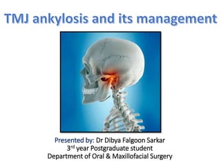
- 1. Presented by: Dr Dibya Falgoon Sarkar 3rd year Postgraduate student Department of Oral & Maxillofacial Surgery
- 2. Introduction • Temporomandibular joint ankylosis is a pathologic condition where the mandible is fused to the fossa by bony/ fibrotic tissues. (Movahed R, Mercuri LG. Management of temporomandibular joint ankylosis. Oral and Maxillofacial Surgery Clinics. 2015 Feb 1;27(1):27-35.) • Ankylosis is a Greek word that literally means “stiff joint”. • TMJ ankylosis interferes with: 1. Mastication 2. Speech 3. Oral hygiene 4. Growth & development • Ankylosis can be potentially life- threatening when we want to acquire an airway during an emergency.
- 3. Epidemiology of TMJ ankylosis in India • Overall prevalence: 0.46 per 1000 in the 3-15 years’ age group. • Age distribution: 3-63 years. • Most common age group: 10-15 years. • Male : Female: 1:9 • Birth/ childhood trauma was the major causative factor. (Gupta VK, Mehrotra D, Malhotra S, Kumar S, Agarwal GG, Pal US. An epidemiological study of temporomandibular joint ankylosis. National journal of maxillofacial surgery. 2012 Jan;3(1):25.)
- 4. Etiology of Ankylosis of TMJ 1. Trauma : • At birth (forceps delivery) • Haemarthrosis • Condylar fracture 2. Infections • Chronic suppurative otitis media • Abscess around joint • Osteomyelitis of jaw • Actinomycosis • Parotitis 3. Inflammation: • Rheumatoid arthritis • Osteoarthritis • Septic arthritis 4. Systemic diseases: • Small pox • Ankylosis spondylitis • Beri beri 5. Other causes: • Bifid condyle • Prolonged trismus • Burns
- 5. Trauma- The predominant cause of TMJ ankylosis • Rowe’s series of 46 cases, out of which 31 resulted from trauma • Sada et al study of 146 cases, out of which 58% accounted from trauma • Sawhney’s series of 70 cases, out of which 69 were the result of trauma. Factors contributing to ankylosis: 1. Age of patient 2. Site and type of condylar fracture 3. Length of immobilization 4. Damage to the articular disc
- 6. How trauma leads to TMJ ankylosis
- 7. Classification of TMJ ankylosis • Kazanjian’s classification: • Rowe classified TMJ ankylosis according to: • Topazian’s classification: True ankylosis False ankylosis When there are fibrous/ osseous adhesions between the surface of the TMJ When the ankylosis is due to other pathologies not directly related to the joint. Location of problem Type of tissues involved Extent of fusion Intra-articular Osseous Complete Extra-articular Fibrous Incomplete Both Type I Type II Type III Affects the condyle only Intermediate type Affects condyle. coronoid and cranial base
- 8. Sawhney’s grading of TMJ ankylosis (1986) (Sawhney CP. Bony ankylosis of the temporomandibular joint: follow-up of 70 patients treated with arthroplasty and acrylic spacer interposition. Plastic and reconstructive surgery. 1986 Jan;77(1):29-40.)
- 9. History: • Accurate history is important to differentiate between true and pseudoankylosis • History of trauma directly to the joint or indirectly to the chin • History of ear infection • History of forceps delivery • Extracapsular causes like zygomatic arch fractures, OSMF should be ruled out • Clinical examination: • Restricted mouth opening with difficulty in mastication and jaw movements. • Patients often have associated obstructive sleep apnoea. • Patients are often seen with craniofacial syndromes, skeletal deformities and psychological distress. • Patients often come with a scar over chin. Diagnosis of TMJ ankylosis
- 10. Unilateral ankylosis Bilateral ankylosis Obvious facial asymmetry Mandible is symmetrical but micrognatic Deviation of chin to affected side Bird-face deformity Flattening and elongation of unaffected side Retrognathic mandible Well defined antegonial notch Prominent antegonial notch bilaterally Absence of condylar movements on affected side Inability to open mouth Unilateral posterior cross-bite B/L Class II malocclusion, multiple carious teeth, crowding ± anterior open bite Reduced mouth opening Increased chin throat angle
- 11. Radiographic investigations • Orthopantomograph- will show both the joints which can be compared in unilateral cases • Lateral oblique view- will show anteroposterior dimensions of the ankylotic mass. Elongation of coronoid process also seen • Posteroanterior view- will show the mediolateral extent of the ankylotic mass and also highlights asymmetry in unilateral cases. • Lateral Cephalogram- Helpful to evaluate the associated skeletal deformities. • CT scan- Helpful guide as it gives the picture of the ankylotic mass in 3D and its proximity to other anatomical structures.
- 12. Management of TMJ ankylosis Infancy Childhood (Growing age group) Adults Cause: • Agenesis of the TMJ (rare) • Birth trauma • Infection Cause: • Trauma • Infection Cause: • Trauma • Infection Aim of treatment: • Restore movement and function • Reconstruction may be done later Aim of treatment: • Restore movement and function • Reconstruction of the TMJ • Correction of occlusal and cosmetic deformity: Early teen: Distraction osteogenesis, myofunctional appliance Late teen: Orthognathic surgery, Distraction or both Aim of treatment: • Restore movement and function • Reconstruction of the TMJ • If OSA present, distraction may be done at first followed by release of ankylosis and TMJ reconstruction.
- 14. Bony Components Components of TMJ Soft-tissue components • Condyle of mandible • Articular eminence • Glenoid fossa • Articular disc • Joint capsule • Ligaments • Muscles
- 15. Unique features of TMJ • Its articular surface covered by fibrocartilage instead of hyaline cartilage. • The right and left TMJ form a bicondylar articulation and functions together as one unit. • Only joint in human body to have a rigid endpoint of closure that of the teeth making occlusal contact. • In contrast to other diarthrodial joints TMJ is last joint to start develop, in about 7th week in utero.
- 16. Important structures related to TMJ Nerves: 1. Facial nerve 2. Auriculotemporal nerve 3. Inferior alveolar nerve Vascular structures: • Middle meningeal artery • Internal maxillary artery • Superficial temporal artery • Masseteric artery
- 17. Facial Nerve • The main trunk of the facial nerve exits from the skull at the stylomastoid foramen. • Surgical landmarks for identification of facial nerve: • Tragal pointer - The main trunk of the facial nerve is located 1 cm anteroinferior and 1 cm deep to the tip of the tragal cartilage. • Digastric ridge- The main trunk is just superior to the attachment of the posterior belly of the digastric muscle to the digastric groove. This landmark also marks the approximate depth of the facial nerve. • Tympanomastoid suture line - The nerve lies 6 to 8 mm deep to the inferior end of the tympanomastoid suture line.
- 18. Facial Nerve • Surgical landmarks for identification of facial nerve: • Stylomastoid foramen - The base of the styloid process is 5 to 8 mm deep to the tympanomastoid suture line. The facial nerve can be identified as it exits the stylomastoid foramen and passes over the posterolateral aspect of the styloid process. • Mastoid - For revision cases, extensive tumors or, as a last resort, a mastoidectomy can be performed to locate the vertical segment of the facial nerve, which can then be followed as it exits the mastoid.
- 19. Relation of VII nerve with TMJ • Approximately 1.3 cm of the facial nerve is visible to surgeon until it divides into temporofacial and cervicofacial branches • According to Alkayat & Bramley’s classic article (1979)
- 20. Radiologic study by Miloro et al
- 21. Applied Aspects • The temporal branch of the facial nerve emerges from the parotid gland and crosses the zygoma under the temporoparietal fascial to innervate the frontalis muscle • Postsurgical palsy manifests as an inability to raise the eyebrow, close the eyelid and ptosis of the brow. • Damage to the zygomatic branch results in temporary or permanent paralysis to the orbicularis oculi and may require temporary patching of the eye to prevent corneal desiccation and abrasion.
- 22. Auriculotemporal Nerve • first branch of the mandibular nerve after it exits the foramen ovale. • supplies sensation to the skin in the temporal and preauricular region, the anterior external meatus • The auriculotemporal nerve provides most of the innervation to the capsule of the temporomandibular joint itself • Branch of mandibular nerve, post division • Radiographic assessment of the exact course of this nerve through the mandible is necessary if screws will be placed through the ramus as is necessary for prosthetic joint replacement Inferior Alveolar Nerve
- 23. Vascular anatomy of TMJ • Superficial temporal artery and vein: are routinely ligated during preauricular approaches. • Internal maxillary artery: is usually not encountered unless a condylectomy is performed. If a condylectomy is performed, Dunn‐Dautrey retractors are used as the internal maxillary artery normally runs-approximately 3 mm medial from the midsigmoid notch.
- 24. Talebzadeh N, Rosenstein TP, Pogrel MA. Anatomy of the structures medial to the temporomandibular joint. Oral Surgery, Oral Medicine, Oral Pathology, Oral Radiology and Endodontics. 1999 Dec 1;88(6):674-8.
- 25. Masseteric artery A careful dissection of 16 intact human cadaveric head specimens was done. The location of the masseteric artery was then determined in relation to 3 points process: 1 ) the anterior-superior aspect of the condylar neck = 10.3 mm; 2 ) the most inferior aspect of the articular tubercle = 11.4 mm; 3 ) the inferior aspect of the sigmoid notch = 3 mm. Journal of Oral and Maxillofacial Surgery. 2009;67 (2) : 369–371
- 26. Surgical Approaches to TMJ 1. Preauricular : Dingman’s, Blair’s, Thoma’s, Al-kayat and Bramley’s, Popowitch’s 2. Postauricular 3. Endaural approach 4. Post ramal/ Hind’s approach 5. Submandibular/Risdon’s approach 6. Hemicoronal 7. Bicoronal
- 27. Preauricular Approach • Basic incision was given by Dingman (1951) • Blair & Ivy modification– “Inverted hockey stick “ incision. Facilitate exposure of arch along with condylar area. • Thoma in 1958 Angulated vertical incision. -carried out across zygomatic arch infront of ear to avoid main trunk of facial nerve
- 28. Alkayat and Bramley Modified Preauricular Incision (Al‐Kayat A, Bramley P. (1980) A modified preauricular approach to the temporomandibular joint and malar arch. Br J Oral Surg, 17:91.) 1. Reduced haemorrhage 2. Provision of donor site for temporalis fascia 3. Better cosmetic result 4.Lesser chance of facial nerve injury
- 29. Facial nerve branches are not damaged
- 30. Post auricular approach • Introduced by Bockenheimer • Advantage: 1. Excellent exposure of the entire joint and the ability to camouflage the scar in patients who have a tendency to form keloids. 2. Better posterior exposure of TMJ • Disadvantage: 1. Auricular stenosis 2. Should not be used in the presence of joint infection or chronic otitis externa
- 32. Endaural Approach • Lempart (1938) • Short facial skin incision extending in to ext. auditory meatus • Advantage: Excellent cosmetics and posterolateral exposure • Disadvantage: 1. Meatal stenosis or chondritis 2. Injury to the branches of the facial nerve • Care must be taken not to incise the tragal cartilage because a perichondritis or poor wound healing may result.
- 33. Post-ramal/ Hind’s Approach • E.C Hinds & Girotin (1967) • Indications – 1. Condylar neck fractures 2. Condylotomy 3. Vertical ramus osteotomy • Incision- 1cm behind ramus of mand. and extends 1cm below the lobe of ear. • Advantages: Highly cosmetic, excellent visibility and accessibility. • Note: Injury may occur to posterior facial vein and main trunk of facial nerve.
- 34. Structures encountered during Hind’s approach
- 35. Combination of Retromandibular + Endaural Approach • Indication: For additional access to the temporomandibular joint for open fracture reduction, costochondral grafting, total alloplastic joint reconstruction, or tumor resection. • When combining both incisions, the surgeon must leave an intervening bridge of tissue that extends inferiorly at least 3 cm from the lowest point of the bony external auditory canal to protect VII nerve
- 36. Submandibular Risdon’s Approach •Risdon (1934) •Mainly used for neck of condyle & ramus region •Incision: Placed one‐finger breadth below the angle of the mandible ensuring avoidance of the marginal mandibular nerve
- 38. Relations of marginal mandibular nerve (Dingman RO, Grabb WC. Surgical anatomy of the mandibular ramus of the facial nerve based on the dissection of 100 facial halves. Plastic and Reconstructive Surgery. 1962 Mar 1;29(3):266-72.) • The marginal mandi- bular(MMN) branch of the facial nerve, posterior to the facial artery, passed above the inferior border of the mandible in 81% of dissections • It ran superficial to the facial vein in 98% cases • Damage to MMN: The patient is unable to depress the lower lip and show the mandibular anterior teeth.
- 39. Surgical techniques • Surgical goals for correction of ankylosis: 1. To free up the ankylosis 2. To return the patient to function 3. To reconstruct the joint and restore the vertical height of the ramus 4. To prevent recurrence 5. To restore normal facial growth pattern 6. To improve esthetics and rehabilitate the patient Release of ankylosis is done by 2 methods: Condylectomy: Only the condyle is detached and removed. Osteoarthrotomy (Gap arthroplasty): Excision of the entire joint and/ an adequate amount of bone below the joint.
- 40. Anaesthesia considerations during TMJ ankylosis surgery • Airway often gets compromised in TMJ ankylosis cases • Fiberoptic intubation/ elective tracheostomy is preferred. • Hypotensive anaesthesia is preferred (Mean intraop arterial pressure: 55-60mm of Hg) • Avoid long acting muscle relaxants until access to the mandible is gained. This will help in identification of the facial nerve branches.
- 41. Condylectomy • Condylectomy consists of excision of the condyle only. Indications: • Severe osteoarthritis • Various other types of arthritis e.g: Rheumatoid arthritis • Malunited condylar fractures with limitation of motion and pain during mastication • Fibrous ankylosis cases where the articular space has not been completely eliminated. Disadvantages of condylectomy: 1. Loss of vertical height of ramus 2. In bilateral condylectomy cases, there will be anterior open bite 3. In unilateral cases: Deviation of chin to affected side on mouth opening.
- 42. Osteoarthrotomy (Gap arthroplasty) • Creates an anatomical gap in the ankylosed segment to form an artificial joint space. • Commonly done in complete ankylosis cases • Key points: 1. A gap of about 1-1.5cm is created to and is not interposed with any material. 2. Bony landmarks to be identified: Zygomatic arch first followed by neck of the condyle and sigmoid notch/ anterior border of ascending ramus (if ankylotic mass is large). 3. Dunn retractor may be engaged at the neck of the condyle to protect the surrounding tissues and maxillary artery. 4. Two horizontal bony cuts are made in the most superior aspect of the ramus. 5. Inferior osteotomy cut should be made first followed by the superior cut. 6. Medial portion of ankylotic mass must be completely removed.
- 43. Complications of Gap arthroplasty: • Excessive gap may lead to anterior open bite • Reankylosis if physiotherapy is not done postopertively
- 44. Interpositional arthroplasty • Principle: An interpositional material is placed between the bone ends after excision of the ankylotic mass to avoid contact and minimize chances of reankylosis. • Interpositional material can be: a) Autogenous: Temporalis muscle/fascia, costochondral graft, dermis graft, fascia lata, sternoclavicular grafts. b) Alloplastic: 1. Metallic: Titanium, gold 2. Nonmetallic: Silastic, acrylic, etc. • Complications: 1. Second surgical site morbidity 2. Foreign body reactions seen with alloplasts
- 45. Kaban’s protocol 1. Aggressive excision of the fibrous/ bony ankylotic mass 2. Coronoidectomy on affected side 3. Coronoidectomy on opposite side if steps 1 and 2 do not result in maximum interincisal opening of >35mm 4. Lining of joint with temporalis fascia/ the native disc 5. Condylar reconstruction with either transport distraction osteogenesis/ costochondral graft with rigid fixation 6. Early mobilization; if DO used; if CCG used, early mobilization with minimal intermaxillary fixation (not >10 days) 7. Aggressive physiotherapy Kaban LB, Bouchard C, Troulis MJ. A protocol for management of temporomandibular joint ankylosis in children. Journal of Oral and Maxillofacial Surgery. 2009 Sep 1;67(9):1966-78.
- 46. Postoperative physiotherapy • Physiotherapy is as important as the surgery itself. • The patient should be encouraged to start active active exercises as soon as it can be tolerated. • Initially: Pressure with finger/ simple finger exercises • Later: Ice-cream sticks, tongue blade, Heister’s jaw opener for forceful mouth openin • Medications can be given to relieve pain and enable movements. • Heat application to the region prior to exercise permits easy movement to relieve mu spasm.
- 47. Reconstruction of Acquired Temporomandibular Joint Defects • Factors to be considered during selection of reconstruction technique: 1. Age of the patient 2. Severity of the problem 3. Surgeon’s experience 4. Socioeconomic factors 5. Ability to perform postoperative physiotherapy TMJ reconstruction Autogenous grafts Nonvascularized: Costochondral graft Sternoclavicular joint Iliac crest graft Coronoid process Vascularized: Free fibula flap Second Metatarsophalangeal joint Alloplastic reconstruction Types: Stock prosthesis Custom-made prosthesis
- 48. Costochondral graft • Most widely used autogenous graft for TMJ reconstruction Advantages: 1.Most widely used 2. Has a cartilage cap, mimicking both the bone and cartilaginous components 3. Has intrinsic growth potential (Useful in children) 4. Easy accessibility and adaptation 5. Gross anatomic similarity to the mandibular condyle • Limitations: 1. Unpredictable growth 2. Poor bone quality 3. Possible separation of cartilage from bone 4. Possible donor-site complications: pleural tear, pneumothorax, pleural effusion, atelectasis, empyema. 5. Reankylosis: 5-39% of cases
- 49. Procedure • Ideal rib for harvesting: Right sixth rib • Reasons for preferring right 6th rib: 1. Aesthic: Incision can be placed in the inframammary crease 2. Least muscle attachment present compared to other ribs 3. Length is satisfactory 4. Postoperative pain is less likely to be confused with cardiogenic pain
- 50. Sternoclavicular joint grafts • Advantages: 1. Better anatomic and physiologic characteristics than CCG 2. Contains a cartilaginous cap 3. Has the potential for growth (Useful in children) 5. Probability of regeneration at donor site • Limitations: 1. Unacceptable location of surgical scar 2. Donor-site complications: damage to the great vessels, instability of the shoulder, clavicle fracture
- 51. Iliac crest grafts • Anterior iliac crest grafts are preferred as it can be harvested without changing patient position. • Advantages: 1. Has a cartilage cap, mimicking both the bone and cartilaginous component 2. Has potential for growth (Useful in children) 3. More suitable for large mandibular defects • Limitations: 1. Donor-site complications: altered gait, poor scar/bone contour, herniation of abdominal contents, ilium fracture, peritonitis, retroperitoneal hematoma 2. Second surgical site
- 52. Coronoid process • Many times we perform removal of the elongated coronoid process in TMJ ankylosis. • Advantage: 1. Avoidance of secondary surgical site and associated donor complications 2. Less evidence of graft resorption • Limitations: 1. Relatively pointed architecture 2. No long-term studies are found 3. No growth center
- 53. Transport distraction osteogenesis • Pre-requisite: Patient must have a large enough residual bone to allow placement of 2 screws to control and transport the DO segment. • Advantage: 1. No need for interpositional material 2. No secondary donor site morbidity 3. Patient can function during distraction osteogenesis 4. Simultaneous correction of secondary deformities • Limitations: 1. Lengthy procedure 2. Patient cooperation is a must • Complications: • Failure of hardware • Lack of consolidation
- 54. Vascularized bone grafts • Second metatarsophalangeal joint free flap: • Advantages: 1. Combination of articular cartilage & bone 2. Fitting anatomy because of small size 3. Has potential for growth • Limitations: a) Donor-site complications: esthetic loss of a toe b) Simple hinge joint does not follow the same movements as the TMJ (Potter JK, Dierks EJ. Vascularized options for reconstruction of the mandibular condyle. InSeminars in plastic surgery 2008 Aug (Vol. 22, No. 03, pp. 156-160). © Thieme Medical Publishers.)
- 55. Vascularized bone grafts (Potter JK, Dierks EJ. Vascularized options for reconstruction of the mandibular condyle. InSeminars in plastic surgery 2008 Aug (Vol. 22, No. 03, pp. 156-160). © Thieme Medical Publishers.) • Free fibula flap: • Not reported for condyle reconstruction in ankylosis cases • Potter & Dierks recommend fibula as a good option for defects of mandibular condyle when: 1. Wound beds are compromised 2. Nonvascularized bone grafts are contraindicated. • Limitations: 1. Lacks articular cartilage 2. Donor-site complications: 3. ankle stiffness, instability and weakness, numbness of the lateral side of the leg, pedal ischemia, foot edema
- 56. Vascularised options for reconstruction of Mandibular condyle (Potter JK, Dierks EJ. Vascularized options for reconstruction of the mandibular condyle. InSeminars in plastic surgery 2008 Aug (Vol. 22, No. 03, pp. 156-160). © Thieme Medical Publishers.) Type of defect Description Choice of vascularized graft Class I Defect of the mandibular condyle only. The condylar replacement would articulate against the undersurface of the intact articular disk. Contoured fibular flap Class II Defect of the condyle & the articular disk. A reconstruction of the condyle would articulate against the fibrocartilage lining of the glenoid fossa and articular eminence. 1. Need for simultaneous proximal mandible reconstruction?- Contoured fibular flap with auricular cartilage interposition graft 2. Reconstruction of TMJ structures only?- Second metatarsal transfer Class III Defect that includes the condyle, disk and glenoid fossa of the temporal bone. A reconstruction of the floor of the middle cranial fossa as well as a condylar replacement may be necessary to restore the temporomandibular articulation. • Need for simultaneous proximal mandible recon- struction? Nonvascularized osseous reconstruction of glenoid fossa: interpositional soft tissue graft; contoured fibular flap Second metatarsal transfer with fibular flap - • Reconstruction of TMJ structures only? - Second metatarsal transfer
- 57. Alloplastic reconstruction of TMJ (Quinn PD. Alloplastic reconstruction of the temporomandibular joint. University of Texas at Dallas, Southwestern Medical Center; 1999) • Surgeons often use stereolithographic models for construction of custom-fitted total joint prostheses in a two- staged surgical procedure
- 58. Alloplastic reconstruction of TMJ (Quinn PD. Alloplastic reconstruction of the temporomandibular joint. University of Texas at Dallas, Southwestern Medical Center; 1999)
- 59. Stock prostheses Custom-made Prostheses Make fit Made to fit Lower cost Higher cost Shorter treatment time frames Longer treatment time frames Removal of bone No or minimal removal of bone More difficult to obtain primary stability Easier to obtain primary stability Potential micromovement No micromovement Placement versatility Less placement versatility Potential for longer surgical time Potential for less surgical time Limited use for large defects Excellent for large or difficult anatomic defects
- 60. Importance of fat graft in prevention of heterotopic bone formation • In 1992, Wolford developed the technique of placing autogenous fat grafts around the TMJ prostheses to prevent postsurgical heterotopic bone and fibrosis development. • Pedicled buccal fat grafts can also be used. • Rationale: To obliterate the dead space around the joint prosthesis, thus preventing the formation and subsequent organization of a blood clot/ hematoma (which calcifies later). (Wolford LM, Karras SC. Autologous fat transplantation around temporomandibular joint total joint prostheses: preliminary treatment outcomes. Journal of oral and maxillofacial surgery. 1997 Mar 1;55(3):245-51.)
- 61. Factors which prevent reankylosis • Radical resection of the ankylotic mass. • Condylar and disc reconstruction. Various studies have proved that restoration of the vertical ramus height by condylar and disc reconstruction with different interpositional materials significantly reduces possibility of recurrence. • Ipsilateral and/or contralateral coronoidectomy. These operations are often necessary according to the passive maximal mouth opening intraoperatively because the coronoid process may be too large to affect the mouth opening in long-standing cases. • Filling the remaining dead space with buccal fat pad. Hematoma formation and ossification in the dead space of the surgical site may contribute to the re-ankylosis (Wolford et al., 2008). • In certain long-standing cases, extended elevation or even cutoff of masseteric muscle is required. • Early postoperative exercises and appropriate physiotherapy are critical for preventing reoccurrence of ankylosis.
- 62. One-stage vs Multistage treatment in TMJ ankylosis • Post-ankylotic facial deformities include: a) Micrognathia, reduced facial height, poor jaw–neck definition, chin shift to the affected side, and occlusal discrepancies. b) In unilateral cases, a cant of the occlusal plane is frequently observed due to mandibular hypoplasia on the affected side, and hence a secondary ipsilateral vertical deficiency in the maxilla is seen. (Mehrotra D, Vishwakarma K, Chellapa AL, Mahajan N. Pre-arthroplasty simultaneous maxillomandibular distraction osteogenesis for the correction of post-ankylotic dentofacial deformities. International journal of oral and maxillofacial surgery. 2016 Jul 1;45(7):820-7.) Correction of TMJ ankylosis and facial deformities can be done in: 1. One-stage surgery provides simultaneous release of ankylosis and correction of skeletal deformities. 2. Multi-staged treatment protocol involves, releasing of ankylosis first. This is followed by orthodontic treatment and correction of secondary deformities at a later stage or vice versa.
- 63. One-stage vs Multistage treatment in TMJ ankylosis Treatment protocols Multistage More preferred in severe skeletal and dental deformities. e.g: Bird face deformities, medical condition,etc. Release of ankylosis first followed by correction of facial deformity Correction of facial deformities first by simultaneous maxillomandibular distraction followed by arthroplasty One stage Preferred in patients with mild to moderate preop malocclusion and skeletal deformities. Eg: Unilateral ankylosis Simultaneous release of ankylosis and correction of skeletal deformities.
- 64. One stage treatment Multi-staged treatment Advantages: • Provides more immediate and complete satisfaction to the patients. • Psychological benefits • Less expensive Advantages: Gives the clinician ample opportunity to monitor relapse or malocclusion development. Provides a more stable postsurgical outcome in the appropriately selected patient population Facilitate early and active postoperative exercise of mouth opening. Useful in obstructive sleep apnoea Disadvantages: • Often results in an unstable proximal condylar segment as a remaining problem during the distraction process • Intermaxillary fixation is often necessary for bone healing, which hampers mouth opening exercises and increases the incidence of recurrence. • Longer operation time. Disadvantages: More prolonged span of treatment period More expensive Zhu S, Wang D, Yin Q, Hu J. Treatment guidelines for temporomandibular joint ankylosis with secondary dentofacial deformities in adults. Journal of Cranio- Maxillofacial Surgery. 2013 Oct 1;41(7):e117-27.
- 67. Management of TMJ ankylosis in patients with Obstructive Sleep Apnoea (OSA) • Many patients present with a triad of TMJ ankylosis, micrognathia and OSA. • Incidence of reankylosis due to non-compliance for active jaw physiotherapy has been high in this group of patients with OSA. • Reason for higher incidence of reankylosis in this group: 1. Patients generally have severe AHI (>30) and PAS <4mm 2. This leads to a choking sensation due to blockage of the airway during jaw opening exercise which reduces patient compliance • Thus, in these patients with reduced PAS, severe AHI and OSA, it is better to perform distraction osteogenesis of the retrognathic mandible at first followed by release of TMJ ankylosis. (Andrade NN, Kalra R, Shetye SP. New protocol to prevent TMJ reankylosis and potentially life threatening complications in triad patients. International journal of oral and maxillofacial surgery. 2012 Dec 1;41(12):1495-500.)
- 68. (Andrade NN, Kalra R, Shetye SP. New protocol to prevent TMJ reankylosis and potentially life threatening complications in triad patients. International journal of oral and maxillofacial surgery. 2012 Dec 1;41(12):1495-500.)
- 69. Complications of TMJ Surgery • During anaesthesia: 1. Difficult intubation due to restricted mouth opening. 2. Aspiration of blood during extubation. We must place a throat pack immediately after releasing the ankylosis. 3. Danger of tongue falling back and obstructing the airway after extubation. We should consider delayed extubation or use oropharyngeal airway. • During postop follow up: 1. Infection 2. Open bite 3. Reankylosis 4. Facial nerve palsy 5. Prosthesis failure 6. Graft resorption
- 70. Complications of TMJ Surgery • During surgery: 1. Haemorrhage due to damage to blood vessels: Internal maxillary artery Superficial temporal vessels Inferior alveolar vessels 2. Damage to external auditory meatus 3. Nerve injury: Zygomatic and temporal branches of facial nerve, auriculotemporal nerve 4. Damage to glenoid fossa and accidental entry into the middle cranial fossa.
Editor's Notes
- Intracapsular condylar fractures are more prone to TMJ ankylosis
- Type 1: Flattening/ deformity of the condyle with little joint space seen on radiograph. Extensive fibrous adhesions are seen during operations Type 2: Bony fusion of the outer edges of the articular surfaces with no fusion in the deeper areas of the joint Type 3: A bridge of bone is seen between ramus of the mandible and ZM arch Type 4: entire joint is replaced by a mass of bone
- Usually there is no history of pain Craniofacial syndromes: Goldenhar syndrome, Pierre-Robbin sequence. Prominent antegonial notch: Due to pull of the suprahyoid depressor muscles Anterior open bite seen in vertical growers
- . By incising the superficial layer of the temporalis fascia and the periosteum over the arch inside the 8 mm boundary, surgeons can prevent damage to the branches of the upper trunk Distance from the lowest point of the external bony auditory canal to the bifurcation was found to be 1.5–2.8 cm (mean, 2.3 cm) Distance from the postglenoid tubercle to the bifurcation was 2.4–3.5 cm (mean, 3.0 cm) Distance between the most anterior concavity of the bony external auditory canal the point on the lateral surface of the malar arch midway between its upper and lower border, where the most posterior twig of the temporal ramus of the facial nerve crosses the arch was 8-35mm
- There is minimal bleeding and less sensory loss. The posterior placement of the skin incision and its wide backwards and upwards sweep spares the main branches of vessels and nerves. Fascial places are easily identified. There is excellent visibility. This is partly due to the large flap and partly because the unyielding temporal fascia is not reflected with the skin as in the approach described by Rowe and Killey (1968). The potential complications of muscle herniation and fibrosis are avoided. The muscle is never exposed and the superficial layer of temporal fascia can be closed without tension. (5) There is remarkably little post-operative discomfort or swelling. (6) A good cosmetic result is achieved except in the very bald. (7) The technique is easily teachable and speedily execute
- In ankylosis it is used for application of traction force for downward pulling of the condyle and ramus during osteotomy & reconstruction
- Goals of TMJ reconstruction:
- potter
- Indications: Preferred when the defect is >6cm in length Preferred in previously irradiated cases
- Theoretically, a reliable autogenous joint replacement would be the procedure of choice, both for skeletally immature and skeletally mature patients because it would obviate the inevitable revision surgeries that alloplastic joint implants require. Physiotherapy can be started early using alloplastic implants
- Severe mandibular hypoplasia requires distraction as we cannot get stable results/ or perform mandibular lengtheninig of >10mm with orthognathic surgeries( SSRO/Inverted L osteotomy)
- A 19-year-old female patient presented with TMJ ankylosis in the left side and moderate facial asymmetry (Fig. 6). This patient had a mouth opening of 20 mm before surgery, which made it possible to perform pre-surgical-orthodontic treatment. One-stage treatment protocol was used to release the ankylosic TMJ and correct facial asymmetry simultaneously.
- A 23-year-old female patient presented with bilateral TMJ ankylosis companied by severe bird face deformity and malocclusion. This patient could not open the mouth completely before surgery. Staged treatment protocol was determined for this patient. Release the ankylotic TMJ by bilateral interpositional arthroplasty with temporal fascia flap was performed as the first-step of surgery. After 1.5 year, the pre-surgical-orthodontic treatment was finished. Then the second-step of surgery, bilateral inverted “L” osteotomy with iliac crest bone graft and doublestaged advancement genioplasty, was performed to correct facial deformity. Significant improvements in facial appearance and occlusion were achieved.