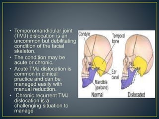
• Temporomandibular joint
(TMJ) dislocation is an
uncommon but debilitating
condition of the facial
skeleton.
• The condition may be
acute or chronic.
• Acute TMJ dislocation is
common in clinical
practice and can be
managed easily with
manual reduction.
• Chronic recurrent TMJ
dislocation is a
challenging situation to
manage
• The temporomandibular
joint (TMJ) is a
specialized joint
between the mandible
and the temporal bone
of the skull.
• The condyle of the
mandible articulates
bilaterally in a concavity
known as the glenoid
fossa or the mandibular
fossa.
• Biomechanics of the TMJ is under
neuromuscular control, comprising-
the muscles of mastication,
the ligaments associated with it, and
neural transmission carried by the
mandibular division of the trigeminal
nerve.
• The most common TMJ dislocation is anterior
dislocation.
• The other types such as-
medial,
 lateral,
superior into the middle cranial fossa,
posterior
are rare and are mostly associated with trauma.
• The pathophysiology of dislocation is the
movement of the condylar process in front of the
articular eminence and an inability to descend
back to its normal position.
• It can be
partial (subluxation) or complete (luxation),
bilateral or unilateral,
acute, and chronic protracted/chronic
recurrent.
Alteration in the
neuromuscular function
• occurs due to
laxity of the articular disc and
the capsular ligament,
 long-standing internal
derangement, and
spasm of the lateral
pterygoid muscles.
 Age and changes in the
dentition also play definite
role in dislocation.
Structural deficit involves
• arthritic changes in the
condyle, i.e.,
 flattening or narrowing,
decrease in the height of the
articular eminence,
morphological changes of
the glenoid fossa, zygomatic
arch, and squamotympanic
fissure.
• It causes over function. i.e.,forceful wide opening of the
mouth while yawning, laughing, vomiting, or seizures,
dental treatments like third molar extractions or root canal
treatments, or oendotracheal intubation, laryngoscopy,
and trans oral fiber optic bronchoscopy.
• Certain antipsychotic medications may also lead to
dislocation.
• Some syndromes are also associated with it such as the
Ehlers-Danlos syndrome, orofacial dystonia, and the Mar
fan syndrome.
• The most common
clinical symptom is the
inability to close the oral
cavity, i.e., “open lock,”
• difficulty in speech,
• drooling of saliva, and
• lip incompetency.
• In acute dislocation,
pain in the pre
auricular region is
present, but chronic
recurrent dislocation is
rarely associated with it.
• Usually bilateral and
at times unilateral
dislocation may lead
to deviation of the
chin to the
contralateral side.
• Palpation over the
preauricular region
may suggest
emptiness in the
joint space.
• The patient may look
anxious
• Clinical history and
examination are the most
important tools in diagnosing
TMJ dislocation.
• Other confirmatory diagnostic
aids include plain and
panoramic radiographies,
showing the location of the
condylar head anterior to the
articular eminence.
• Three-dimensional computed
tomography is the best in
terms of its perfection to show
this entity.
• On the basis of the clinico-radiological
evaluation, TMJ dislocation is classified into the
following three types:
• Type I - the head of the condyle is directly below
the tip of the eminence
• Type II - the head of the condyle is in front of the
tip of the eminence
• Type III - the head of the condyle is high-up in
front of the base of the eminence.
• Acute dislocation
• It is a very painful clinical condition, but easy to manage.
The conservative methods in its management include
symptomatic pain relief with analgesics and manual
reduction
• . Sometimes, the manual reduction is complicated by the
secondary reflex spasm of the lateral pterygoid muscle,
followed by painful stimuli from the joint capsule. As the
condition is very painful, it is always better to perform
manual reduction under local anesthesia.
• Induction of the gag reflex by probing the soft palate
creates a reflex neuromuscular action that resulted in the
reduction.
Temporomandibular joint dislocation

Temporomandibular joint dislocation

  • 2.
    • Temporomandibular joint (TMJ)dislocation is an uncommon but debilitating condition of the facial skeleton. • The condition may be acute or chronic. • Acute TMJ dislocation is common in clinical practice and can be managed easily with manual reduction. • Chronic recurrent TMJ dislocation is a challenging situation to manage
  • 5.
    • The temporomandibular joint(TMJ) is a specialized joint between the mandible and the temporal bone of the skull. • The condyle of the mandible articulates bilaterally in a concavity known as the glenoid fossa or the mandibular fossa.
  • 6.
    • Biomechanics ofthe TMJ is under neuromuscular control, comprising- the muscles of mastication, the ligaments associated with it, and neural transmission carried by the mandibular division of the trigeminal nerve.
  • 7.
    • The mostcommon TMJ dislocation is anterior dislocation. • The other types such as- medial,  lateral, superior into the middle cranial fossa, posterior are rare and are mostly associated with trauma.
  • 8.
    • The pathophysiologyof dislocation is the movement of the condylar process in front of the articular eminence and an inability to descend back to its normal position. • It can be partial (subluxation) or complete (luxation), bilateral or unilateral, acute, and chronic protracted/chronic recurrent.
  • 9.
    Alteration in the neuromuscularfunction • occurs due to laxity of the articular disc and the capsular ligament,  long-standing internal derangement, and spasm of the lateral pterygoid muscles.  Age and changes in the dentition also play definite role in dislocation. Structural deficit involves • arthritic changes in the condyle, i.e.,  flattening or narrowing, decrease in the height of the articular eminence, morphological changes of the glenoid fossa, zygomatic arch, and squamotympanic fissure.
  • 10.
    • It causesover function. i.e.,forceful wide opening of the mouth while yawning, laughing, vomiting, or seizures, dental treatments like third molar extractions or root canal treatments, or oendotracheal intubation, laryngoscopy, and trans oral fiber optic bronchoscopy. • Certain antipsychotic medications may also lead to dislocation. • Some syndromes are also associated with it such as the Ehlers-Danlos syndrome, orofacial dystonia, and the Mar fan syndrome.
  • 11.
    • The mostcommon clinical symptom is the inability to close the oral cavity, i.e., “open lock,” • difficulty in speech, • drooling of saliva, and • lip incompetency. • In acute dislocation, pain in the pre auricular region is present, but chronic recurrent dislocation is rarely associated with it.
  • 12.
    • Usually bilateraland at times unilateral dislocation may lead to deviation of the chin to the contralateral side. • Palpation over the preauricular region may suggest emptiness in the joint space. • The patient may look anxious
  • 13.
    • Clinical historyand examination are the most important tools in diagnosing TMJ dislocation. • Other confirmatory diagnostic aids include plain and panoramic radiographies, showing the location of the condylar head anterior to the articular eminence. • Three-dimensional computed tomography is the best in terms of its perfection to show this entity.
  • 14.
    • On thebasis of the clinico-radiological evaluation, TMJ dislocation is classified into the following three types: • Type I - the head of the condyle is directly below the tip of the eminence • Type II - the head of the condyle is in front of the tip of the eminence • Type III - the head of the condyle is high-up in front of the base of the eminence.
  • 15.
    • Acute dislocation •It is a very painful clinical condition, but easy to manage. The conservative methods in its management include symptomatic pain relief with analgesics and manual reduction
  • 16.
    • . Sometimes,the manual reduction is complicated by the secondary reflex spasm of the lateral pterygoid muscle, followed by painful stimuli from the joint capsule. As the condition is very painful, it is always better to perform manual reduction under local anesthesia. • Induction of the gag reflex by probing the soft palate creates a reflex neuromuscular action that resulted in the reduction.