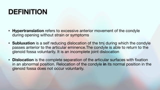
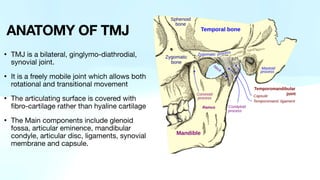
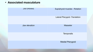
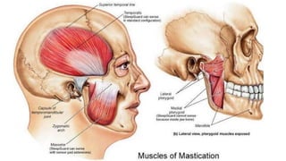
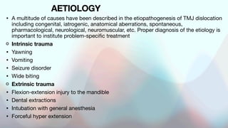
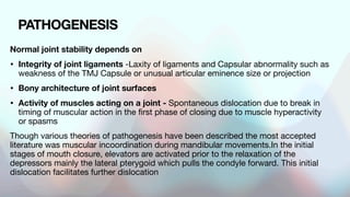
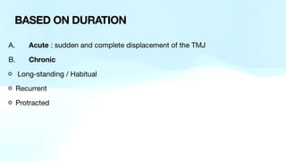
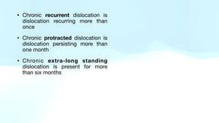
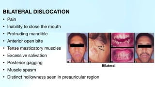
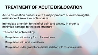
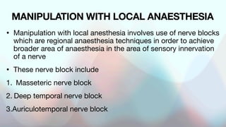
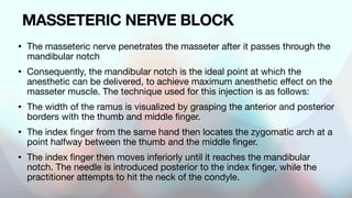
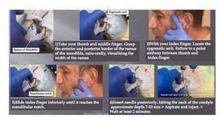
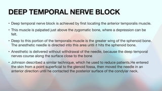
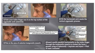
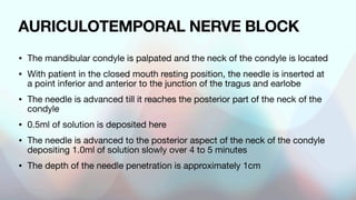
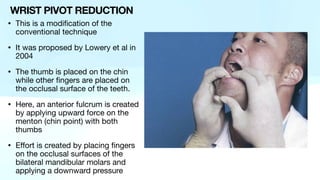
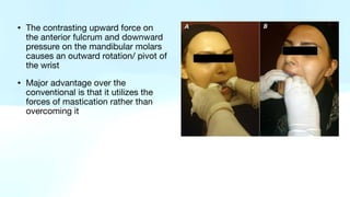
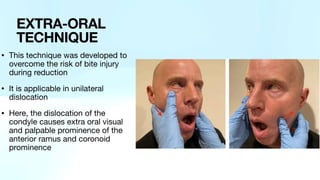
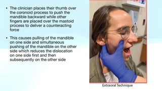
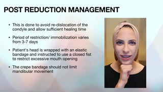
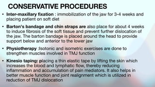
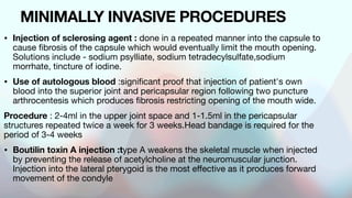
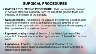
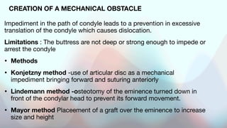
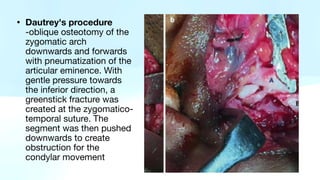
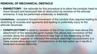
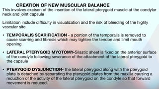
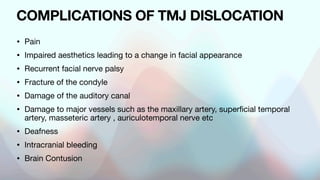
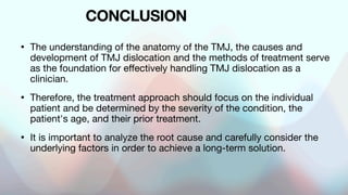
SlideShare a Scribd company logo
DR. BIMMA NWEZE ( HO MFU NHA)
TMJ DISLOCATION
OUTLINE
• INTRODUCTION
• DEFINITION
• EPIDERMOLOGY
• CLASSIFICATION
• AETIOLOGY
• PATHOGENESIS
• CLINICAL PRESENTATION
• DIAGNOSIS
• MANAGEMENT
• COMPLICATIONS
• CONCLUSION
• REFERENCES
• Temporomandibular joint (TMJ)
dislocation is an uncommon but
debilitating condition of the facial
skeleton
• It is the displacement of the
mandibular condyle from the
articular groove of the temporal
bone
• Hippocrates- described
Dislocation and it's treatment in
5th century BC
INTRODUCTION
DEFINITION
• Hypertranslation refers to excessive anterior movement of the condyle
during opening without strain or symptoms
• Subluxation is a self reducing dislocation of the tmj during which the condyle
passes anterior to the articular eminence.The condyle is able to return to the
glenoid fossa voluntarily. It is an incomplete joint dislocation
• Dislocation is the complete separation of the articular surfaces with fixation
in an abnormal position. Relocation of the condyle in its normal position in the
glenoid fossa does not occur voluntarily.
• TMJ is a bilateral, ginglymo-diathrodial,
synovial joint.
• It is a freely mobile joint which allows both
rotational and transitional movement
• The articulating surface is covered with
fibro-cartilage rather than hyaline cartilage
• The Main components include glenoid
fossa, articular eminence, mandibular
condyle, articular disc, ligaments, synovial
membrane and capsule.
ANATOMY OF TMJ
• Ginglymoathrodial joint : Superior component for translation and
Inferior component for rotation
• Mandibular condyle
• Articular cartilage : covered by perichondrium type II collagen
• Articular part of temporal bone : Concave surface of mean thickness
of 0.9mm
• Articular disc : It is composed of fibrocartilaginous tissue and divided
into an anterior zone, intermediate zone and posterior band. The disc
divides the joint into the upper compartment which allows translational
movement and lower compartment allowing rotational movement.
• Synovial cavity
• Synovial fluid
• Ligaments :
A. Functional - Collateral, Capsular , Temporomandibular
B. Accessory ligaments - Sphenomandibular and Stylomandibular
• Vascular supply-
A. Anterior - Superficial temporal and maxillary arteries
B. Posterior -Masseteric artery
• Innervation- Auriculotemporal nerve, masseteric nerve, posterior
deep temporal nerve
• Associated musculature
JAW OPENING Suprahyoid muscles - Rotation
Lateral Pterygoid -Translation
Jaw elevation Masseter
Temporalis
Medial Pterygoid
EPIDERMOLOGY
• Uncommon condition
• Incidence - 3%-7% of Gen population
• More common in females
• Uncommon in extremes of age
• Staz- Incidence of recurrent dislocation of 7% of 240 cases of
TMDs
• More common Bilateral
• Most commonly occurs in the anterior direction
AETIOLOGY
• A multitude of causes have been described in the etiopathogenesis of TMJ dislocation
including congenital, iatrogenic, anatomical aberrations, spontaneous,
pharmacological, neurological, neuromuscular, etc. Proper diagnosis of the etiology is
important to institute problem-specific treatment
Intrinsic trauma
• Yawning
• Vomiting
• Seizure disorder
• Wide biting
Extrinsic trauma
• Flexion-extension injury to the mandible
• Dental extractions
• Intubation with general anesthesia
• Forceful hyper extension
Connective tissue disorders
• Ehlers-Danios syndrome
• Marfan syndrome
Anatomic
• Steep eminence
• Abnormal condylar shape
• Internal derangement
Psychogenic
• Epilepsy
• Parkinson's disease
• Muscular dystrophy
Drug induced: -Extrapyramidal reactions to prochlorperazine -All Antipsychotic drugs
PATHOGENESIS
Normal joint stability depends on
• Integrity of joint ligaments -Laxity of ligaments and Capsular abnormality such as
weakness of the TMJ Capsule or unusual articular eminence size or projection
• Bony architecture of joint surfaces
• Activity of muscles acting on a joint - Spontaneous dislocation due to break in
timing of muscular action in the first phase of closing due to muscle hyperactivity
or spasms
Though various theories of pathogenesis have been described the most accepted
literature was muscular incoordination during mandibular movements.In the initial
stages of mouth closure, elevators are activated prior to the relaxation of the
depressors mainly the lateral pterygoid which pulls the condyle forward. This initial
dislocation facilitates further dislocation
ON THE BASIS OF
• Duration
• Direction of displacement
• Site of displacement
• Clinico-radiological evaluation
CLASSIFICATION
A. Acute : sudden and complete displacement of the TMJ
B. Chronic
Long-standing / Habitual
Recurrent
Protracted
BASED ON DURATION
• Chronic recurrent dislocation is
dislocation recurring more than
once
• Chronic protracted dislocation is
dislocation persisting more than
one month
• Chronic extra-long standing
dislocation is present for more
than six months
• Anterior
• Posterior
• Medial
• Lateral
• Superior
Direction of displacement
• Anterior dislocation is the most
common type of dislocation due to
weakness of the capsule in the
anterior region
• Posterior dislocation usually follows
trauma to the external auditory canal
and skull base
• Superior dislocation results when the
condyle is pushed into the middle
cranial fossa accompanied by glenoid
fossa fracture
• Lateral dislocation is rare and also
associated with high energy trauma
Based on the position of the head
of the condyle to the articular
eminence seen on clinico-
radiological evaluation
Type I - the head of condyle is
directly below the tip of the
eminence
Type II - the head of condyle is in
front of the tip of the eminence
Type Ill -the head of condyle is
high up in front of the base of the
eminence.
BASED ON CLINICO-
RADIOLOGICAL EVALUATION
• UNILATERAL : TMJ dislocation affecting only one side of the jaw.
• BILATERAL : More common . This occurs when there is dislocation of
both TMJs and affecting both sides of the jaw.
POSITION OF DISPLACEMENT
• Deviation of the chin to the contra
lateral side
• Profuse drooling of saliva
• Difficulty in speech
• Difficulty in mastication and
swallowing
• The mouth is partly open and the
affected condyle cannot be
palpable
CLINICAL PRESENTATION
UNILATERAL ACUTE DISLOCATION
BILATERAL DISLOCATION
• Pain
• Inability to close the mouth
• Protruding mandible
• Anterior open bite
• Tense masticatory muscles
• Excessive salivation
• Posterior gagging
• Muscle spasm
• Distinct hollowness seen in preauricular region
• History : to determine cause, onset/
duration.Important to note previous
history of local joint laxity, TMJD or use
of antipsychotic drugs
• Clinical Examination : Neurological and
musculoskeletal disorders
• Investigation : Radiological examination
• Treatment
MANAGEMENT
• Investigations• Orthopantomogram (OPG) (open and closed) This is the
commonly used screening modality for the examination of TMJ. Morphology of
condyle, articular eminence, and joint space can be evaluated. Open mouth OPG
shows the position of the condyle in relation to the articular eminence.•
• TMJ tomogram Open and closed mouth TMJ images can be obtained in different
slices.
• Computed tomography(CT)Evaluation of the morphology of osseous TMJ
compo-nents—condyle, articular eminence and the glenoid fossa—are better
assessed with CT.•
• Cone beam computed tomography (CBCT) facilitates accurate measurement of
condylar height, width, and length as well as inclination of articular eminence.•
• MRI demonstrates the soft tissue morphology, particu-larly disc shape,
displacement, and effusion of the joint frequently associated with dislocation.•
• Electromyography(EMG) evaluates the activity of the muscles which may be
hypoactive, normoactive, or hyperactive.•
• Ultrasonography :Thickness and length of the muscles can be evaluated both at
rest and clench
Diagnosis
Evaluation and treatment methods for TMJ dislocation have
continued to evolve due to varied aetiology and presentations,
as different types of dislocation can result from traumatic and
non-traumatic causes.
To decide the treatment plan, the aetiology and duration of the
dislocation must be understood
TREATMENT OF ACUTE DISLOCATION
Acute dislocation presents with a major problem of overcoming the
resistance of severe muscle spasm.
Immediate attention for relief of pain and anxiety in order to
minimize damage to the joint structure
This can be achieved by
• Manipulation without any form of anaesthesia
• Manipulation with local anaesthesia
• Manipulation under general anesthesia/ sedation with muscle relaxants
MANIPULATION WITH LOCAL ANAESTHESIA
• Manipulation with local anesthesia involves use of nerve blocks
which are regional anaesthesia techniques in order to achieve
broader area of anaesthesia in the area of sensory innervation
of a nerve
• These nerve block include
1. Masseteric nerve block
2. Deep temporal nerve block
3.Auriculotemporal nerve block
MASSETERIC NERVE BLOCK
• The masseteric nerve penetrates the masseter after it passes through the
mandibular notch
• Consequently, the mandibular notch is the ideal point at which the
anesthetic can be delivered, to achieve maximum anesthetic effect on the
masseter muscle. The technique used for this injection is as follows:
• The width of the ramus is visualized by grasping the anterior and posterior
borders with the thumb and middle finger.
• The index finger from the same hand then locates the zygomatic arch at a
point halfway between the thumb and the middle finger.
• The index finger then moves inferiorly until it reaches the mandibular
notch. The needle is introduced posterior to the index finger, while the
practitioner attempts to hit the neck of the condyle.
DEEP TEMPORAL NERVE BLOCK
• Deep temporal nerve block is achieved by
fi
rst locating the anterior temporalis muscle.
• This muscle is palpated just above the zygomatic bone, where a depression can be
felt.
• Deep to this portion of the temporalis muscle is the greater wing of the sphenoid bone.
The anesthetic needle is directed into this area until it hits the sphenoid bone.
• Anesthetic is delivered without withdrawal of the needle, because the deep temporal
nerves course along the surface close to the bone
• Johnson described a similar technique, which he used to reduce patients.He entered
the skin from a point super
fi
cial to the glenoid fossa, then moved the needle in an
anterior direction until he contacted the posterior surface of the condylar neck.
AURICULOTEMPORAL NERVE BLOCK
• The mandibular condyle is palpated and the neck of the condyle is located
• With patient in the closed mouth resting position, the needle is inserted at
a point inferior and anterior to the junction of the tragus and earlobe
• The needle is advanced till it reaches the posterior part of the neck of the
condyle
• 0.5ml of solution is deposited here
• The needle is advanced to the posterior aspect of the neck of the condyle
depositing 1.0ml of solution slowly over 4 to 5 minutes
• The depth of the needle penetration is approximately 1cm
• Conventional technique
( Hippocratic/Nealton's method)
• Wrist pivot technique
• Extra oral technique
• Gag reflex
TECHNIQUES
This is the most widely described and
successful technique
It is a bimanual reduction of dislocated
joints
It may be performed with or without
sedation
This technique involves placing gauze-
wrapped thumbs over the external
oblique ridge of the mandible(or the
molars)
HIPPOCRATES
MANEUVER
• First a downward directed force
is applied to distract the condyle
down the anterior slope of the
eminence
• Followed by a posteriorly/
backward and superior
distracted force to reposition the
condyle past the peak of the
eminence back into its normal
resting position into the glenoid
fossa
• This is a modification of the
conventional technique
• It was proposed by Lowery et al in
2004
• The thumb is placed on the chin
while other fingers are placed on
the occlusal surface of the teeth.
• Here, an anterior fulcrum is created
by applying upward force on the
menton (chin point) with both
thumbs
• Effort is created by placing fingers
on the occlusal surfaces of the
bilateral mandibular molars and
applying a downward pressure
WRIST PIVOT REDUCTION
• The contrasting upward force on
the anterior fulcrum and downward
pressure on the mandibular molars
causes an outward rotation/ pivot of
the wrist
• Major advantage over the
conventional is that it utilizes the
forces of mastication rather than
overcoming it
• This technique was developed to
overcome the risk of bite injury
during reduction
• It is applicable in unilateral
dislocation
• Here, the dislocation of the
condyle causes extra oral visual
and palpable prominence of the
anterior ramus and coronoid
prominence
EXTRA-ORAL
TECHNIQUE
• The clinician places their thumb over
the coronoid process to push the
mandible backward while other
fingers are placed over the mastoid
process to deliver a counteracting
force
• This causes pulling of the mandible
on one side and simultaneous
pushing of the mandible on the other
side which reduces the dislocation
on one side first and then
subsequently on the other side
• Gag reflex is induced by probing the soft palate using a mouth mirror or
tongue depressor
• This reflex relaxes the lateral pterygoid through a series of coordinated
neuromuscular activities likely by relaxing the spastic elevator muscles
while simultaneously triggering the depressor muscles
• This reduces dislocation in a natural way
GAG REFLEX
POST REDUCTION MANAGEMENT
• This is done to avoid re-dislocation of the
condyle and allow sufficient healing time
• Period of restriction/ immobilization varies
from 3-7 days
• Patient's head is wrapped with an elastic
bandage and instructed to use a closed fist
to restrict excessive mouth opening
• The crepe bandage should not limit
mandibular movement
MANAGEMENT OF CHRONIC DISLOCATION
• Chronic recurrent dislocation are repeated episodes of dislocation
where there is abnormal anterior excursion of the condyles beyond the
articular eminence but the patient is able to manipulate back into its
normal position
• The condylar head moves unassisted,forward and backward over the
articular eminence
• The recurrent, incomplete, self reducing, habitual dislocation is termed
hyper mobility or chronic subluxation of the TMJ
• The triad : - ligamentous and capsular flaccidity -eminential erosion
-flattening and trauma
• Seen in the acts of yawning, vomiting, laughing, severe epilepsy,
dystrophia and ehlers-danlos syndrome
• The types of management can be classified
based on the degree of invasiveness
• Conservative methods include
1. Physiotherapy
2. Inter-maxillary fixation
3. Chin straps
4. Kinesio taping
5. Barton's bandage
• Minimally invasive methods
include
1. Injection of sclerosing
agents
2. Autologous blood injection
3. Prolotherapy
4. Boutilin toxin injection
• Surgical procedures
1. Capsular tightening procedure
Capsulorrhaphy
2. Creation of mechanical obstacle
Dautrey's procedure
Glenotemporal osteotomy
3. Removal of mechanical obstacle
Eminectomy
Condylectomy
4. Creation of new muscular balance
Temporalis scarification
Lateral pterygoid myotomy
Pterygoid dysjunction
CONSERVATIVE PROCEDURES
• Inter-maxillary fixation : immobilization of the jaw for 3-4 weeks and
placing patient on soft diet
• Barton's bandage and chin straps are also place for about 4 weeks
to induce fibrosis of the soft tissue and prevent further dislocation of
the jaw. The barton bandage is placed around the head to provide
support below and anterior to the lower jaw
• Physiotherapy :Isotonic and isometric exercises are done to
strengthen muscles involved in TMJ function
• Kinesio taping: placing a thin elastic tape by lifting the skin which
increases the blood and lymphatic flow, thereby reducing
inflammation and accumulation of pain mediators. It also helps in
better muscle function and joint realignment which is utilized in
reduction of TMJ dislocation
KINESIO TAPING
Barton's bandage
MINIMALLY INVASIVE PROCEDURES
• Injection of sclerosing agent : done in a repeated manner into the capsule to
cause fibrosis of the capsule which would eventually limit the mouth opening.
Solutions include - sodium psylliate, sodium tetradecylsulfate,sodium
morrhate, tincture of iodine.
• Use of autologous blood :significant proof that injection of patient's own
blood into the superior joint and pericapsular region following two puncture
arthrocentesis which produces fibrosis restricting opening of the mouth wide.
Procedure : 2-4ml in the upper joint space and 1-1.5ml in the pericapsular
structures repeated twice a week for 3 weeks.Head bandage is required for the
period of 3-4 weeks
• Boutilin toxin A injection :type A weakens the skeletal muscle when injected
by preventing the release of acetylcholine at the neuromuscular junction.
Injection into the lateral pterygoid is the most effective as it produces forward
movement of the condyle
SURGICAL PROCEDURES
CAPSULE TIGHTENING PROCEDURE : TMJ is completely covered
in capsule attached superiorly from the rim of the glenoid fossa and
inferiorly till the neck of the condyle.
• Capsulorrhaphy - Shortening the capsule by removing a section and
suturing it to make it tight. Modifications include suturing of the
capsule to the zygomatic arch or temporalis fasica and overlapping of
the capsule to act as a reinforcement
• Ligamentorraphy - surgical fixation of the lateral ligament of the
capsule to the periosteum of the zygomatic arch followed IMF for one
week
• Limitations : Effective over a short period, Violation of the
intracapsular space causing complications such as hemarthrosis or
degenerative changes.
CREATION OF A MECHANICAL OBSTACLE
Impediment in the path of condyle leads to a prevention in excessive
translation of the condyle which causes dislocation.
Limitations : The buttress are not deep or strong enough to impede or
arrest the condyle
• Methods
• Konjetzny method -use of articular disc as a mechanical
impediment bringing forward and suturing anteriorly
• Lindemann method -osteotomy of the eminence turned down in
front of the condylar head to prevent its forward movement.
• Mayor method Placeement of a graft over the eminence to increase
size and height
• Dautrey's procedure
-oblique osteotomy of the
zygomatic arch
downwards and forwards
with pneumatization of the
articular eminence. With
gentle pressure towards
the inferior direction, a
greenstick fracture was
created at the zygomatico-
temporal suture. The
segment was then pushed
downwards to create
obstruction for the
condylar movement
REMOVAL OF MECHANICAL OBSTACLE
• EMINECTOMY : the rationale for this procedure is to allow the condylar head to
move forward and backward free of obstruction by excision of the articular
eminence. It may be performed unilaterally or bilaterally.
Limitations : excessive forward movement of the condyle than required leading to
stretching of muscles and ligaments and leading to potentially injury to the
articular structures
• CONDYLECTOMY is done by excision of the of the condyle head above the
attachment of the lateral pterygoid muscle.This allows free translation of the
condyle along the articular eminence.It may high or low depending on the
portion (either superior or inferior)of the condyle used.High condylectomy is a
more conservative approach. Condylectomy is the last option as it results in
facial and occlusal deformity.
CREATION OF NEW MUSCULAR BALANCE
This involves excison of the insertion of the lateral pterygoid muscle at the condylar
neck and joint capsule
Limitation include difficulty in visualization and the risk of bleeding of the highly
vascular site
• TEMPORALIS SCARIFICATION - a portion of the temporalis is removed to
cause scarring and fibrosis which may tighten the tendon and limit mouth
opening
• LATERAL PTERYGOID MYOTOMY-Silastic sheet is fixed on the anterior surface
of the condyle following severance of the attachment of the lateral pterygoid to
the capsule
• PTERYGOID DYSJUNCTION- the lateral pterygoid along with the pterygoid
plate is detached by separating the pterygoid plates from the maxilla causing a
reduction of the activity of the lateral pterygoid on the condyle so that forward
movement is reduced.
COMPLICATIONS OF TMJ DISLOCATION
• Pain
• Impaired aesthetics leading to a change in facial appearance
• Recurrent facial nerve palsy
• Fracture of the condyle
• Damage of the auditory canal
• Damage to major vessels such as the maxillary artery, superficial temporal
artery, masseteric artery , auriculotemporal nerve etc
• Deafness
• Intracranial bleeding
• Brain Contusion
CONCLUSION
• The understanding of the anatomy of the TMJ, the causes and
development of TMJ dislocation and the methods of treatment serve
as the foundation for effectively handling TMJ dislocation as a
clinician.
• Therefore, the treatment approach should focus on the individual
patient and be determined by the severity of the condition, the
patient's age, and their prior treatment.
• It is important to analyze the root cause and carefully consider the
underlying factors in order to achieve a long-term solution.
REFERENCES
• Krishnakumar Raja, V.B. (2021). Temporomandibular dislocation.
• Okeson, J.P. (2021). Management of temporomandibular disorders and
occlusion (6th ed.).
• Shorey, C.W., Campbell, J.H., et al. (2000). Dislocation of the
temporomandibular joint. Oral med Oral path Oral Radiol, 89, 662-668.
• Fonesca, R.J., Marcani, R.D., Turvey, T.A. (Year). Oral and Maxillofacial
surgery (2nd ed.).
MANAGEMENT OF TEMPEROMANDIBULAR JOINT DISLOCATION

More Related Content

Similar to MANAGEMENT OF TEMPEROMANDIBULAR JOINT DISLOCATION

condylar dislocation- etiopathogenesis & treatment
condylar dislocation- etiopathogenesis & treatmentcondylar dislocation- etiopathogenesis & treatment
condylar dislocation- etiopathogenesis & treatment
AshokKp4
 
Disorders of TMJ
Disorders of TMJDisorders of TMJ
Disorders of TMJ
Dr Nandika Babele
 
TMJ DISORDERS - PART 2.ppt
TMJ DISORDERS - PART 2.pptTMJ DISORDERS - PART 2.ppt
TMJ DISORDERS - PART 2.ppt
DentalYoutube
 
BASICS OF Temporomandibular joint..ppttt
BASICS OF Temporomandibular joint..pptttBASICS OF Temporomandibular joint..ppttt
BASICS OF Temporomandibular joint..ppttt
aknawaz5591
 
Tandem stenosis
Tandem stenosisTandem stenosis
Tandem stenosis
Mirant Dave
 
Temporomandibular joint ankylosis and its management
Temporomandibular joint ankylosis and its managementTemporomandibular joint ankylosis and its management
Temporomandibular joint ankylosis and its management
Dibya Falgoon Sarkar
 
CURRENT MANAGEMENT OF ANKLE INJURIES.pptx
CURRENT MANAGEMENT OF ANKLE INJURIES.pptxCURRENT MANAGEMENT OF ANKLE INJURIES.pptx
CURRENT MANAGEMENT OF ANKLE INJURIES.pptx
EnejoJoseph
 
Myositis ossificans
Myositis ossificansMyositis ossificans
Myositis ossificans
Ponnilavan Ponz
 
TMD-FL.pdf
TMD-FL.pdfTMD-FL.pdf
TMD-FL.pdf
SamarElgendi
 
TMJ.pptx
TMJ.pptxTMJ.pptx
TMJ.pptx
AmjadShalan1
 
Neck of Femur, IT and Subtrochanteric fracture- Dr Sundar Ortho.pptx
Neck of Femur, IT and Subtrochanteric fracture- Dr Sundar Ortho.pptxNeck of Femur, IT and Subtrochanteric fracture- Dr Sundar Ortho.pptx
Neck of Femur, IT and Subtrochanteric fracture- Dr Sundar Ortho.pptx
Dr. Sundar Karki
 
Subluxation and dislocation of temporo mandibular joint
Subluxation and dislocation of temporo mandibular jointSubluxation and dislocation of temporo mandibular joint
Subluxation and dislocation of temporo mandibular joint
Dr. Akshay Shah
 
Tmj final.pptx...........................
Tmj final.pptx...........................Tmj final.pptx...........................
Tmj final.pptx...........................
anweshagarg49
 
Humeral shaft fractures
Humeral shaft fracturesHumeral shaft fractures
Humeral shaft fractures
Supun Dhanasekara
 
shaharukh ahamd
shaharukh ahamdshaharukh ahamd
shaharukh ahamd
Shahrukh Ahamd
 
Syndromic cranial synostosis
Syndromic cranial synostosisSyndromic cranial synostosis
Syndromic cranial synostosis
Jamil Kifayatullah
 
Surgical approach intercondylar/ supracondylar humerus fracture
Surgical approach intercondylar/ supracondylar humerus fractureSurgical approach intercondylar/ supracondylar humerus fracture
Surgical approach intercondylar/ supracondylar humerus fracture
Khadijah Nordin
 
MUSCLES OF MASTICATION.pptx
MUSCLES OF MASTICATION.pptxMUSCLES OF MASTICATION.pptx
MUSCLES OF MASTICATION.pptx
Dr K. Abhilasha
 
Mandible fracture symposium march-13
Mandible fracture   symposium march-13Mandible fracture   symposium march-13
Mandible fracture symposium march-13
Narendra Markad
 
MANDIBULAR FRACTURES MANAGEMENT PROTOCO
MANDIBULAR  FRACTURES MANAGEMENT PROTOCOMANDIBULAR  FRACTURES MANAGEMENT PROTOCO
MANDIBULAR FRACTURES MANAGEMENT PROTOCO
EUROUNDISA
 

Similar to MANAGEMENT OF TEMPEROMANDIBULAR JOINT DISLOCATION (20)

condylar dislocation- etiopathogenesis & treatment
condylar dislocation- etiopathogenesis & treatmentcondylar dislocation- etiopathogenesis & treatment
condylar dislocation- etiopathogenesis & treatment
 
Disorders of TMJ
Disorders of TMJDisorders of TMJ
Disorders of TMJ
 
TMJ DISORDERS - PART 2.ppt
TMJ DISORDERS - PART 2.pptTMJ DISORDERS - PART 2.ppt
TMJ DISORDERS - PART 2.ppt
 
BASICS OF Temporomandibular joint..ppttt
BASICS OF Temporomandibular joint..pptttBASICS OF Temporomandibular joint..ppttt
BASICS OF Temporomandibular joint..ppttt
 
Tandem stenosis
Tandem stenosisTandem stenosis
Tandem stenosis
 
Temporomandibular joint ankylosis and its management
Temporomandibular joint ankylosis and its managementTemporomandibular joint ankylosis and its management
Temporomandibular joint ankylosis and its management
 
CURRENT MANAGEMENT OF ANKLE INJURIES.pptx
CURRENT MANAGEMENT OF ANKLE INJURIES.pptxCURRENT MANAGEMENT OF ANKLE INJURIES.pptx
CURRENT MANAGEMENT OF ANKLE INJURIES.pptx
 
Myositis ossificans
Myositis ossificansMyositis ossificans
Myositis ossificans
 
TMD-FL.pdf
TMD-FL.pdfTMD-FL.pdf
TMD-FL.pdf
 
TMJ.pptx
TMJ.pptxTMJ.pptx
TMJ.pptx
 
Neck of Femur, IT and Subtrochanteric fracture- Dr Sundar Ortho.pptx
Neck of Femur, IT and Subtrochanteric fracture- Dr Sundar Ortho.pptxNeck of Femur, IT and Subtrochanteric fracture- Dr Sundar Ortho.pptx
Neck of Femur, IT and Subtrochanteric fracture- Dr Sundar Ortho.pptx
 
Subluxation and dislocation of temporo mandibular joint
Subluxation and dislocation of temporo mandibular jointSubluxation and dislocation of temporo mandibular joint
Subluxation and dislocation of temporo mandibular joint
 
Tmj final.pptx...........................
Tmj final.pptx...........................Tmj final.pptx...........................
Tmj final.pptx...........................
 
Humeral shaft fractures
Humeral shaft fracturesHumeral shaft fractures
Humeral shaft fractures
 
shaharukh ahamd
shaharukh ahamdshaharukh ahamd
shaharukh ahamd
 
Syndromic cranial synostosis
Syndromic cranial synostosisSyndromic cranial synostosis
Syndromic cranial synostosis
 
Surgical approach intercondylar/ supracondylar humerus fracture
Surgical approach intercondylar/ supracondylar humerus fractureSurgical approach intercondylar/ supracondylar humerus fracture
Surgical approach intercondylar/ supracondylar humerus fracture
 
MUSCLES OF MASTICATION.pptx
MUSCLES OF MASTICATION.pptxMUSCLES OF MASTICATION.pptx
MUSCLES OF MASTICATION.pptx
 
Mandible fracture symposium march-13
Mandible fracture   symposium march-13Mandible fracture   symposium march-13
Mandible fracture symposium march-13
 
MANDIBULAR FRACTURES MANAGEMENT PROTOCO
MANDIBULAR  FRACTURES MANAGEMENT PROTOCOMANDIBULAR  FRACTURES MANAGEMENT PROTOCO
MANDIBULAR FRACTURES MANAGEMENT PROTOCO
 

Recently uploaded

CHEMOTHERAPY_RDP_CHAPTER 6_Anti Malarial Drugs.pdf
CHEMOTHERAPY_RDP_CHAPTER 6_Anti Malarial Drugs.pdfCHEMOTHERAPY_RDP_CHAPTER 6_Anti Malarial Drugs.pdf
CHEMOTHERAPY_RDP_CHAPTER 6_Anti Malarial Drugs.pdf
rishi2789
 
Post-Menstrual Smell- When to Suspect Vaginitis.pptx
Post-Menstrual Smell- When to Suspect Vaginitis.pptxPost-Menstrual Smell- When to Suspect Vaginitis.pptx
Post-Menstrual Smell- When to Suspect Vaginitis.pptx
FFragrant
 
SENSORY NEEDS B.SC. NURSING SEMESTER II.
SENSORY NEEDS B.SC. NURSING SEMESTER II.SENSORY NEEDS B.SC. NURSING SEMESTER II.
SENSORY NEEDS B.SC. NURSING SEMESTER II.
KULDEEP VYAS
 
vonoprazan A novel drug for GERD presentation
vonoprazan A novel drug for GERD presentationvonoprazan A novel drug for GERD presentation
vonoprazan A novel drug for GERD presentation
Dr.pavithra Anandan
 
NARCOTICS- POLICY AND PROCEDURES FOR ITS USE
NARCOTICS- POLICY AND PROCEDURES FOR ITS USENARCOTICS- POLICY AND PROCEDURES FOR ITS USE
NARCOTICS- POLICY AND PROCEDURES FOR ITS USE
Dr. Ahana Haroon
 
LOW BIRTH WEIGHT. PRETERM BABIES OR SMALL FOR DATES BABIES
LOW BIRTH WEIGHT. PRETERM BABIES OR SMALL FOR DATES BABIESLOW BIRTH WEIGHT. PRETERM BABIES OR SMALL FOR DATES BABIES
LOW BIRTH WEIGHT. PRETERM BABIES OR SMALL FOR DATES BABIES
ShraddhaTamshettiwar
 
Demystifying Fallopian Tube Blockage- Grading the Differences and Implication...
Demystifying Fallopian Tube Blockage- Grading the Differences and Implication...Demystifying Fallopian Tube Blockage- Grading the Differences and Implication...
Demystifying Fallopian Tube Blockage- Grading the Differences and Implication...
FFragrant
 
CHEMOTHERAPY_RDP_CHAPTER 2 _LEPROSY.pdf1
CHEMOTHERAPY_RDP_CHAPTER 2 _LEPROSY.pdf1CHEMOTHERAPY_RDP_CHAPTER 2 _LEPROSY.pdf1
CHEMOTHERAPY_RDP_CHAPTER 2 _LEPROSY.pdf1
rishi2789
 
Lecture 6 -- Memory 2015.pptlearning occurs when a stimulus (unconditioned st...
Lecture 6 -- Memory 2015.pptlearning occurs when a stimulus (unconditioned st...Lecture 6 -- Memory 2015.pptlearning occurs when a stimulus (unconditioned st...
Lecture 6 -- Memory 2015.pptlearning occurs when a stimulus (unconditioned st...
AyushGadhvi1
 
Cell Therapy Expansion and Challenges in Autoimmune Disease
Cell Therapy Expansion and Challenges in Autoimmune DiseaseCell Therapy Expansion and Challenges in Autoimmune Disease
Cell Therapy Expansion and Challenges in Autoimmune Disease
Health Advances
 
How to choose the best dermatologists in Indore.
How to choose the best dermatologists in Indore.How to choose the best dermatologists in Indore.
How to choose the best dermatologists in Indore.
Gokuldas Hospital
 
Know the difference between Endodontics and Orthodontics.
Know the difference between Endodontics and Orthodontics.Know the difference between Endodontics and Orthodontics.
Know the difference between Endodontics and Orthodontics.
Gokuldas Hospital
 
Recent advances on Cervical cancer .pptx
Recent advances on Cervical cancer .pptxRecent advances on Cervical cancer .pptx
Recent advances on Cervical cancer .pptx
DrGirishJHoogar
 
NAVIGATING THE HORIZONS OF TIME LAPSE EMBRYO MONITORING.pdf
NAVIGATING THE HORIZONS OF TIME LAPSE EMBRYO MONITORING.pdfNAVIGATING THE HORIZONS OF TIME LAPSE EMBRYO MONITORING.pdf
NAVIGATING THE HORIZONS OF TIME LAPSE EMBRYO MONITORING.pdf
Rahul Sen
 
Nano-gold for Cancer Therapy chemistry investigatory project
Nano-gold for Cancer Therapy chemistry investigatory projectNano-gold for Cancer Therapy chemistry investigatory project
Nano-gold for Cancer Therapy chemistry investigatory project
SIVAVINAYAKPK
 
CHEMOTHERAPY_RDP_CHAPTER 3_ANTIFUNGAL AGENT.pdf
CHEMOTHERAPY_RDP_CHAPTER 3_ANTIFUNGAL AGENT.pdfCHEMOTHERAPY_RDP_CHAPTER 3_ANTIFUNGAL AGENT.pdf
CHEMOTHERAPY_RDP_CHAPTER 3_ANTIFUNGAL AGENT.pdf
rishi2789
 
pharmacology for dummies free pdf download.pdf
pharmacology for dummies free pdf download.pdfpharmacology for dummies free pdf download.pdf
pharmacology for dummies free pdf download.pdf
KerlynIgnacio
 
June 2024 Oncology Cartoons By Dr Kanhu Charan Patro
June 2024 Oncology Cartoons By Dr Kanhu Charan PatroJune 2024 Oncology Cartoons By Dr Kanhu Charan Patro
June 2024 Oncology Cartoons By Dr Kanhu Charan Patro
Kanhu Charan
 
CHEMOTHERAPY_RDP_CHAPTER 4_ANTI VIRAL DRUGS.pdf
CHEMOTHERAPY_RDP_CHAPTER 4_ANTI VIRAL DRUGS.pdfCHEMOTHERAPY_RDP_CHAPTER 4_ANTI VIRAL DRUGS.pdf
CHEMOTHERAPY_RDP_CHAPTER 4_ANTI VIRAL DRUGS.pdf
rishi2789
 
Cosmetology and Trichology Courses at Kosmoderma Academy PRP (Hair), DR Growt...
Cosmetology and Trichology Courses at Kosmoderma Academy PRP (Hair), DR Growt...Cosmetology and Trichology Courses at Kosmoderma Academy PRP (Hair), DR Growt...
Cosmetology and Trichology Courses at Kosmoderma Academy PRP (Hair), DR Growt...
Kosmoderma Academy Of Aesthetic Medicine
 

Recently uploaded (20)

CHEMOTHERAPY_RDP_CHAPTER 6_Anti Malarial Drugs.pdf
CHEMOTHERAPY_RDP_CHAPTER 6_Anti Malarial Drugs.pdfCHEMOTHERAPY_RDP_CHAPTER 6_Anti Malarial Drugs.pdf
CHEMOTHERAPY_RDP_CHAPTER 6_Anti Malarial Drugs.pdf
 
Post-Menstrual Smell- When to Suspect Vaginitis.pptx
Post-Menstrual Smell- When to Suspect Vaginitis.pptxPost-Menstrual Smell- When to Suspect Vaginitis.pptx
Post-Menstrual Smell- When to Suspect Vaginitis.pptx
 
SENSORY NEEDS B.SC. NURSING SEMESTER II.
SENSORY NEEDS B.SC. NURSING SEMESTER II.SENSORY NEEDS B.SC. NURSING SEMESTER II.
SENSORY NEEDS B.SC. NURSING SEMESTER II.
 
vonoprazan A novel drug for GERD presentation
vonoprazan A novel drug for GERD presentationvonoprazan A novel drug for GERD presentation
vonoprazan A novel drug for GERD presentation
 
NARCOTICS- POLICY AND PROCEDURES FOR ITS USE
NARCOTICS- POLICY AND PROCEDURES FOR ITS USENARCOTICS- POLICY AND PROCEDURES FOR ITS USE
NARCOTICS- POLICY AND PROCEDURES FOR ITS USE
 
LOW BIRTH WEIGHT. PRETERM BABIES OR SMALL FOR DATES BABIES
LOW BIRTH WEIGHT. PRETERM BABIES OR SMALL FOR DATES BABIESLOW BIRTH WEIGHT. PRETERM BABIES OR SMALL FOR DATES BABIES
LOW BIRTH WEIGHT. PRETERM BABIES OR SMALL FOR DATES BABIES
 
Demystifying Fallopian Tube Blockage- Grading the Differences and Implication...
Demystifying Fallopian Tube Blockage- Grading the Differences and Implication...Demystifying Fallopian Tube Blockage- Grading the Differences and Implication...
Demystifying Fallopian Tube Blockage- Grading the Differences and Implication...
 
CHEMOTHERAPY_RDP_CHAPTER 2 _LEPROSY.pdf1
CHEMOTHERAPY_RDP_CHAPTER 2 _LEPROSY.pdf1CHEMOTHERAPY_RDP_CHAPTER 2 _LEPROSY.pdf1
CHEMOTHERAPY_RDP_CHAPTER 2 _LEPROSY.pdf1
 
Lecture 6 -- Memory 2015.pptlearning occurs when a stimulus (unconditioned st...
Lecture 6 -- Memory 2015.pptlearning occurs when a stimulus (unconditioned st...Lecture 6 -- Memory 2015.pptlearning occurs when a stimulus (unconditioned st...
Lecture 6 -- Memory 2015.pptlearning occurs when a stimulus (unconditioned st...
 
Cell Therapy Expansion and Challenges in Autoimmune Disease
Cell Therapy Expansion and Challenges in Autoimmune DiseaseCell Therapy Expansion and Challenges in Autoimmune Disease
Cell Therapy Expansion and Challenges in Autoimmune Disease
 
How to choose the best dermatologists in Indore.
How to choose the best dermatologists in Indore.How to choose the best dermatologists in Indore.
How to choose the best dermatologists in Indore.
 
Know the difference between Endodontics and Orthodontics.
Know the difference between Endodontics and Orthodontics.Know the difference between Endodontics and Orthodontics.
Know the difference between Endodontics and Orthodontics.
 
Recent advances on Cervical cancer .pptx
Recent advances on Cervical cancer .pptxRecent advances on Cervical cancer .pptx
Recent advances on Cervical cancer .pptx
 
NAVIGATING THE HORIZONS OF TIME LAPSE EMBRYO MONITORING.pdf
NAVIGATING THE HORIZONS OF TIME LAPSE EMBRYO MONITORING.pdfNAVIGATING THE HORIZONS OF TIME LAPSE EMBRYO MONITORING.pdf
NAVIGATING THE HORIZONS OF TIME LAPSE EMBRYO MONITORING.pdf
 
Nano-gold for Cancer Therapy chemistry investigatory project
Nano-gold for Cancer Therapy chemistry investigatory projectNano-gold for Cancer Therapy chemistry investigatory project
Nano-gold for Cancer Therapy chemistry investigatory project
 
CHEMOTHERAPY_RDP_CHAPTER 3_ANTIFUNGAL AGENT.pdf
CHEMOTHERAPY_RDP_CHAPTER 3_ANTIFUNGAL AGENT.pdfCHEMOTHERAPY_RDP_CHAPTER 3_ANTIFUNGAL AGENT.pdf
CHEMOTHERAPY_RDP_CHAPTER 3_ANTIFUNGAL AGENT.pdf
 
pharmacology for dummies free pdf download.pdf
pharmacology for dummies free pdf download.pdfpharmacology for dummies free pdf download.pdf
pharmacology for dummies free pdf download.pdf
 
June 2024 Oncology Cartoons By Dr Kanhu Charan Patro
June 2024 Oncology Cartoons By Dr Kanhu Charan PatroJune 2024 Oncology Cartoons By Dr Kanhu Charan Patro
June 2024 Oncology Cartoons By Dr Kanhu Charan Patro
 
CHEMOTHERAPY_RDP_CHAPTER 4_ANTI VIRAL DRUGS.pdf
CHEMOTHERAPY_RDP_CHAPTER 4_ANTI VIRAL DRUGS.pdfCHEMOTHERAPY_RDP_CHAPTER 4_ANTI VIRAL DRUGS.pdf
CHEMOTHERAPY_RDP_CHAPTER 4_ANTI VIRAL DRUGS.pdf
 
Cosmetology and Trichology Courses at Kosmoderma Academy PRP (Hair), DR Growt...
Cosmetology and Trichology Courses at Kosmoderma Academy PRP (Hair), DR Growt...Cosmetology and Trichology Courses at Kosmoderma Academy PRP (Hair), DR Growt...
Cosmetology and Trichology Courses at Kosmoderma Academy PRP (Hair), DR Growt...
 

MANAGEMENT OF TEMPEROMANDIBULAR JOINT DISLOCATION

  • 1. DR. BIMMA NWEZE ( HO MFU NHA) TMJ DISLOCATION
  • 2. OUTLINE • INTRODUCTION • DEFINITION • EPIDERMOLOGY • CLASSIFICATION • AETIOLOGY • PATHOGENESIS • CLINICAL PRESENTATION • DIAGNOSIS • MANAGEMENT • COMPLICATIONS • CONCLUSION • REFERENCES
  • 3. • Temporomandibular joint (TMJ) dislocation is an uncommon but debilitating condition of the facial skeleton • It is the displacement of the mandibular condyle from the articular groove of the temporal bone • Hippocrates- described Dislocation and it's treatment in 5th century BC INTRODUCTION
  • 4. DEFINITION • Hypertranslation refers to excessive anterior movement of the condyle during opening without strain or symptoms • Subluxation is a self reducing dislocation of the tmj during which the condyle passes anterior to the articular eminence.The condyle is able to return to the glenoid fossa voluntarily. It is an incomplete joint dislocation • Dislocation is the complete separation of the articular surfaces with fixation in an abnormal position. Relocation of the condyle in its normal position in the glenoid fossa does not occur voluntarily.
  • 5. • TMJ is a bilateral, ginglymo-diathrodial, synovial joint. • It is a freely mobile joint which allows both rotational and transitional movement • The articulating surface is covered with fibro-cartilage rather than hyaline cartilage • The Main components include glenoid fossa, articular eminence, mandibular condyle, articular disc, ligaments, synovial membrane and capsule. ANATOMY OF TMJ
  • 6. • Ginglymoathrodial joint : Superior component for translation and Inferior component for rotation • Mandibular condyle • Articular cartilage : covered by perichondrium type II collagen • Articular part of temporal bone : Concave surface of mean thickness of 0.9mm • Articular disc : It is composed of fibrocartilaginous tissue and divided into an anterior zone, intermediate zone and posterior band. The disc divides the joint into the upper compartment which allows translational movement and lower compartment allowing rotational movement. • Synovial cavity • Synovial fluid
  • 7. • Ligaments : A. Functional - Collateral, Capsular , Temporomandibular B. Accessory ligaments - Sphenomandibular and Stylomandibular • Vascular supply- A. Anterior - Superficial temporal and maxillary arteries B. Posterior -Masseteric artery • Innervation- Auriculotemporal nerve, masseteric nerve, posterior deep temporal nerve
  • 8. • Associated musculature JAW OPENING Suprahyoid muscles - Rotation Lateral Pterygoid -Translation Jaw elevation Masseter Temporalis Medial Pterygoid
  • 9.
  • 10. EPIDERMOLOGY • Uncommon condition • Incidence - 3%-7% of Gen population • More common in females • Uncommon in extremes of age • Staz- Incidence of recurrent dislocation of 7% of 240 cases of TMDs • More common Bilateral • Most commonly occurs in the anterior direction
  • 11. AETIOLOGY • A multitude of causes have been described in the etiopathogenesis of TMJ dislocation including congenital, iatrogenic, anatomical aberrations, spontaneous, pharmacological, neurological, neuromuscular, etc. Proper diagnosis of the etiology is important to institute problem-specific treatment Intrinsic trauma • Yawning • Vomiting • Seizure disorder • Wide biting Extrinsic trauma • Flexion-extension injury to the mandible • Dental extractions • Intubation with general anesthesia • Forceful hyper extension
  • 12. Connective tissue disorders • Ehlers-Danios syndrome • Marfan syndrome Anatomic • Steep eminence • Abnormal condylar shape • Internal derangement Psychogenic • Epilepsy • Parkinson's disease • Muscular dystrophy Drug induced: -Extrapyramidal reactions to prochlorperazine -All Antipsychotic drugs
  • 13. PATHOGENESIS Normal joint stability depends on • Integrity of joint ligaments -Laxity of ligaments and Capsular abnormality such as weakness of the TMJ Capsule or unusual articular eminence size or projection • Bony architecture of joint surfaces • Activity of muscles acting on a joint - Spontaneous dislocation due to break in timing of muscular action in the first phase of closing due to muscle hyperactivity or spasms Though various theories of pathogenesis have been described the most accepted literature was muscular incoordination during mandibular movements.In the initial stages of mouth closure, elevators are activated prior to the relaxation of the depressors mainly the lateral pterygoid which pulls the condyle forward. This initial dislocation facilitates further dislocation
  • 14. ON THE BASIS OF • Duration • Direction of displacement • Site of displacement • Clinico-radiological evaluation CLASSIFICATION
  • 15. A. Acute : sudden and complete displacement of the TMJ B. Chronic Long-standing / Habitual Recurrent Protracted BASED ON DURATION
  • 16. • Chronic recurrent dislocation is dislocation recurring more than once • Chronic protracted dislocation is dislocation persisting more than one month • Chronic extra-long standing dislocation is present for more than six months
  • 17. • Anterior • Posterior • Medial • Lateral • Superior Direction of displacement
  • 18. • Anterior dislocation is the most common type of dislocation due to weakness of the capsule in the anterior region • Posterior dislocation usually follows trauma to the external auditory canal and skull base • Superior dislocation results when the condyle is pushed into the middle cranial fossa accompanied by glenoid fossa fracture • Lateral dislocation is rare and also associated with high energy trauma
  • 19. Based on the position of the head of the condyle to the articular eminence seen on clinico- radiological evaluation Type I - the head of condyle is directly below the tip of the eminence Type II - the head of condyle is in front of the tip of the eminence Type Ill -the head of condyle is high up in front of the base of the eminence. BASED ON CLINICO- RADIOLOGICAL EVALUATION
  • 20. • UNILATERAL : TMJ dislocation affecting only one side of the jaw. • BILATERAL : More common . This occurs when there is dislocation of both TMJs and affecting both sides of the jaw. POSITION OF DISPLACEMENT
  • 21. • Deviation of the chin to the contra lateral side • Profuse drooling of saliva • Difficulty in speech • Difficulty in mastication and swallowing • The mouth is partly open and the affected condyle cannot be palpable CLINICAL PRESENTATION UNILATERAL ACUTE DISLOCATION
  • 22. BILATERAL DISLOCATION • Pain • Inability to close the mouth • Protruding mandible • Anterior open bite • Tense masticatory muscles • Excessive salivation • Posterior gagging • Muscle spasm • Distinct hollowness seen in preauricular region
  • 23. • History : to determine cause, onset/ duration.Important to note previous history of local joint laxity, TMJD or use of antipsychotic drugs • Clinical Examination : Neurological and musculoskeletal disorders • Investigation : Radiological examination • Treatment MANAGEMENT
  • 24. • Investigations• Orthopantomogram (OPG) (open and closed) This is the commonly used screening modality for the examination of TMJ. Morphology of condyle, articular eminence, and joint space can be evaluated. Open mouth OPG shows the position of the condyle in relation to the articular eminence.• • TMJ tomogram Open and closed mouth TMJ images can be obtained in different slices. • Computed tomography(CT)Evaluation of the morphology of osseous TMJ compo-nents—condyle, articular eminence and the glenoid fossa—are better assessed with CT.• • Cone beam computed tomography (CBCT) facilitates accurate measurement of condylar height, width, and length as well as inclination of articular eminence.• • MRI demonstrates the soft tissue morphology, particu-larly disc shape, displacement, and effusion of the joint frequently associated with dislocation.• • Electromyography(EMG) evaluates the activity of the muscles which may be hypoactive, normoactive, or hyperactive.• • Ultrasonography :Thickness and length of the muscles can be evaluated both at rest and clench
  • 25.
  • 26. Diagnosis Evaluation and treatment methods for TMJ dislocation have continued to evolve due to varied aetiology and presentations, as different types of dislocation can result from traumatic and non-traumatic causes. To decide the treatment plan, the aetiology and duration of the dislocation must be understood
  • 27. TREATMENT OF ACUTE DISLOCATION Acute dislocation presents with a major problem of overcoming the resistance of severe muscle spasm. Immediate attention for relief of pain and anxiety in order to minimize damage to the joint structure This can be achieved by • Manipulation without any form of anaesthesia • Manipulation with local anaesthesia • Manipulation under general anesthesia/ sedation with muscle relaxants
  • 28. MANIPULATION WITH LOCAL ANAESTHESIA • Manipulation with local anesthesia involves use of nerve blocks which are regional anaesthesia techniques in order to achieve broader area of anaesthesia in the area of sensory innervation of a nerve • These nerve block include 1. Masseteric nerve block 2. Deep temporal nerve block 3.Auriculotemporal nerve block
  • 29. MASSETERIC NERVE BLOCK • The masseteric nerve penetrates the masseter after it passes through the mandibular notch • Consequently, the mandibular notch is the ideal point at which the anesthetic can be delivered, to achieve maximum anesthetic effect on the masseter muscle. The technique used for this injection is as follows: • The width of the ramus is visualized by grasping the anterior and posterior borders with the thumb and middle finger. • The index finger from the same hand then locates the zygomatic arch at a point halfway between the thumb and the middle finger. • The index finger then moves inferiorly until it reaches the mandibular notch. The needle is introduced posterior to the index finger, while the practitioner attempts to hit the neck of the condyle.
  • 30.
  • 31. DEEP TEMPORAL NERVE BLOCK • Deep temporal nerve block is achieved by fi rst locating the anterior temporalis muscle. • This muscle is palpated just above the zygomatic bone, where a depression can be felt. • Deep to this portion of the temporalis muscle is the greater wing of the sphenoid bone. The anesthetic needle is directed into this area until it hits the sphenoid bone. • Anesthetic is delivered without withdrawal of the needle, because the deep temporal nerves course along the surface close to the bone • Johnson described a similar technique, which he used to reduce patients.He entered the skin from a point super fi cial to the glenoid fossa, then moved the needle in an anterior direction until he contacted the posterior surface of the condylar neck.
  • 32.
  • 33. AURICULOTEMPORAL NERVE BLOCK • The mandibular condyle is palpated and the neck of the condyle is located • With patient in the closed mouth resting position, the needle is inserted at a point inferior and anterior to the junction of the tragus and earlobe • The needle is advanced till it reaches the posterior part of the neck of the condyle • 0.5ml of solution is deposited here • The needle is advanced to the posterior aspect of the neck of the condyle depositing 1.0ml of solution slowly over 4 to 5 minutes • The depth of the needle penetration is approximately 1cm
  • 34.
  • 35. • Conventional technique ( Hippocratic/Nealton's method) • Wrist pivot technique • Extra oral technique • Gag reflex TECHNIQUES
  • 36. This is the most widely described and successful technique It is a bimanual reduction of dislocated joints It may be performed with or without sedation This technique involves placing gauze- wrapped thumbs over the external oblique ridge of the mandible(or the molars) HIPPOCRATES MANEUVER
  • 37. • First a downward directed force is applied to distract the condyle down the anterior slope of the eminence • Followed by a posteriorly/ backward and superior distracted force to reposition the condyle past the peak of the eminence back into its normal resting position into the glenoid fossa
  • 38. • This is a modification of the conventional technique • It was proposed by Lowery et al in 2004 • The thumb is placed on the chin while other fingers are placed on the occlusal surface of the teeth. • Here, an anterior fulcrum is created by applying upward force on the menton (chin point) with both thumbs • Effort is created by placing fingers on the occlusal surfaces of the bilateral mandibular molars and applying a downward pressure WRIST PIVOT REDUCTION
  • 39. • The contrasting upward force on the anterior fulcrum and downward pressure on the mandibular molars causes an outward rotation/ pivot of the wrist • Major advantage over the conventional is that it utilizes the forces of mastication rather than overcoming it
  • 40. • This technique was developed to overcome the risk of bite injury during reduction • It is applicable in unilateral dislocation • Here, the dislocation of the condyle causes extra oral visual and palpable prominence of the anterior ramus and coronoid prominence EXTRA-ORAL TECHNIQUE
  • 41. • The clinician places their thumb over the coronoid process to push the mandible backward while other fingers are placed over the mastoid process to deliver a counteracting force • This causes pulling of the mandible on one side and simultaneous pushing of the mandible on the other side which reduces the dislocation on one side first and then subsequently on the other side
  • 42. • Gag reflex is induced by probing the soft palate using a mouth mirror or tongue depressor • This reflex relaxes the lateral pterygoid through a series of coordinated neuromuscular activities likely by relaxing the spastic elevator muscles while simultaneously triggering the depressor muscles • This reduces dislocation in a natural way GAG REFLEX
  • 43. POST REDUCTION MANAGEMENT • This is done to avoid re-dislocation of the condyle and allow sufficient healing time • Period of restriction/ immobilization varies from 3-7 days • Patient's head is wrapped with an elastic bandage and instructed to use a closed fist to restrict excessive mouth opening • The crepe bandage should not limit mandibular movement
  • 44. MANAGEMENT OF CHRONIC DISLOCATION • Chronic recurrent dislocation are repeated episodes of dislocation where there is abnormal anterior excursion of the condyles beyond the articular eminence but the patient is able to manipulate back into its normal position • The condylar head moves unassisted,forward and backward over the articular eminence • The recurrent, incomplete, self reducing, habitual dislocation is termed hyper mobility or chronic subluxation of the TMJ • The triad : - ligamentous and capsular flaccidity -eminential erosion -flattening and trauma • Seen in the acts of yawning, vomiting, laughing, severe epilepsy, dystrophia and ehlers-danlos syndrome
  • 45. • The types of management can be classified based on the degree of invasiveness • Conservative methods include 1. Physiotherapy 2. Inter-maxillary fixation 3. Chin straps 4. Kinesio taping 5. Barton's bandage
  • 46. • Minimally invasive methods include 1. Injection of sclerosing agents 2. Autologous blood injection 3. Prolotherapy 4. Boutilin toxin injection
  • 47. • Surgical procedures 1. Capsular tightening procedure Capsulorrhaphy 2. Creation of mechanical obstacle Dautrey's procedure Glenotemporal osteotomy 3. Removal of mechanical obstacle Eminectomy Condylectomy 4. Creation of new muscular balance Temporalis scarification Lateral pterygoid myotomy Pterygoid dysjunction
  • 48. CONSERVATIVE PROCEDURES • Inter-maxillary fixation : immobilization of the jaw for 3-4 weeks and placing patient on soft diet • Barton's bandage and chin straps are also place for about 4 weeks to induce fibrosis of the soft tissue and prevent further dislocation of the jaw. The barton bandage is placed around the head to provide support below and anterior to the lower jaw • Physiotherapy :Isotonic and isometric exercises are done to strengthen muscles involved in TMJ function • Kinesio taping: placing a thin elastic tape by lifting the skin which increases the blood and lymphatic flow, thereby reducing inflammation and accumulation of pain mediators. It also helps in better muscle function and joint realignment which is utilized in reduction of TMJ dislocation
  • 50. MINIMALLY INVASIVE PROCEDURES • Injection of sclerosing agent : done in a repeated manner into the capsule to cause fibrosis of the capsule which would eventually limit the mouth opening. Solutions include - sodium psylliate, sodium tetradecylsulfate,sodium morrhate, tincture of iodine. • Use of autologous blood :significant proof that injection of patient's own blood into the superior joint and pericapsular region following two puncture arthrocentesis which produces fibrosis restricting opening of the mouth wide. Procedure : 2-4ml in the upper joint space and 1-1.5ml in the pericapsular structures repeated twice a week for 3 weeks.Head bandage is required for the period of 3-4 weeks • Boutilin toxin A injection :type A weakens the skeletal muscle when injected by preventing the release of acetylcholine at the neuromuscular junction. Injection into the lateral pterygoid is the most effective as it produces forward movement of the condyle
  • 51. SURGICAL PROCEDURES CAPSULE TIGHTENING PROCEDURE : TMJ is completely covered in capsule attached superiorly from the rim of the glenoid fossa and inferiorly till the neck of the condyle. • Capsulorrhaphy - Shortening the capsule by removing a section and suturing it to make it tight. Modifications include suturing of the capsule to the zygomatic arch or temporalis fasica and overlapping of the capsule to act as a reinforcement • Ligamentorraphy - surgical fixation of the lateral ligament of the capsule to the periosteum of the zygomatic arch followed IMF for one week • Limitations : Effective over a short period, Violation of the intracapsular space causing complications such as hemarthrosis or degenerative changes.
  • 52. CREATION OF A MECHANICAL OBSTACLE Impediment in the path of condyle leads to a prevention in excessive translation of the condyle which causes dislocation. Limitations : The buttress are not deep or strong enough to impede or arrest the condyle • Methods • Konjetzny method -use of articular disc as a mechanical impediment bringing forward and suturing anteriorly • Lindemann method -osteotomy of the eminence turned down in front of the condylar head to prevent its forward movement. • Mayor method Placeement of a graft over the eminence to increase size and height
  • 53. • Dautrey's procedure -oblique osteotomy of the zygomatic arch downwards and forwards with pneumatization of the articular eminence. With gentle pressure towards the inferior direction, a greenstick fracture was created at the zygomatico- temporal suture. The segment was then pushed downwards to create obstruction for the condylar movement
  • 54. REMOVAL OF MECHANICAL OBSTACLE • EMINECTOMY : the rationale for this procedure is to allow the condylar head to move forward and backward free of obstruction by excision of the articular eminence. It may be performed unilaterally or bilaterally. Limitations : excessive forward movement of the condyle than required leading to stretching of muscles and ligaments and leading to potentially injury to the articular structures • CONDYLECTOMY is done by excision of the of the condyle head above the attachment of the lateral pterygoid muscle.This allows free translation of the condyle along the articular eminence.It may high or low depending on the portion (either superior or inferior)of the condyle used.High condylectomy is a more conservative approach. Condylectomy is the last option as it results in facial and occlusal deformity.
  • 55.
  • 56. CREATION OF NEW MUSCULAR BALANCE This involves excison of the insertion of the lateral pterygoid muscle at the condylar neck and joint capsule Limitation include difficulty in visualization and the risk of bleeding of the highly vascular site • TEMPORALIS SCARIFICATION - a portion of the temporalis is removed to cause scarring and fibrosis which may tighten the tendon and limit mouth opening • LATERAL PTERYGOID MYOTOMY-Silastic sheet is fixed on the anterior surface of the condyle following severance of the attachment of the lateral pterygoid to the capsule • PTERYGOID DYSJUNCTION- the lateral pterygoid along with the pterygoid plate is detached by separating the pterygoid plates from the maxilla causing a reduction of the activity of the lateral pterygoid on the condyle so that forward movement is reduced.
  • 57. COMPLICATIONS OF TMJ DISLOCATION • Pain • Impaired aesthetics leading to a change in facial appearance • Recurrent facial nerve palsy • Fracture of the condyle • Damage of the auditory canal • Damage to major vessels such as the maxillary artery, superficial temporal artery, masseteric artery , auriculotemporal nerve etc • Deafness • Intracranial bleeding • Brain Contusion
  • 58. CONCLUSION • The understanding of the anatomy of the TMJ, the causes and development of TMJ dislocation and the methods of treatment serve as the foundation for effectively handling TMJ dislocation as a clinician. • Therefore, the treatment approach should focus on the individual patient and be determined by the severity of the condition, the patient's age, and their prior treatment. • It is important to analyze the root cause and carefully consider the underlying factors in order to achieve a long-term solution.
  • 59. REFERENCES • Krishnakumar Raja, V.B. (2021). Temporomandibular dislocation. • Okeson, J.P. (2021). Management of temporomandibular disorders and occlusion (6th ed.). • Shorey, C.W., Campbell, J.H., et al. (2000). Dislocation of the temporomandibular joint. Oral med Oral path Oral Radiol, 89, 662-668. • Fonesca, R.J., Marcani, R.D., Turvey, T.A. (Year). Oral and Maxillofacial surgery (2nd ed.).