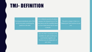
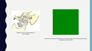
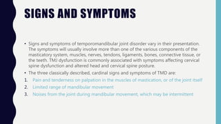
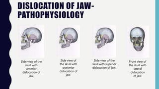
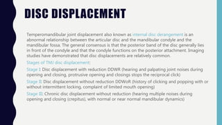
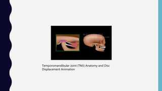
The temporomandibular joint (TMJ) connects the jawbone to the skull and allows for opening and closing of the mouth. It contains articular discs that divide the joint into compartments. TMJ disorders can cause pain, limited jaw movement, and joint noises. Common causes of pain include muscle issues, abnormal disc positioning, arthritis, and infections. Dislocated jaws may occur from trauma and cause difficulties opening the mouth. Displaced discs involve abnormal relationships between discs and bones. Mandibular fractures from falls or hits result in jaw pain and malocclusion.
This seminar explains about the development, relations, ligaments, various attachments, vascular and nervous supply and various surgical approaches and its modifications to TMJ
this presentation describes the detail anatomy of Temporo-mandibular joint with respect to its articulating surfaces, ligaments, muscles and blood and nerve supply.
A Brief description of the causes and clinical manifestations of the internal derangement of the temporomandibular joint , with particular emphasis on Disc Displacements .
The temporomandibular joint is the joint that connects jaw to skull. When this joint is injured or damaged, it can lead to a localized pain disorder called temporomandibular joint (TMJ) syndrome or temporomandibular disorder (TMD), also said as TMJ Arthritis as it related to inflammation of joint. The prognosis of this case is good. Some patient able to get this disorder resolve by some treatment and home remedies. Only a few of them need to get the surgery.
Anoverview of TMD'S categories and main types of interocclusal appliances( occlusal splints ) used during the management of these musculoskeletal disorders .
TMJ is a ginglymo-diarthroidal joint that is freely mobile with superior and inferior joint spaces separated by articular disc.
The type of imaging technique depends upon the clinical problems associated, so either imaging of hard tissue (OSSEOUS) or soft tissue is desired.
Certain protocols are to be taken care before the imaging procedure:
the amount of diagnostic information available from particular imaging modality.
The cost of examination
The radiation dose
history, classification, types of veneers, indications and contraindications, working procedure, preparation, ipmpression taking for veneers, surface treatment and cementation, veneers vs crowns
drug abuse- what is it? most common stimulants, cocaine abuse, aderall abuse, meth abuse, physiology of drug abuse, physical signs of drug abuse, treatment
Lung Cancer: Artificial Intelligence, Synergetics, Complex System Analysis, S...Oleg Kshivets
RESULTS: Overall life span (LS) was 2252.1±1742.5 days and cumulative 5-year survival (5YS) reached 73.2%, 10 years – 64.8%, 20 years – 42.5%. 513 LCP lived more than 5 years (LS=3124.6±1525.6 days), 148 LCP – more than 10 years (LS=5054.4±1504.1 days).199 LCP died because of LC (LS=562.7±374.5 days). 5YS of LCP after bi/lobectomies was significantly superior in comparison with LCP after pneumonectomies (78.1% vs.63.7%, P=0.00001 by log-rank test). AT significantly improved 5YS (66.3% vs. 34.8%) (P=0.00000 by log-rank test) only for LCP with N1-2. Cox modeling displayed that 5YS of LCP significantly depended on: phase transition (PT) early-invasive LC in terms of synergetics, PT N0—N12, cell ratio factors (ratio between cancer cells- CC and blood cells subpopulations), G1-3, histology, glucose, AT, blood cell circuit, prothrombin index, heparin tolerance, recalcification time (P=0.000-0.038). Neural networks, genetic algorithm selection and bootstrap simulation revealed relationships between 5YS and PT early-invasive LC (rank=1), PT N0—N12 (rank=2), thrombocytes/CC (3), erythrocytes/CC (4), eosinophils/CC (5), healthy cells/CC (6), lymphocytes/CC (7), segmented neutrophils/CC (8), stick neutrophils/CC (9), monocytes/CC (10); leucocytes/CC (11). Correct prediction of 5YS was 100% by neural networks computing (area under ROC curve=1.0; error=0.0).
CONCLUSIONS: 5YS of LCP after radical procedures significantly depended on: 1) PT early-invasive cancer; 2) PT N0--N12; 3) cell ratio factors; 4) blood cell circuit; 5) biochemical factors; 6) hemostasis system; 7) AT; 8) LC characteristics; 9) LC cell dynamics; 10) surgery type: lobectomy/pneumonectomy; 11) anthropometric data. Optimal diagnosis and treatment strategies for LC are: 1) screening and early detection of LC; 2) availability of experienced thoracic surgeons because of complexity of radical procedures; 3) aggressive en block surgery and adequate lymph node dissection for completeness; 4) precise prediction; 5) adjuvant chemoimmunoradiotherapy for LCP with unfavorable prognosis.
Factory Supply Best Quality Pmk Oil CAS 28578–16–7 PMK Powder in Stockrebeccabio
Factory Supply Best Quality Pmk Oil CAS 28578–16–7 PMK Powder in Stock
Telegram: bmksupplier
signal: +85264872720
threema: TUD4A6YC
You can contact me on Telegram or Threema
Communicate promptly and reply
Free of customs clearance, Double Clearance 100% pass delivery to USA, Canada, Spain, Germany, Netherland, Poland, Italy, Sweden, UK, Czech Republic, Australia, Mexico, Russia, Ukraine, Kazakhstan.Door to door service
Hot Selling Organic intermediates
These simplified slides by Dr. Sidra Arshad present an overview of the non-respiratory functions of the respiratory tract.
Learning objectives:
1. Enlist the non-respiratory functions of the respiratory tract
2. Briefly explain how these functions are carried out
3. Discuss the significance of dead space
4. Differentiate between minute ventilation and alveolar ventilation
5. Describe the cough and sneeze reflexes
Study Resources:
1. Chapter 39, Guyton and Hall Textbook of Medical Physiology, 14th edition
2. Chapter 34, Ganong’s Review of Medical Physiology, 26th edition
3. Chapter 17, Human Physiology by Lauralee Sherwood, 9th edition
4. Non-respiratory functions of the lungs https://academic.oup.com/bjaed/article/13/3/98/278874
- Video recording of this lecture in English language: https://youtu.be/lK81BzxMqdo
- Video recording of this lecture in Arabic language: https://youtu.be/Ve4P0COk9OI
- Link to download the book free: https://nephrotube.blogspot.com/p/nephrotube-nephrology-books.html
- Link to NephroTube website: www.NephroTube.com
- Link to NephroTube social media accounts: https://nephrotube.blogspot.com/p/join-nephrotube-on-social-media.html
Title: Sense of Taste
Presenter: Dr. Faiza, Assistant Professor of Physiology
Qualifications:
MBBS (Best Graduate, AIMC Lahore)
FCPS Physiology
ICMT, CHPE, DHPE (STMU)
MPH (GC University, Faisalabad)
MBA (Virtual University of Pakistan)
Learning Objectives:
Describe the structure and function of taste buds.
Describe the relationship between the taste threshold and taste index of common substances.
Explain the chemical basis and signal transduction of taste perception for each type of primary taste sensation.
Recognize different abnormalities of taste perception and their causes.
Key Topics:
Significance of Taste Sensation:
Differentiation between pleasant and harmful food
Influence on behavior
Selection of food based on metabolic needs
Receptors of Taste:
Taste buds on the tongue
Influence of sense of smell, texture of food, and pain stimulation (e.g., by pepper)
Primary and Secondary Taste Sensations:
Primary taste sensations: Sweet, Sour, Salty, Bitter, Umami
Chemical basis and signal transduction mechanisms for each taste
Taste Threshold and Index:
Taste threshold values for Sweet (sucrose), Salty (NaCl), Sour (HCl), and Bitter (Quinine)
Taste index relationship: Inversely proportional to taste threshold
Taste Blindness:
Inability to taste certain substances, particularly thiourea compounds
Example: Phenylthiocarbamide
Structure and Function of Taste Buds:
Composition: Epithelial cells, Sustentacular/Supporting cells, Taste cells, Basal cells
Features: Taste pores, Taste hairs/microvilli, and Taste nerve fibers
Location of Taste Buds:
Found in papillae of the tongue (Fungiform, Circumvallate, Foliate)
Also present on the palate, tonsillar pillars, epiglottis, and proximal esophagus
Mechanism of Taste Stimulation:
Interaction of taste substances with receptors on microvilli
Signal transduction pathways for Umami, Sweet, Bitter, Sour, and Salty tastes
Taste Sensitivity and Adaptation:
Decrease in sensitivity with age
Rapid adaptation of taste sensation
Role of Saliva in Taste:
Dissolution of tastants to reach receptors
Washing away the stimulus
Taste Preferences and Aversions:
Mechanisms behind taste preference and aversion
Influence of receptors and neural pathways
Impact of Sensory Nerve Damage:
Degeneration of taste buds if the sensory nerve fiber is cut
Abnormalities of Taste Detection:
Conditions: Ageusia, Hypogeusia, Dysgeusia (parageusia)
Causes: Nerve damage, neurological disorders, infections, poor oral hygiene, adverse drug effects, deficiencies, aging, tobacco use, altered neurotransmitter levels
Neurotransmitters and Taste Threshold:
Effects of serotonin (5-HT) and norepinephrine (NE) on taste sensitivity
Supertasters:
25% of the population with heightened sensitivity to taste, especially bitterness
Increased number of fungiform papillae
MANAGEMENT OF ATRIOVENTRICULAR CONDUCTION BLOCK.pdfJim Jacob Roy
Cardiac conduction defects can occur due to various causes.
Atrioventricular conduction blocks ( AV blocks ) are classified into 3 types.
This document describes the acute management of AV block.
The prostate is an exocrine gland of the male mammalian reproductive system
It is a walnut-sized gland that forms part of the male reproductive system and is located in front of the rectum and just below the urinary bladder
Function is to store and secrete a clear, slightly alkaline fluid that constitutes 10-30% of the volume of the seminal fluid that along with the spermatozoa, constitutes semen
A healthy human prostate measures (4cm-vertical, by 3cm-horizontal, 2cm ant-post ).
It surrounds the urethra just below the urinary bladder. It has anterior, median, posterior and two lateral lobes
It’s work is regulated by androgens which are responsible for male sex characteristics
Generalised disease of the prostate due to hormonal derangement which leads to non malignant enlargement of the gland (increase in the number of epithelial cells and stromal tissue)to cause compression of the urethra leading to symptoms (LUTS
2. TMJ- DEFINITION
The temporomandibular joints (TMJ)
are the two joints connecting the
jawbone to the skull.
It is a bilateral synovial articulation
between the temporal bone of the
skull above and the mandible below;
it is from these bones that its name
is derived.
This joint is unique in that it is a
bilateral joint that functions as one
unit.
Since the TMJ is connected to the
mandible, the right and left joints
must function together and
therefore are not independent of
each other
3. STRUCTURE
The main components are the joint capsule,
articular disc, mandibular condyles, articular
surface of the temporal bone,
temporomandibular ligament, stylomandibular
ligament, sphenomandibular ligament, and
lateral pterygoid muscle.
4.
5. FUNCTION
Each temporomandibular joint is classed as a "ginglymoarthrodial" joint since it is both a ginglymus (hinging joint)
and an arthrodial (sliding) joint. The condyle of the mandible articulates with the temporal bone in the mandibular
fossa. The mandibular fossa is a concave depression in the squamous portion of the temporal bone.
These two bones are actually separated by an articular disc, which divides the joint into two distinct compartments.
The inferior compartment allows for rotation of the condylar head around an instantaneous axis of rotation,
corresponding to the first 20mm or so of the opening of the mouth. After the mouth is open to this extent, the mouth
can no longer open without the superior compartment of the temporomandibular joints becoming active.
6.
7. FUNCTION
At this point, if the mouth continues to open, not only are the condylar heads rotating within the lower compartment of the temporomandibular joints, but the
entire apparatus (condylar head and articular disc) translates.
Although this had traditionally been explained as a forward and downward sliding motion, on the anterior concave surface of the mandibular fossa and the
posterior convex surface of the articular eminence, this translation actually amounts to a rotation around another axis. This effectively produces an evolute which
can be termed the resultant axis of mandibular rotation, which lies in the vicinity of the mandibular foramen, allowing for a low-tension environment for the
vasculature and innervation of the mandible.
The necessity of translation to produce further opening past that which can be accomplished with sole rotation of the condyle can be demonstrated by placing a
resistant fist against the chin and trying to open the mouth more than 20 or so mm.
The resting position of the temporomandibular joint is not with the teeth biting together. Instead, the muscular balance and proprioceptive feedback allow a
physiologic rest for the mandible, an interocclusal clearance or freeway space, which is 2 to 4 mm between the teeth.
8. JAW MOVEMENT
Normal full jaw opening is 40-50 millimeters as
measured from edge of lower front teeth to
edge of upper front teeth.
When measuring the vertical range of motion,
the measurement must be adjusted for the
overbite. For example, if the measurement
from the edge of the lower front teeth to the
edge of the upper front teeth is 40 millimeters
and the overbite is 3 millimeters, then the jaw
opening is 43 millimeters.
During jaw movements, only the mandible
moves.
Normal movements of the mandible during
function, such as mastication, or chewing, are
known as excursions. There are two lateral
excursions (left and right) and the forward
excursion, known as protrusion. The reversal of
protrusion is retrusion.
9. Sagittal section of the articulation of
the mandible
Dynamics of temporomandibular joint during voluntary mouth opening and closing
visualized by real-time MRI
10. CLINICAL SIGNIFICANCE- PAIN
• Temporomandibular joint pain is generally due to one of four reasons:
1. Myofascial pain dysfunction syndrome, primarily involving the muscles of
mastication. This is the most common cause.
2. Internal derangements, an abnormal relationship of the disc to any of the other
components of the joint. Disc displacement is an example of internal derangement.
3. Osteoarthritis of the temporomandibular joint, a degenerative joint disease of the
articular surfaces.
4. Temporal arteritis, for which it is considered a reliable diagnostic criteria.
11. TMD
• Pain or dysfunction of the temporomandibular joint (TMJ) is sometimes referred to as
temporomandibular joint dysfunction or temporomandibular joint disorder (TMD). This
term is used to refer to a group of problems involving the temporomandibular joints
and the muscles, tendons, ligaments, blood vessels, and other tissues associated with
them.
• Although rare, other pathologic conditions may also affect the function of
temporomandibular joints, causing pain and swelling. These conditions include
chondrosarcoma, osteosarcoma, giant cell tumor, and aneurysmal bone cyst.
12. EXAMINATION
• The temporomandibular joints can be felt in front of or within the external acoustic
meatus during movements of the mandible. Auscultation of the joint can also be done.
13. SIGNS AND SYMPTOMS
• Signs and symptoms of temporomandibular joint disorder vary in their presentation.
The symptoms will usually involve more than one of the various components of the
masticatory system, muscles, nerves, tendons, ligaments, bones, connective tissue, or
the teeth. TMJ dysfunction is commonly associated with symptoms affecting cervical
spine dysfunction and altered head and cervical spine posture.
• The three classically described, cardinal signs and symptoms of TMD are:
1. Pain and tenderness on palpation in the muscles of mastication, or of the joint itself
2. Limited range of mandibular movement
3. Noises from the joint during mandibular movement, which may be intermittent
14. OTHER SIGNS AND SYMPTOMS
Headache e.g. pain in
the occipital region or
the forehead
Pain elsewhere, such
as the teeth or neck
hearing loss
Tinnitus Dizziness
Sensation of
malocclusion
15. TMD CLASSIFICATION
MUSCULAR:
• Hyperactivity, spasm, and trismus
• Inflammation (myositis)
• Trauma
• Myofascial pain and fibromyalgia
• Atrophy or hypertrophy
ARTHROGENIC:
• Disc displacement (internal derangement)
• Hypomobility of the disc (adhesions or
scars)
• Dislocation and subluxation
• Arthritis
• Infections
• Metabolic disease (gout,
chondrocalcinosis)
• Capsulitis, synovitis
• Ankylosis (fibrous or bony)
• Fracture
• Absent, large, or small condyloid process
16. DISLOCATION OF JAW
Dislocations occur when two bones that originally met at the joint detach. Dislocations should not be confused with
Subluxation. Subluxation is when the joint is still partially attached to the bone.
When a person has a dislocated jaw it is difficult to open and close the mouth. Dislocation can occur following a series
of events if the jaw locks while open or unable to close. If the jaw is dislocated, it may cause an extreme headache or
inability to concentrate. When the muscle's alignment is out of sync, a pain will occur due to unwanted rotation of the
jaw.
If the pain remains constant, it may require surgery to realign the jaw. Depending on the severity of the jaw's
dislocation, pain relief such as paracetamol may assist to alleviate the initial chronic pain. If the pain relief is taken for
an extended period of time, it may negatively affect the person while talking, eating, drinking, etc.
17. DISLOCATION OF JAW- SYMPTOMS
The symptoms can be numerous depending on the severity of the dislocation injury and how long the person is inflicted with the injury. Symptoms of a dislocated jaw include a
bite that feels “off” or abnormal, hard time talking or moving jaw, not able to close mouth completely, drooling due to not being able to shut mouth completely, teeth feel they are
out of alignment, and a pain that becomes unbearable
The immediate symptom can be a loud crunch noise occurring right up against the ear drum. This is instantly followed by excruciating pain, particularly in the side where the
dislocation occurred.[citation needed]
Short-term symptoms can range from mild to chronic headaches, muscle tension or pain in the face, jaw and neck.
Long-term symptoms can result in sleep deprivation, tiredness/lethargy, frustration, bursts of anger or short fuse, difficulty performing everyday tasks, depression, social issues
relating to difficulty talking, hearing sensitivity (particularly to high pitched sounds), tinnitus and pain when seated associated with posture while at a computer and reading books
from general pressure on the jaw and facial muscles when tilting head down or up. And possible causing subsequent facial asymmetry.
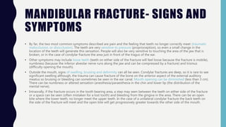
In contrast, symptoms of a fractured jaw include bleeding coming from the mouth, unable to open the mouth wide without pain, bruising and swelling of the face, difficulty eating
due to the constant pain, loss of feeling in the face (more specifically the lower lip) and lacks full range of motion of the jaw
18. DISLOCATION OF JAW-
PATHOPHYSIOLOGY
• There are four different positions of jaw dislocation: posterior, anterior, superior and lateral. The most common position is
anterior, while the other types are rare.
• Anterior dislocation shifts the lower jaw forward if the mouth excessively opens. This type of dislocation may happen
bilaterally or unilaterally after yawning. The muscles that are affected during anterior jaw dislocation are the masseter and
temporalis which pull up on the mandible and the lateral pterygoid which relaxes the mandibular condyle. The condyle can
get locked in front of the articular eminence.
• Posterior dislocation is possible for people who get injured by being punched in the chin. This dislocation will push the jaw
back affecting the alignment of the mandibular condyle and mastoid. The external auditory canal may be fractured.
• Superior dislocations occur after being punched below the mandibular ramus as the mouth remains half-open. Since great
force occurs in a punch, the angle of the jaw will be forced upward moving towards the condylar head. This can result in a
fracture of the glenoid fossa and displacement of the condyle into the middle cranial fossa, potentially injuring the facial and
vestibulocochlear nerves and the temporal lobe.
• Lateral dislocations move the mandibular condyle away from the skull and are likely to happen together with jaw fractures.
• Posterior, superior and lateral dislocations are uncommon injuries and usually result from high-energy trauma to the chin. By
contrast, anterior dislocations are more often the result of low-energy trauma (e.g. tooth extraction) or secondary to a
medical condition that affects the stability of the joint (e.g. seizures, ligamentous laxity, degeneration of joint capsule).
19. DISLOCATION OF JAW-
PATHOPHYSIOLOGY
Side view of the
skull with
anterior
dislocation of
jaw.
Side view of
the skull with
posterior
dislocation of
jaw.
Side view of the
skull with superior
dislocation of jaw.
Front view of
the skull with
lateral
dislocation
of jaw.
20. DISC DISPLACEMENT
Temperomandibular joint displacement also known as internal disc derangement is an
abnormal relationship between the articular disc and the mandibular condyle and the
mandibular fossa. The general consensus is that the posterior band of the disc generally lies
in front of the condyle and that the condyle functions on the posterior attachment. Imaging
studies have demonstrated that disc displacements are relatively common.
Stages of TMJ disc displacement:
Stage I: Disc displacement with reduction DDWR (hearing and palpating joint noises during
opening and closing, protrusive opening and closings stops the reciprocal click)
Stage II: Disc displacement without reduction DDWoR (history of clicking and popping with or
without intermittent locking, complaint of limited mouth opening)
Stage III: Chronic disc displacement without reduction (hearing multiple noises during
opening and closing (crepitus), with normal or near normal mandibular dynamics)
23. MANDIBULAR FRACTURE
Mandibular fracture, also known as
fracture of the jaw, is a break through
the mandibular bone. In about 60% of
cases the break occurs in two places. It
may result in a decreased ability to fully
open the mouth.
Mandibular fractures are typically the
result of trauma. This can include a fall
onto the chin or a hit from the side.
Rarely they may be due to
osteonecrosis or tumors in the bone.
24. MANDIBULAR FRACTURE- SIGNS AND
SYMPTOMS
• By far, the two most common symptoms described are pain and the feeling that teeth no longer correctly meet (traumatic
malocclusion, or disocclusion). The teeth are very sensitive to pressure (proprioception), so even a small change in the
location of the teeth will generate this sensation. People will also be very sensitive to touching the area of the jaw that is
broken, or in the case of condylar fracture the area just in front of the tragus of the ear.
• Other symptoms may include loose teeth (teeth on either side of the fracture will feel loose because the fracture is mobile),
numbness (because the inferior alveolar nerve runs along the jaw and can be compressed by a fracture) and trismus
(difficulty opening the mouth).
• Outside the mouth, signs of swelling, bruising and deformity can all be seen. Condylar fractures are deep, so it is rare to see
significant swelling although, the trauma can cause fracture of the bone on the anterior aspect of the external auditory
meatus so bruising or bleeding can sometimes be seen in the ear canal. Mouth opening can be diminished (less than 3 cm).
There can be numbness or altered sensation (anesthesia/paraesthesia in the chin and lower lip (the distribution of the
mental nerve).
• Intraorally, if the fracture occurs in the tooth bearing area, a step may seen between the teeth on either side of the fracture
or a space can be seen (often mistaken for a lost tooth) and bleeding from the gingiva in the area. There can be an open
bite where the lower teeth, no longer meet the upper teeth. In the case of a unilateral condylar fracture the back teeth on
the side of the fracture will meet and the open bite will get progressively greater towards the other side of the mouth.