Downloaded 1,051 times
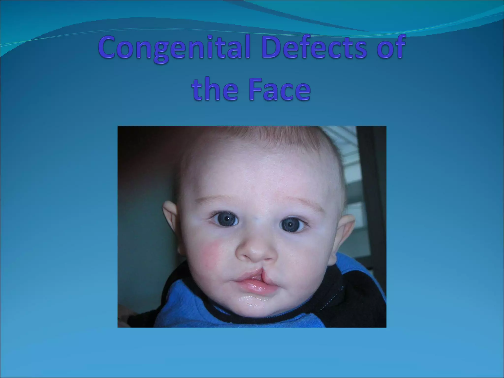


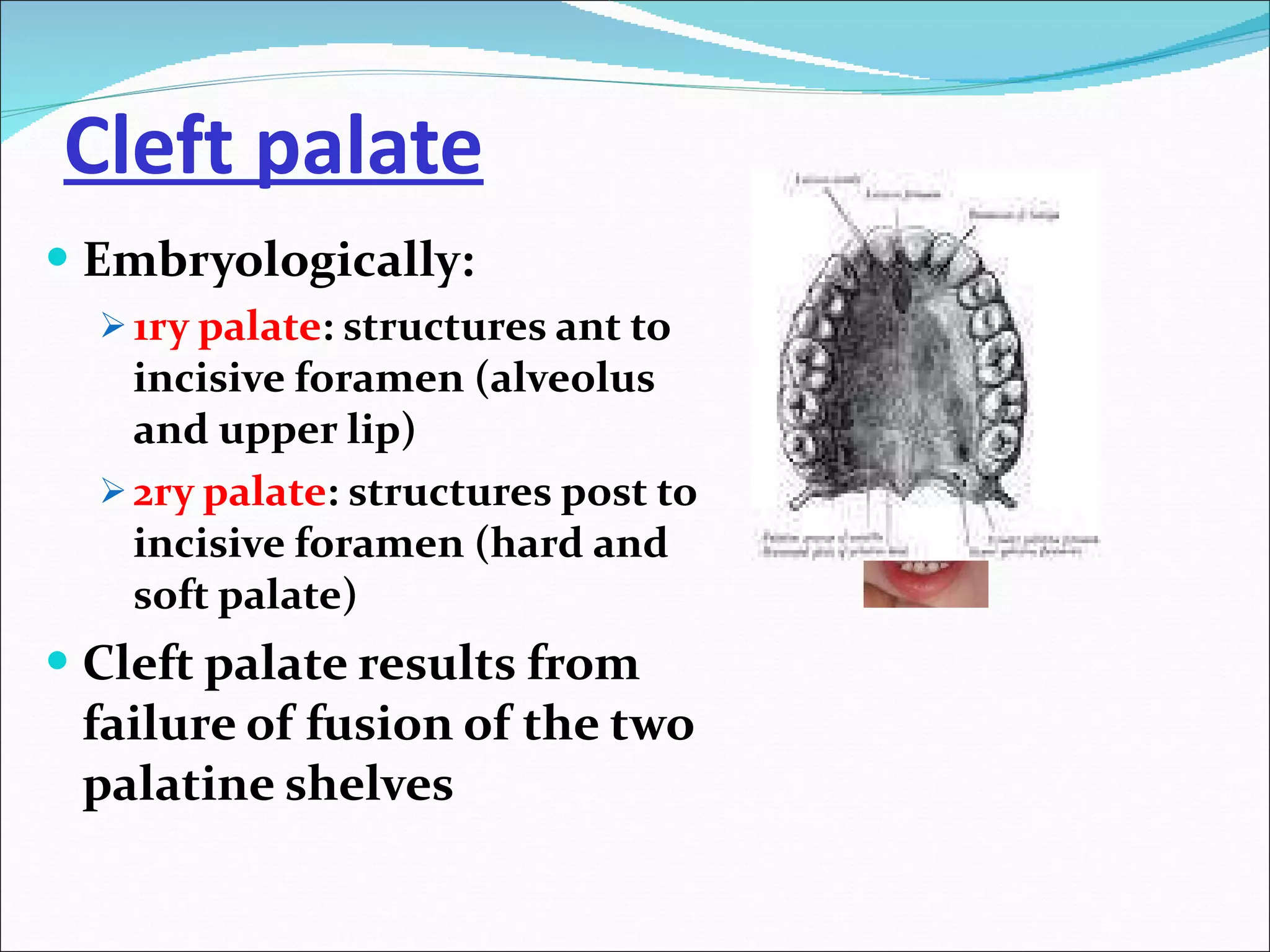


Cleft lip and palate is a congenital abnormality caused by failure of fusion of structures like the lip, alveolus, hard and soft palate during embryonic development. It has an incidence of 1 in 600 live births for cleft lip and palate, and 1 in 1000 for isolated cleft palate. Treatment involves surgical repair of clefts as well as management of associated issues like feeding problems, hearing loss, dental abnormalities, and speech and facial growth defects. Secondary surgery may be needed to improve cosmetic or functional outcomes.


