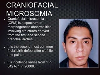
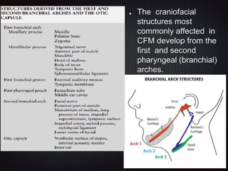
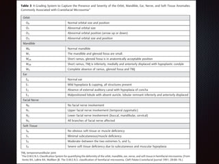
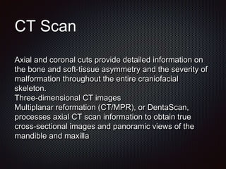
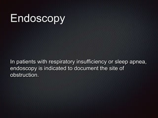
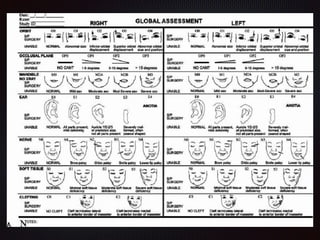
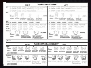
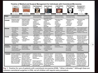
CRANIOFACIAL
MICROSOMIA and PARRY-
ROMBERG SYNDROME
Dr. Aditi Sharma
Senior Resident
Department of Plastic Surgery
J.N.M.C.H., Aligarh
CRANIOFACIAL
MICROSOMIA
Craniofacial microsomia
(CFM) is a spectrum of
morphogenetic abnormalities
involving structures derived
from the first and second
branchial arches.
It is the second most common
facial birth defect after cleft lip
and palate.
It’s incidence varies from 1 in
642 to 1 in 26000.
TERMINOLOGY
• Hemifacial microsomia
• Oculo-auriculo-vertebral spectrum
• Goldenhar syndrome
• First and second branchial arch syndrome
• Otomandibular dysostosis
• Facio-auriculo-vertebral syndrome
• Lateral facial dysplasia
EMBRYOLOGY
The head and neck originate from six embryonic
structures ,the brachial arches.
In the fourth week of gestation, neural crest cells
migrate from the neural tube to begin the development
of the pharyngeal arch ectomesenchyme.
Each arch has three layers (endoderm, mesenchyme
from ectomesenchyme and mesoderm, and ectoderm),
which produce the four primordial components: muscle,
artery, nerve, and cartilage.
The craniofacial
structures most
commonly affected in
CFM develop from the
first and second
pharyngeal (branchial)
arches.
During the first trimester, all brachial arches have their
own aortic arch.
In the 3rd week of gestation, ICA & ECA develop from 3rd
brachial arch.
In 4th week,1st aortic arch disappears ,while 2nd aortic
arch forms stapedial artery , which forms anastomoses
between ICA &ECA.
In 5th week,stapedial artery atrophies and disappears.
By day 40, stapedial artery disappears completely with
ECA supplying the 1st and 2nd brachial arch.
Etiology and
pathophysiology
The exact pathogenesis is yet to be determined.
Theory1:- Hemorrhage caused by stapedial artery
malformations in first 6 weeks of gestation.
Poswillo et al. reproduced in mice some of the
phenotypic anomalies seen in CFM phenotype by
administering teratogens (triazine) that caused a
hematoma of the stapedial artery and resulted in local
and regional necrosis
Theory2:- Disruption of neural crest cell migration.
Genetic transmission
Kaye et al. performed segregation analysis on 74 families of
probands with CFM and rejected the hypothesis that
genetic transmission is not a causative factor. The evidence
favored autosomal- dominant inheritance; however,
recessive and polygenic models were not distinguishable.
Despite the suggestion of autosomal-dominant
transmission, they found only a 2–3% overall recurrence
rate in first-degree relatives. This figure compares to the
10% recurrence risk in first- and second- degree relatives
reported by the same group in an earlier study of 294
individuals with CFM.
PRESENTATION
Right side of the face is more commonly
affected(60%).
Male predominance(65%).
Unilateral involvement more common.
Various structures of face are involved to different
extents.
Mandible
Mandibular hypoplasia is the most obvious finding as well
as the most common finding(90%).
Ramus and condyle are affected more commonly than
body or I/L parasymphysis.
Mandibular hypoplasia may range from mild hypoplasia or
flattening of the condylar head to complete agenesis of
the condyle, ramus, and glenoid fossa
A wide array of temporomandibular joint abnormalities
results from the variable mandibular hypoplasia, leading
to deformities ranging from mild malpositioning with
aberrant cranial base articulation to complete obliteration.
Maxilla
Historically, it was believed that the mandibular
deformity was associated with a maxillary hypoplasia.
Recent findings suggest that the maxillary anomaly
is not a true volumetric deficiency. The maxillary cant
observed is secondary to vertical growth inhibition by
the pathologically small mandible.
Craniofacial skeleton
• Craniofacial bones other than the mandible or maxilla
can be involved, especially the tympanic and mastoid
portions of the temporal bone; the petrous portion
usually is remarkably spared.
• The styloid process is frequently smaller on the
affected side.
• The mastoid process can have a flattened
appearance, and there can be partial or complete
lack of pneumatization of the mastoid air cells.
• The zygoma can be underdeveloped in all its
dimensions, with flattening of the malar eminence.
• A decrease in the span of the zygomatic arch
results in a decrease in the length of the lateral
canthal–tragal line on the affected side.
• Disparities in the vertical axis of the orbit can be
seen, with or without evidence of microphthalmos.
Muscles of mastication
• Muscle function, especially that of the lateral pterygoid
muscle, is impaired .
• A severe limitation of protrusive and lateral movements
secondary to hypoplasia of the lateral pterygoid muscle is
observed.
• When the patient opens the mouth, the deviation toward
the affected side is produced .
• In many cases, the coronoid process is absent, and there
is reduction in the size of the temporalis muscle.
• The associated masseter and medial pterygoid muscles
are also grossly deficient.
Ear
• Auricular malformations seen in CFM are as diverse
as those demonstrated by the syndrome’s other
component features.
• The ear anomalies associated with CFM can be
categorized into external ear malformations (e.g.,
microtia), middle ear malformations and atresia, and
the presence of branchial remnants and sinus tracts.
Nervous system
Cranial nerve abnormalities are frequent in CFM and
can include arhinencephaly of the unilateral and
bilateral type, unilateral agenesis and hypoplasia of the
optic nerve with secondary changes in the lateral
geniculate body and visual cortex, congenital
ophthalmoplegia and Duane retraction syndrome,
hypoplasia of the trochlear and abducens nuclei and
nerves, congenital trigeminal anesthesia, and aplasia of
the trigeminal nerve and motor and sensory nucleus.
The most common cranial nerve anomaly is facial
paralysis secondary to agenesis of the facial nerve in
the temporal bone or hypoplasia of the intracranial
portion of the facial nerve and facial nucleus in the
brainstem.
Soft tissue
This deficiency is mostly evident in the malar and
masseteric region of the face as well as in the region
of the external ear, orbit, and the temporal region.
Lack of soft-tissue bulk contributes to a characteristic
malar flattening and temporal hollowing that is best
appreciated when viewed from a submental
perspective.
Extracraniofacial anomalies
A vast range of associated extracraniofacial
anomalies has been reported in the craniofacial
microsomia literature,including skeletal, cardiac,
renal, gastrointestinal, and pulmonary malformations.
For instance, the constellation of hemifacial
hypoplasia, epibulbar lipodermoids, and vertebral
anomalies, including fused and/or hemivertebrae,
definesone of the subsets of CFM named Goldenhar
syndrome.
CLASSIFICATION
David et al. propounded the most comprehensive classification from the
Australian Cranio-Facial Unit (ACFU) of Adelaide, Australia.
Three categories were sought in each patient — skeletal, auricular, and
soft tissue (SAT).
The five skeletal categories are
S1 — Small mandible of normal shape
S2 — Condyle, ramus, and the sigmoid notch identifiable but grossly
distorted. Mandible strikingly different in size and shape from normal
S3 — Mandible severely malformed, ranging from poorly identifiable
ramal component to complete agenesis of ramus
S4 — S3 mandible + orbital involvement — gross posterior recession of
lateral and posterior orbital rims
S5 — S4 defect + orbital dystopia, hypoplasia and asymmetrical
neuroranium and a flat temporal fossa.
The four auricular
categories are
A0 — Normal
A1 — Small malformed
auricle, retaining all
features
A2 — Rudimentary
auricle, with hook at
cranial end corresponding
to the helix
A3 — Malformed lobule +
absent rest of pinna.
And the three soft tissue
categories are
T1 — Minimal contour
defect with no cranial
nerve involvement
T2 — Moderate defect
T3 — Major defect,
obvious facial scoliosis,
severe hypoplasia of
cranial nerves, parotid,
muscles of mastication,
eye involvement + facial
clefts.
Diagnosis
When comparing the SAT and OMENS systems,
Cousley recommended the following minimal
diagnostic criteria:
1. Ipsilateral mandibular and ear defects.
2. Asymmetrical mandibular or ear defects in
association with:
a. Two or more indirectly associated anomalies, and
b. A positive family history of CFM.
Pre-operative assessment
• COMPLETE HISTORY
• PHYSICAL EXAMINATION
• PHOTOGRAPHS (Frontal, Lateral,oblique,
Submental Vertex, And Occlusal Views)
• CEPHALOGRAMS (posteroanterior, lateral, and
basilar)
• THREE DIMENSIONAL CT SCAN
• ENDOSCOPY
• SLEEP STUDIES
Photography
• Baseline photography
• Standardized records should
include full face, submental
vertex, lateral, oblique smile,
and occlusal views.
• Three-dimensional camera
systems
Cephalometrics
Grayson et al. described the technique of multiplane
cephalometryWith lateral, coronal, and basilar radiographs, skeletal
landmarks can be identified in three coronal and three axial planes
and used to construct an estimation of the midline for each plane.
These midlines are compared with the mid- sagittal plane, which is
determined by relatively stable bilateral structures such as the
occipital condyles, the center of the foramen magnum, and the
medial axis of the spheno-occipital synchondrosis.
By use of this technique, a phenomenon termed warping can be
observed within the skeleton of the patient with CFM. The midline
constructs deviate progressively laterally as one passes anteriorly
from the skull base to the piriform rim in the coronal plane and
inferiorly from the orbits to the mandible in the axial plane.
CT Scan
Axial and coronal cuts provide detailed information on
the bone and soft-tissue asymmetry and the severity of
malformation throughout the entire craniofacial
skeleton.
Three-dimensional CT images
Multiplanar reformation (CT/MPR), or DentaScan,
processes axial CT scan information to obtain true
cross-sectional images and panoramic views of the
mandible and maxilla
Sleep studies
In patients with obstructive sleep apnea, sleep studies
(polysomnography) can define the degree of the
respiratory dysfunction and are invaluable, along with
interpretation of the clinical symptoms and endoscopic
findings, in determining whether surgical intervention,
i.e., mandibular distraction, is indicated.
Endoscopy
In patients with respiratory insufficiency or sleep apnea,
endoscopy is indicated to document the site of
obstruction.
Principles of treatment
• Multidisciplinary treatment team
• Timing of osteotomy – different school of thoughts
• Clinician is usually dealing with a composite skeletal and
soft-tissue deformity – “bone carpentry” alone is often
insufficient in the global treatment of the patient with
craniofacial microsomia
• The surgical reconstructive requirements are dependent on
the individual anatomic and functional deficiencies.
• The skeletal reconstructive efforts have traditionally been
directed at correction of the mandibular deficiency, usually
involving the ramus and body, as well as the condyle and
temporomandibular joint.
Surgical interventions are designed to restore the
patient’s craniofacial form and function and must
account for the expected facial growth pattern, timing of
dental eruption, schedule for school and extracurricular
activities, along with otherpsychosocialfactors.
Forexample,interventions such as orthognathic
surgery are likely most effective if postponed until
completion of facial growth. However, infants may
require timely treatment for any upper airway
obstruction with mandible distraction or tracheostomy.
Communication among team members is paramount to
coordinate timing of surgical interventions.
Management
The UCLA Craniofacial Clinic Protocol and the ACFU Protocol are aimed at
maximizing results and minimizing the number of procedures. The timing
and types of procedures may vary depending on the severity of the
deformity and the individual patient. In general, corrections include many of
the following:
Preauricular skin tags (age under 1 year): Excision;
Macrostomia or wide mouth (age under 1 year): Commisuroplasty;
Mandibular hypoplasia (5-8 years of age): Internal distraction osteogenesis
is used to lengthen the lower jaw (for severe cases with absence of
mandibular condyle and ramus) a rib graft may be necessary.
External ear deformity or absence (6-8 years of age): Staged ear
reconstruction with a rib graft framework, elevation, lobule (ear lobe) and
tragus (front of ear) reconstruction are performed: Staged ear
reconstruction of microtia (a) using costo-chondral graft framework (b).
Final result after lobule transposition and elevation .
Orbital dystopia (asymmetric eyes) (6-11 years of age):
Although rarely required, repositioning of the orbit and/or
advancement of the forehead and brow (fronto-orbital
advancement) may be performed.
Jaw asymmetry (15-18 years or age of skeletal maturity):
Preoperative orthodontics, followed by jaw (orthognathic)
surgery with Le Fort I (upper jaw) and mandibular sagittal-
split (lower jaw) osteotomies are often necessary.
Soft tissue asymmetry (after jaw surgery): Final facial
contouring with autogenous fat grafting, dermal fat grafts or
even a fascial-fat free flap from the upper back are often
necessary after other corrections.
TREATMENT
Tracheostomy and gastrostomy
Commissuroplasty or closure of the lateral facial
cleft
Mandibular distraction can be employed at any
age from the neonate to the adult
Rib or iliac bone grafting has been the traditional
method of reconstructing the type III mandibular
skeletal defect (absence of the ramus and
condyle).
mandibular distraction can be repeated
(secondary distraction)
TREATMENT cont.
An alternative technique is the reconstruction of
the missing ramus/condyle with a microvascular
fibula free flap with or without a skin “paddle” to
correct the soft tissue deficiency.
Autogenous fat injections for contour deformity- It
should also be noted that fat injections also
improve the quality of the overlying skin (dermal
thickness and cutaneous contour)
Microvascular free flap- It is wise to defer the free
flap until the underlying skeletal deficiencies have
been corrected
TREATMENT cont.
Auricular reconstruction is required in patients with
microtia.
The multistaged reconstruction is usually deferred
until the child is at least 8 years of age .
Maxillomandibular orthognathic
surgery
In the skeletally mature patient, traditional
maxillomandibular orthognathic surgery is indicated.
The mandibular osteotomies include the bilateral
sagittal split of the ramus and the vertical or oblique
osteotomy of the ramus.
Obwegeser combined the Le Fort I maxillary
osteotomy with bilateral sagittal split of the
mandibular ramus and genioplasty to ensure leveling
of the occlusal plane and establishment of the optimal
occlusal relationships.
Genioplasty, usually in three planes, completes the
procedure .
TREATMENT
ALGORITHM
YOUNGER THAN TWO YEARS OF AGE
Excision of the preauricular skin tags and cartilage
remnants
Correction of macrostomia
Patient with involvement of the fronto-orbital region,
characterized by severe retrusion of the supraorbital bar
and frontal bone, a fronto-orbital advancement cranial
vault remodeling can be performed as a combined
craniofacial surgical procedure.
Mandibular distraction for patients with sleep apneoa
TREATMENT ALGORITHM Cont
TWO TO SIX YEARS OF AGE
No treatment is required for Pruzansky type I
mandible and a horizontal occlusal plane distraction
osteogenesis is required for Pruzansky II
In the patient with a Pruzansky type III a preliminary
costochondral rib graft reconstruction should be
performed at approximately age 4 years.
TREATMENT ALGORITHM cont.
BETWEEN 6 AND 14 YEARS OF AGE
Orthodontic treatment.
Ear reconstruction.(rib cartilage is large enough at 6
years).
Soft-tissue augmentation, often by free-tissue
transfer.(subcutaneous volumizers, dermal fillers, fat
grafts , local flaps and microvascular free flaps etc.
TREATMENT ALGORITHM Cont.
AFTER 14 YEARS OF AGE
Bone grafting to deficient areas of the facial skeleton.
Orthognathic surgery.
Parry Romberg syndrome
• It is a rare Craniofacial disorder characterised by
hemifacial atrophy of skin, subcutaneous tissue,fat
and in severe cases of the underlying muscle and
bone.
• It was first described by Parry in 1825 and by
Romberg in 1846.
• Eulenburg described the entity as "progressive facial
hemi atrophy" in 1871.
• Onset is typically within first 20 yrs of life, but may
occur late.
• Symptoms progress over a 2-10 year period, before
spontaneous stabilisation.
• Extra-cutaneous manifestations include neural ,ocular
and oral pathology.
Etiology and Pathogenesis
Definite pathogenesis is unknown.
Most commonly described as autoimmune process similar
to en coupe de sabre (French for by/in a strike of the
sword due to its resemblance to sword scar),a variant of
localized scleroderma involving the frontoparietal face and
skull.
This pattern is supported by findings of inflammatory
histopathology, serum autoantibodies, coexistent
autoimmune diseases (eg, lupus), and positive response
to immunosuppression in patients with PRS.
Neurologically based theories suggest that PRS may be the
result of disordered developmental migration of neural crest
cells, neurotrophic viral infection, trigeminal peripheral
neuritis, intracranial vascular malformation, or peripheral
sympathetic nervous system dysfunction after traumatic
disruption of the cervical plexus or thoracic sympathetic
trunk.
Hyperactivity of sympathetic nervous system, specifically
inflammation of superior cervical ganglion, causing features
of PHA.
On histology dermal lymphocytic infiltrate centered around
neurovascular bundles in dermis also supports a neurologic
target
The current literature best describes PRS as mild,
moderate, or severe, to stratify disease and guide patient
management.
Mild disease is described as skin and subcutaneous
tissue atrophy limited to a single sensory branch of the
trigeminal nerve.
Moderate disease is described as atrophy limited to 2
branches of the trigeminal nerve.
Severe disease is described as atrophy in a distribution
involving all 3 branches of the trigeminal nerve or any
bone involvement (Fig.).
Disease presentation
Skin and soft tissue involvement
PRS characteristically manifests on the face, caudal to the
forehead.Atrophy begins superficially, although the epidermis is
minimally affected, and may progress to involve the subcutaneous
tissue, fat, fascia, muscle, cartilage, and bone.
Alopecia and hyperpigmentation or depigmentation are commonly
seen overlying the atrophic tissue.
Nearly all cases involve the cheek, periorbital and perioral areas;
more progressive cases may spread to involve the nasolabial fold,
brow, ear, and neck.
Neurologic involvement
PRS has been described as a neurocutaneous
syndrome, because the most common systemic
disease manifestations are neurologically based.
Seizures, migraines, hemiplegia, aneurysms, brain
atrophy, cranial neuropathies (cranial nerves III, V,
VI, and VII), intracranial vascular malformations,
trigeminal neuralgia, cognitive impairment, and
behavioural disorders have been reported in
association with PRS.
Ophthalmologic involvement
The most commonly reported ocular finding is enophthalmos
secondary to orbital, malar, and maxillary subcutaneous
tissue deficiency.
Other ophthalmologic manifestations include
ophthalmoplegia, strabismus, iris heterochromia, and uveitis.
Patients with neurologic symptoms may experience optic
nerve dysfunction, neuroretinopathy, or Horner syndrome.
In cases of eyelid soft tissue atrophy and retraction, exposure
keratopathy is a concern, and intervention is critical to prevent
permanent corneal scarring and preserve vision.
Oral involvement
PRS with oral cavity involvement most frequently
affects the tongue, gingiva, and soft palate.
Delayed tooth eruption and dental root exposure or
resorption are relatively common, whereas more
extensive tooth or mandibular atrophy may be seen in
severe cases.
Temporomandibular joint involvement often manifests
as pain with mastication and difficulty or complete
inability to open the mouth or perform normal jaw
movements.
Differential diagnosis
• ECDS
• Progeria, Dunnigan syndrome, and Kobberling
syndrome.
• Lipoatrophy in endocrine disorders, such as
hyperthyroidism and diabetes,
• Autoimmune diseases - systemic sclerosis and
dermatomyositis,
• Drug-induced atrophy - protease inhibitors for
treatment of HIV.
• Hemifacial microsomia.
Clinical Evaluation and
Diagnosis
Effective evaluation of patients with suspected PRS involves 4
major elements:
a complete disease history:-progressive symptom onset and any
systemic disease manifestations.,
a physical examination with attention to the head and neck careful
attention to facial asymmetry, particularly in the maxillary, malar,
and orbital regions,
consideration of potential differential diagnoses, and
attention to the patient’s psychological health.
Additional workup
Computed tomography scanning is indicated in most cases to
assess or confirm bone hypotrophy.
MRI is the study of choice in the evaluation of neurologic
symptoms and often reveals whitematter hyperintense signaling
ipsilateral to soft tissue involvement.
Lumbar puncture and electroencephalography are only indicated
for seizure workup.
Doppler ultrasound studies may be beneficial in the detection of
possible arteriovenous malformation in areas of significant soft
tissue atrophy.
ophthalmology consultation to evaluate the patient’s
visual acuity,
extraocular movements,
intraocular pressure, and
optic disc to prevent eventual visual impairment.
Laboratory studies
• Raised TLC , Eosinophil count and ESR (10-20%)
• Autoantibody – ANA antibody , ssDNA antibody, anti
ds DNA antibody. anticentromere, and anti-Scl-70
antibodies
• ss-DNA and antihistone antibody, have been correlated
with disease severity and progressive disease features
• Age under 10 years was a significant risk factor for
profound skeletal dysplasia.
• Many patients have positive serum antinuclear
antibodies and some may have elevated rheumatoid
factor or anticardiolipin antibodies, but these findings
are nonspecific and have no role in disease
management.
Management
Management of PRS involves stabilization of systemic symptoms and
aesthetic improvement of soft tissue deficiency. This process should
begin with noninvasive medical management at the time of diagnosis
to prevent further disease progression.
Although the condition’s unclear etiology prevents curative or
targeted medical therapy, there are reports of limited success with
trials of D-penicillamine, oral steroids, methotrexate, antimalarial
medications, retinoids, topical steroids, and vitamin D analogs plus
Photochemotherapy with Psoralens and long wave ultraviolet
radiation.
Interdisciplinary management is critical in cases of neurologic,
ophthalmologic, cardiac, or other major system involvement.
The major aesthetic challenge and goal of treatment for patients
with PRS is the restoration of facial contour and symmetry.
There is controversy regarding the ideal timing of reconstructive
intervention. The current literature broadly suggests proceeding
after a 1-to 2-year period of stable disease, but this
recommendation changes with disease extent and desired
procedure.
Reconstruction often begins with autologous or synthetic fillers
and continues with increasingly invasive procedures as indicated
for the extent of tissue involvement.
Surgical options include fat grafting, orthognathic surgery, and free
tissue transfer, with most patients requiring a combination of
various techniques.
Synthetic tissue fillers
Synthetic tissue fillers are the least invasive option for
the correction of facial contour defects, with the
advantage of requiring only a single-stage procedure.
Injectable calcium hydroxyapatite, poly-Llactic acid,
silicone, and polyethylene have been used in successful
correction of mild or moderate soft tissue atrophy.
Feldman and colleagues have recently reported
successful correction of enophthalmos, lagophthalmos,
and exposure keratopathy with hyaluronic acid
injectables.
Limitations of synthetic tissue fillers include postprocedure
resorption and foreign body reaction.
Consistent reapplication, although costly, is required to
maintain optimal facial contour. Foreign body reactions may
involve inflammation, ulceration, or thrombosis at the
injection site.
To date, no foreign material has proven to be completely
safe, stable, nondegradable, and diffusion or migration
resistant.
Fat grafting is often preferred over synthetic injectables
because the procedure carries a lower risk of infection,
seroma, scarring, and migration.
Autologous Fat Grafting
Autologous fat grafting, or lipoinjection, has been shown to provide
exceptional soft tissue restoration in patients with PRS. It is readily available,
relatively inexpensive, and may be harvested repeatedly.
The Coleman technique, also known as structural fat grafting or
LipoStructure, is most commonly used and involves the purification of
harvested fat via centrifugation and reinjection of fat in small aliquots to
ensure proper proximity to a vascular supply.
When performed in stages, deep compartments are grafted first to establish
proper tissue projection, and subsequent sessions focus on superficial
compartments to perfect facial contour.Serial injections alone may be
sufficient for patients with mild or moderate disease, whereas patients with
severe disease often undergo fat grafting in conjunction with other
operations. In these cases, lipoinjection at the time of free tissue transfer
creates a more seamless transition between the host and transplanted tissue
in the immediate postoperative period, often necessitating fewer flap revision
operations.19
Skeletal Reconstruction
Patients who develop PRS early, before puberty or even
as early as preschool age, are at a higher risk of facial
skeleton deformities. Hypoplasia of the maxilla, mandible,
zygoma, temporal bone, and frontal bone are commonly
seen.
Reconstructive options include orthognathic surgery,
bone or cartilage grafting, and implant augmentation.
Patients with significant deviation of the maxilla and
mandible will require orthognathic surgery before soft
tissue transfer to establish a stable skeletal foundation
and optimal occlusion.
A Le Fort I osteotomy with subsequent autogenous bone grafting
to fill the remaining bone gap is often performed in a single stage.
In more severe cases, mandibular lengthening may be indicated
with second-stage Le Fort I or sagittal split osteotomy 6 to 8
months later.
If indicated, osseous genioplasty, in isolation or at the time of
orthognathic surgery, is performed to correct abnormalities or
atrophy of the chin prominence.
Onlay bone graft, with harvest from the rib or calvarium, may be
used at the time of free tissue flap to correct atrophy of the zygoma
or maxillary buttress; however, some degree of postoperative
resorption is expected with the use of nonvascularized bone or
cartilage grafts.
Alternatively, porous polyethylene implants may be used in
reconstruction of the zygoma, maxilla, chin prominence, and
mandible, because they are malleable and provide an optimal
foundation for vascular and soft tissue ingrowth.
Free Tissue
Transplantation
Free tissue transfer is the gold standard for largevolume
reconstruction in patients with severe soft tissue atrophy, providing
superior long-term functional and aesthetic results to previously
mentioned methods.
Satisfactory aesthetic outcomes have been described with the use
of various free flaps, including the greater omentum, groin,
latissimus dorsi, parascapular, dorsal thoracic, and anterolateral
thigh. Advantages and disadvantages of each flap should be
considered.
Free flap transfer, as the most invasive of current PRS management
options, carries the greatest risk of hematoma and infection. Other
postoperative complications include partial flap loss, excess bulk,
atrophy, dehiscence, seroma, and transient facial nerve palsy. The
majority of free tissue transfer procedures invariably require revision
surgeries for flap thinning or resuspension.
The ideal free flap material for facial soft tissue repair should have a texture
similar to that of the subcutaneous tissue, be able to conform to the defect,
and undergominimalpostoperative growth or migration.
Greater omental free flaps were commonly used at the arrival of
microsurgery; however, they are particularly susceptible to postoperative
sagging owing to a lack of inherent structural strength and are not routinely
used.
Groin free flap transplantation was previously the most commonly used
technique in microsurgical reconstruction of PRS-related soft tissue defects,
according to Agostini and colleagues.16 The major advantage that groin flaps
offer is a relatively unnoticeable donor site defect25; however, this advantage
is offset by the significant susceptibility to graft volume increase with
postoperative patient growth and weight gain, almost always requiring staged
revision.
Latissimus dorsi free flap offers excellent compliance, a long vascular pedicle,
abundant tissue, hairless skin, and minimal sagging relative to other
donorsites. Reported disadvantages include poor facial skin color match and
a conspicuous donor site scar. In addition, the flap is not amenable to fine
contour modification with liposuction.
The parascapular flap, based on the descending cutaneous
branch of the circumflex scapular artery, also has a long vascular
pedicle, exceptional structural strength, abundant and compact
subcutaneous fat, and a thick dermis.
The dorsal thoracic adipofascial free flap, also referred to as the
circumflex scapular artery-based adipofascial flap, includes the
fascial components of both scapular and parascapular
fasciocutanous flaps, providing thin but large dimension coverage.
Advantages of this flap include less donor site damage and
scapular scarring in comparison with the scapular or parascapular
flap.
The anterolateral thigh flap offers a large soft tissue volume,
large vascular pedicle, and minimal donor site morbidity; however,
the flap’s substantial weight confers a large risk of postoperative
ptosis. Significant fatty tissue removal with preservation of the
dermis and subdermal plexus should be performed to thin the flap
appropriately before implantation.
The proper timing of microvascular surgery is highly debated.
Manysurgeonspreferto defersurgery until the disease has
completely stabilized; however, there are reports that free
tissue transfer effectively inhibits or slows down disease
progression when performed in cases of active disease.
Initiation of treatment before disease burn-out is also
associated with an increased number of total procedures but
improved overall patient satisfaction and a presumed
reduction in potential psychosocial harm.
Extensive preoperative planning is imperative for all
microsurgical procedures. The design of an appropriate flap
for facial contour correction, while accounting for flap
shrinkage after harvest, is a technically challenging process.
Current literature describes use of facial plaster templates or,
more recently, creation of a facial profile with 3dimensional
laser scanning systems to assist flap construction.
“Thank you .”

craniofacial microsomia

  • 1.
    CRANIOFACIAL MICROSOMIA and PARRY- ROMBERGSYNDROME Dr. Aditi Sharma Senior Resident Department of Plastic Surgery J.N.M.C.H., Aligarh
  • 2.
    CRANIOFACIAL MICROSOMIA Craniofacial microsomia (CFM) isa spectrum of morphogenetic abnormalities involving structures derived from the first and second branchial arches. It is the second most common facial birth defect after cleft lip and palate. It’s incidence varies from 1 in 642 to 1 in 26000.
  • 3.
    TERMINOLOGY • Hemifacial microsomia •Oculo-auriculo-vertebral spectrum • Goldenhar syndrome • First and second branchial arch syndrome • Otomandibular dysostosis • Facio-auriculo-vertebral syndrome • Lateral facial dysplasia
  • 4.
    EMBRYOLOGY The head andneck originate from six embryonic structures ,the brachial arches. In the fourth week of gestation, neural crest cells migrate from the neural tube to begin the development of the pharyngeal arch ectomesenchyme. Each arch has three layers (endoderm, mesenchyme from ectomesenchyme and mesoderm, and ectoderm), which produce the four primordial components: muscle, artery, nerve, and cartilage.
  • 5.
    The craniofacial structures most commonlyaffected in CFM develop from the first and second pharyngeal (branchial) arches.
  • 6.
    During the firsttrimester, all brachial arches have their own aortic arch. In the 3rd week of gestation, ICA & ECA develop from 3rd brachial arch. In 4th week,1st aortic arch disappears ,while 2nd aortic arch forms stapedial artery , which forms anastomoses between ICA &ECA. In 5th week,stapedial artery atrophies and disappears. By day 40, stapedial artery disappears completely with ECA supplying the 1st and 2nd brachial arch.
  • 7.
    Etiology and pathophysiology The exactpathogenesis is yet to be determined. Theory1:- Hemorrhage caused by stapedial artery malformations in first 6 weeks of gestation. Poswillo et al. reproduced in mice some of the phenotypic anomalies seen in CFM phenotype by administering teratogens (triazine) that caused a hematoma of the stapedial artery and resulted in local and regional necrosis Theory2:- Disruption of neural crest cell migration.
  • 8.
    Genetic transmission Kaye etal. performed segregation analysis on 74 families of probands with CFM and rejected the hypothesis that genetic transmission is not a causative factor. The evidence favored autosomal- dominant inheritance; however, recessive and polygenic models were not distinguishable. Despite the suggestion of autosomal-dominant transmission, they found only a 2–3% overall recurrence rate in first-degree relatives. This figure compares to the 10% recurrence risk in first- and second- degree relatives reported by the same group in an earlier study of 294 individuals with CFM.
  • 9.
    PRESENTATION Right side ofthe face is more commonly affected(60%). Male predominance(65%). Unilateral involvement more common. Various structures of face are involved to different extents.
  • 10.
    Mandible Mandibular hypoplasia isthe most obvious finding as well as the most common finding(90%). Ramus and condyle are affected more commonly than body or I/L parasymphysis. Mandibular hypoplasia may range from mild hypoplasia or flattening of the condylar head to complete agenesis of the condyle, ramus, and glenoid fossa A wide array of temporomandibular joint abnormalities results from the variable mandibular hypoplasia, leading to deformities ranging from mild malpositioning with aberrant cranial base articulation to complete obliteration.
  • 11.
    Maxilla Historically, it wasbelieved that the mandibular deformity was associated with a maxillary hypoplasia. Recent findings suggest that the maxillary anomaly is not a true volumetric deficiency. The maxillary cant observed is secondary to vertical growth inhibition by the pathologically small mandible.
  • 12.
    Craniofacial skeleton • Craniofacialbones other than the mandible or maxilla can be involved, especially the tympanic and mastoid portions of the temporal bone; the petrous portion usually is remarkably spared. • The styloid process is frequently smaller on the affected side. • The mastoid process can have a flattened appearance, and there can be partial or complete lack of pneumatization of the mastoid air cells.
  • 13.
    • The zygomacan be underdeveloped in all its dimensions, with flattening of the malar eminence. • A decrease in the span of the zygomatic arch results in a decrease in the length of the lateral canthal–tragal line on the affected side. • Disparities in the vertical axis of the orbit can be seen, with or without evidence of microphthalmos.
  • 14.
    Muscles of mastication •Muscle function, especially that of the lateral pterygoid muscle, is impaired . • A severe limitation of protrusive and lateral movements secondary to hypoplasia of the lateral pterygoid muscle is observed. • When the patient opens the mouth, the deviation toward the affected side is produced . • In many cases, the coronoid process is absent, and there is reduction in the size of the temporalis muscle. • The associated masseter and medial pterygoid muscles are also grossly deficient.
  • 15.
    Ear • Auricular malformationsseen in CFM are as diverse as those demonstrated by the syndrome’s other component features. • The ear anomalies associated with CFM can be categorized into external ear malformations (e.g., microtia), middle ear malformations and atresia, and the presence of branchial remnants and sinus tracts.
  • 16.
    Nervous system Cranial nerveabnormalities are frequent in CFM and can include arhinencephaly of the unilateral and bilateral type, unilateral agenesis and hypoplasia of the optic nerve with secondary changes in the lateral geniculate body and visual cortex, congenital ophthalmoplegia and Duane retraction syndrome, hypoplasia of the trochlear and abducens nuclei and nerves, congenital trigeminal anesthesia, and aplasia of the trigeminal nerve and motor and sensory nucleus. The most common cranial nerve anomaly is facial paralysis secondary to agenesis of the facial nerve in the temporal bone or hypoplasia of the intracranial portion of the facial nerve and facial nucleus in the brainstem.
  • 17.
    Soft tissue This deficiencyis mostly evident in the malar and masseteric region of the face as well as in the region of the external ear, orbit, and the temporal region. Lack of soft-tissue bulk contributes to a characteristic malar flattening and temporal hollowing that is best appreciated when viewed from a submental perspective.
  • 18.
    Extracraniofacial anomalies A vastrange of associated extracraniofacial anomalies has been reported in the craniofacial microsomia literature,including skeletal, cardiac, renal, gastrointestinal, and pulmonary malformations. For instance, the constellation of hemifacial hypoplasia, epibulbar lipodermoids, and vertebral anomalies, including fused and/or hemivertebrae, definesone of the subsets of CFM named Goldenhar syndrome.
  • 19.
  • 22.
    David et al.propounded the most comprehensive classification from the Australian Cranio-Facial Unit (ACFU) of Adelaide, Australia. Three categories were sought in each patient — skeletal, auricular, and soft tissue (SAT). The five skeletal categories are S1 — Small mandible of normal shape S2 — Condyle, ramus, and the sigmoid notch identifiable but grossly distorted. Mandible strikingly different in size and shape from normal S3 — Mandible severely malformed, ranging from poorly identifiable ramal component to complete agenesis of ramus S4 — S3 mandible + orbital involvement — gross posterior recession of lateral and posterior orbital rims S5 — S4 defect + orbital dystopia, hypoplasia and asymmetrical neuroranium and a flat temporal fossa.
  • 23.
    The four auricular categoriesare A0 — Normal A1 — Small malformed auricle, retaining all features A2 — Rudimentary auricle, with hook at cranial end corresponding to the helix A3 — Malformed lobule + absent rest of pinna.
  • 24.
    And the threesoft tissue categories are T1 — Minimal contour defect with no cranial nerve involvement T2 — Moderate defect T3 — Major defect, obvious facial scoliosis, severe hypoplasia of cranial nerves, parotid, muscles of mastication, eye involvement + facial clefts.
  • 25.
    Diagnosis When comparing theSAT and OMENS systems, Cousley recommended the following minimal diagnostic criteria: 1. Ipsilateral mandibular and ear defects. 2. Asymmetrical mandibular or ear defects in association with: a. Two or more indirectly associated anomalies, and b. A positive family history of CFM.
  • 26.
    Pre-operative assessment • COMPLETEHISTORY • PHYSICAL EXAMINATION • PHOTOGRAPHS (Frontal, Lateral,oblique, Submental Vertex, And Occlusal Views) • CEPHALOGRAMS (posteroanterior, lateral, and basilar) • THREE DIMENSIONAL CT SCAN • ENDOSCOPY • SLEEP STUDIES
  • 27.
    Photography • Baseline photography •Standardized records should include full face, submental vertex, lateral, oblique smile, and occlusal views. • Three-dimensional camera systems
  • 28.
    Cephalometrics Grayson et al.described the technique of multiplane cephalometryWith lateral, coronal, and basilar radiographs, skeletal landmarks can be identified in three coronal and three axial planes and used to construct an estimation of the midline for each plane. These midlines are compared with the mid- sagittal plane, which is determined by relatively stable bilateral structures such as the occipital condyles, the center of the foramen magnum, and the medial axis of the spheno-occipital synchondrosis. By use of this technique, a phenomenon termed warping can be observed within the skeleton of the patient with CFM. The midline constructs deviate progressively laterally as one passes anteriorly from the skull base to the piriform rim in the coronal plane and inferiorly from the orbits to the mandible in the axial plane.
  • 29.
    CT Scan Axial andcoronal cuts provide detailed information on the bone and soft-tissue asymmetry and the severity of malformation throughout the entire craniofacial skeleton. Three-dimensional CT images Multiplanar reformation (CT/MPR), or DentaScan, processes axial CT scan information to obtain true cross-sectional images and panoramic views of the mandible and maxilla
  • 30.
    Sleep studies In patientswith obstructive sleep apnea, sleep studies (polysomnography) can define the degree of the respiratory dysfunction and are invaluable, along with interpretation of the clinical symptoms and endoscopic findings, in determining whether surgical intervention, i.e., mandibular distraction, is indicated.
  • 31.
    Endoscopy In patients withrespiratory insufficiency or sleep apnea, endoscopy is indicated to document the site of obstruction.
  • 34.
    Principles of treatment •Multidisciplinary treatment team • Timing of osteotomy – different school of thoughts • Clinician is usually dealing with a composite skeletal and soft-tissue deformity – “bone carpentry” alone is often insufficient in the global treatment of the patient with craniofacial microsomia • The surgical reconstructive requirements are dependent on the individual anatomic and functional deficiencies. • The skeletal reconstructive efforts have traditionally been directed at correction of the mandibular deficiency, usually involving the ramus and body, as well as the condyle and temporomandibular joint.
  • 35.
    Surgical interventions aredesigned to restore the patient’s craniofacial form and function and must account for the expected facial growth pattern, timing of dental eruption, schedule for school and extracurricular activities, along with otherpsychosocialfactors. Forexample,interventions such as orthognathic surgery are likely most effective if postponed until completion of facial growth. However, infants may require timely treatment for any upper airway obstruction with mandible distraction or tracheostomy. Communication among team members is paramount to coordinate timing of surgical interventions.
  • 36.
    Management The UCLA CraniofacialClinic Protocol and the ACFU Protocol are aimed at maximizing results and minimizing the number of procedures. The timing and types of procedures may vary depending on the severity of the deformity and the individual patient. In general, corrections include many of the following: Preauricular skin tags (age under 1 year): Excision; Macrostomia or wide mouth (age under 1 year): Commisuroplasty; Mandibular hypoplasia (5-8 years of age): Internal distraction osteogenesis is used to lengthen the lower jaw (for severe cases with absence of mandibular condyle and ramus) a rib graft may be necessary. External ear deformity or absence (6-8 years of age): Staged ear reconstruction with a rib graft framework, elevation, lobule (ear lobe) and tragus (front of ear) reconstruction are performed: Staged ear reconstruction of microtia (a) using costo-chondral graft framework (b). Final result after lobule transposition and elevation .
  • 37.
    Orbital dystopia (asymmetriceyes) (6-11 years of age): Although rarely required, repositioning of the orbit and/or advancement of the forehead and brow (fronto-orbital advancement) may be performed. Jaw asymmetry (15-18 years or age of skeletal maturity): Preoperative orthodontics, followed by jaw (orthognathic) surgery with Le Fort I (upper jaw) and mandibular sagittal- split (lower jaw) osteotomies are often necessary. Soft tissue asymmetry (after jaw surgery): Final facial contouring with autogenous fat grafting, dermal fat grafts or even a fascial-fat free flap from the upper back are often necessary after other corrections.
  • 38.
    TREATMENT Tracheostomy and gastrostomy Commissuroplastyor closure of the lateral facial cleft Mandibular distraction can be employed at any age from the neonate to the adult Rib or iliac bone grafting has been the traditional method of reconstructing the type III mandibular skeletal defect (absence of the ramus and condyle). mandibular distraction can be repeated (secondary distraction)
  • 39.
    TREATMENT cont. An alternativetechnique is the reconstruction of the missing ramus/condyle with a microvascular fibula free flap with or without a skin “paddle” to correct the soft tissue deficiency. Autogenous fat injections for contour deformity- It should also be noted that fat injections also improve the quality of the overlying skin (dermal thickness and cutaneous contour) Microvascular free flap- It is wise to defer the free flap until the underlying skeletal deficiencies have been corrected
  • 40.
    TREATMENT cont. Auricular reconstructionis required in patients with microtia. The multistaged reconstruction is usually deferred until the child is at least 8 years of age .
  • 41.
    Maxillomandibular orthognathic surgery In theskeletally mature patient, traditional maxillomandibular orthognathic surgery is indicated. The mandibular osteotomies include the bilateral sagittal split of the ramus and the vertical or oblique osteotomy of the ramus. Obwegeser combined the Le Fort I maxillary osteotomy with bilateral sagittal split of the mandibular ramus and genioplasty to ensure leveling of the occlusal plane and establishment of the optimal occlusal relationships. Genioplasty, usually in three planes, completes the procedure .
  • 42.
    TREATMENT ALGORITHM YOUNGER THAN TWOYEARS OF AGE Excision of the preauricular skin tags and cartilage remnants Correction of macrostomia Patient with involvement of the fronto-orbital region, characterized by severe retrusion of the supraorbital bar and frontal bone, a fronto-orbital advancement cranial vault remodeling can be performed as a combined craniofacial surgical procedure. Mandibular distraction for patients with sleep apneoa
  • 43.
    TREATMENT ALGORITHM Cont TWOTO SIX YEARS OF AGE No treatment is required for Pruzansky type I mandible and a horizontal occlusal plane distraction osteogenesis is required for Pruzansky II In the patient with a Pruzansky type III a preliminary costochondral rib graft reconstruction should be performed at approximately age 4 years.
  • 44.
    TREATMENT ALGORITHM cont. BETWEEN6 AND 14 YEARS OF AGE Orthodontic treatment. Ear reconstruction.(rib cartilage is large enough at 6 years). Soft-tissue augmentation, often by free-tissue transfer.(subcutaneous volumizers, dermal fillers, fat grafts , local flaps and microvascular free flaps etc.
  • 45.
    TREATMENT ALGORITHM Cont. AFTER14 YEARS OF AGE Bone grafting to deficient areas of the facial skeleton. Orthognathic surgery.
  • 48.
  • 49.
    • It isa rare Craniofacial disorder characterised by hemifacial atrophy of skin, subcutaneous tissue,fat and in severe cases of the underlying muscle and bone. • It was first described by Parry in 1825 and by Romberg in 1846. • Eulenburg described the entity as "progressive facial hemi atrophy" in 1871. • Onset is typically within first 20 yrs of life, but may occur late. • Symptoms progress over a 2-10 year period, before spontaneous stabilisation. • Extra-cutaneous manifestations include neural ,ocular and oral pathology.
  • 50.
    Etiology and Pathogenesis Definitepathogenesis is unknown. Most commonly described as autoimmune process similar to en coupe de sabre (French for by/in a strike of the sword due to its resemblance to sword scar),a variant of localized scleroderma involving the frontoparietal face and skull. This pattern is supported by findings of inflammatory histopathology, serum autoantibodies, coexistent autoimmune diseases (eg, lupus), and positive response to immunosuppression in patients with PRS.
  • 51.
    Neurologically based theoriessuggest that PRS may be the result of disordered developmental migration of neural crest cells, neurotrophic viral infection, trigeminal peripheral neuritis, intracranial vascular malformation, or peripheral sympathetic nervous system dysfunction after traumatic disruption of the cervical plexus or thoracic sympathetic trunk. Hyperactivity of sympathetic nervous system, specifically inflammation of superior cervical ganglion, causing features of PHA. On histology dermal lymphocytic infiltrate centered around neurovascular bundles in dermis also supports a neurologic target
  • 52.
    The current literaturebest describes PRS as mild, moderate, or severe, to stratify disease and guide patient management. Mild disease is described as skin and subcutaneous tissue atrophy limited to a single sensory branch of the trigeminal nerve. Moderate disease is described as atrophy limited to 2 branches of the trigeminal nerve. Severe disease is described as atrophy in a distribution involving all 3 branches of the trigeminal nerve or any bone involvement (Fig.).
  • 54.
    Disease presentation Skin andsoft tissue involvement PRS characteristically manifests on the face, caudal to the forehead.Atrophy begins superficially, although the epidermis is minimally affected, and may progress to involve the subcutaneous tissue, fat, fascia, muscle, cartilage, and bone. Alopecia and hyperpigmentation or depigmentation are commonly seen overlying the atrophic tissue. Nearly all cases involve the cheek, periorbital and perioral areas; more progressive cases may spread to involve the nasolabial fold, brow, ear, and neck.
  • 55.
    Neurologic involvement PRS hasbeen described as a neurocutaneous syndrome, because the most common systemic disease manifestations are neurologically based. Seizures, migraines, hemiplegia, aneurysms, brain atrophy, cranial neuropathies (cranial nerves III, V, VI, and VII), intracranial vascular malformations, trigeminal neuralgia, cognitive impairment, and behavioural disorders have been reported in association with PRS.
  • 56.
    Ophthalmologic involvement The mostcommonly reported ocular finding is enophthalmos secondary to orbital, malar, and maxillary subcutaneous tissue deficiency. Other ophthalmologic manifestations include ophthalmoplegia, strabismus, iris heterochromia, and uveitis. Patients with neurologic symptoms may experience optic nerve dysfunction, neuroretinopathy, or Horner syndrome. In cases of eyelid soft tissue atrophy and retraction, exposure keratopathy is a concern, and intervention is critical to prevent permanent corneal scarring and preserve vision.
  • 57.
    Oral involvement PRS withoral cavity involvement most frequently affects the tongue, gingiva, and soft palate. Delayed tooth eruption and dental root exposure or resorption are relatively common, whereas more extensive tooth or mandibular atrophy may be seen in severe cases. Temporomandibular joint involvement often manifests as pain with mastication and difficulty or complete inability to open the mouth or perform normal jaw movements.
  • 59.
    Differential diagnosis • ECDS •Progeria, Dunnigan syndrome, and Kobberling syndrome. • Lipoatrophy in endocrine disorders, such as hyperthyroidism and diabetes, • Autoimmune diseases - systemic sclerosis and dermatomyositis, • Drug-induced atrophy - protease inhibitors for treatment of HIV. • Hemifacial microsomia.
  • 60.
    Clinical Evaluation and Diagnosis Effectiveevaluation of patients with suspected PRS involves 4 major elements: a complete disease history:-progressive symptom onset and any systemic disease manifestations., a physical examination with attention to the head and neck careful attention to facial asymmetry, particularly in the maxillary, malar, and orbital regions, consideration of potential differential diagnoses, and attention to the patient’s psychological health.
  • 61.
    Additional workup Computed tomographyscanning is indicated in most cases to assess or confirm bone hypotrophy. MRI is the study of choice in the evaluation of neurologic symptoms and often reveals whitematter hyperintense signaling ipsilateral to soft tissue involvement. Lumbar puncture and electroencephalography are only indicated for seizure workup. Doppler ultrasound studies may be beneficial in the detection of possible arteriovenous malformation in areas of significant soft tissue atrophy.
  • 62.
    ophthalmology consultation toevaluate the patient’s visual acuity, extraocular movements, intraocular pressure, and optic disc to prevent eventual visual impairment.
  • 63.
    Laboratory studies • RaisedTLC , Eosinophil count and ESR (10-20%) • Autoantibody – ANA antibody , ssDNA antibody, anti ds DNA antibody. anticentromere, and anti-Scl-70 antibodies • ss-DNA and antihistone antibody, have been correlated with disease severity and progressive disease features • Age under 10 years was a significant risk factor for profound skeletal dysplasia. • Many patients have positive serum antinuclear antibodies and some may have elevated rheumatoid factor or anticardiolipin antibodies, but these findings are nonspecific and have no role in disease management.
  • 64.
    Management Management of PRSinvolves stabilization of systemic symptoms and aesthetic improvement of soft tissue deficiency. This process should begin with noninvasive medical management at the time of diagnosis to prevent further disease progression. Although the condition’s unclear etiology prevents curative or targeted medical therapy, there are reports of limited success with trials of D-penicillamine, oral steroids, methotrexate, antimalarial medications, retinoids, topical steroids, and vitamin D analogs plus Photochemotherapy with Psoralens and long wave ultraviolet radiation. Interdisciplinary management is critical in cases of neurologic, ophthalmologic, cardiac, or other major system involvement.
  • 65.
    The major aestheticchallenge and goal of treatment for patients with PRS is the restoration of facial contour and symmetry. There is controversy regarding the ideal timing of reconstructive intervention. The current literature broadly suggests proceeding after a 1-to 2-year period of stable disease, but this recommendation changes with disease extent and desired procedure. Reconstruction often begins with autologous or synthetic fillers and continues with increasingly invasive procedures as indicated for the extent of tissue involvement. Surgical options include fat grafting, orthognathic surgery, and free tissue transfer, with most patients requiring a combination of various techniques.
  • 66.
    Synthetic tissue fillers Synthetictissue fillers are the least invasive option for the correction of facial contour defects, with the advantage of requiring only a single-stage procedure. Injectable calcium hydroxyapatite, poly-Llactic acid, silicone, and polyethylene have been used in successful correction of mild or moderate soft tissue atrophy. Feldman and colleagues have recently reported successful correction of enophthalmos, lagophthalmos, and exposure keratopathy with hyaluronic acid injectables.
  • 67.
    Limitations of synthetictissue fillers include postprocedure resorption and foreign body reaction. Consistent reapplication, although costly, is required to maintain optimal facial contour. Foreign body reactions may involve inflammation, ulceration, or thrombosis at the injection site. To date, no foreign material has proven to be completely safe, stable, nondegradable, and diffusion or migration resistant. Fat grafting is often preferred over synthetic injectables because the procedure carries a lower risk of infection, seroma, scarring, and migration.
  • 68.
    Autologous Fat Grafting Autologousfat grafting, or lipoinjection, has been shown to provide exceptional soft tissue restoration in patients with PRS. It is readily available, relatively inexpensive, and may be harvested repeatedly. The Coleman technique, also known as structural fat grafting or LipoStructure, is most commonly used and involves the purification of harvested fat via centrifugation and reinjection of fat in small aliquots to ensure proper proximity to a vascular supply. When performed in stages, deep compartments are grafted first to establish proper tissue projection, and subsequent sessions focus on superficial compartments to perfect facial contour.Serial injections alone may be sufficient for patients with mild or moderate disease, whereas patients with severe disease often undergo fat grafting in conjunction with other operations. In these cases, lipoinjection at the time of free tissue transfer creates a more seamless transition between the host and transplanted tissue in the immediate postoperative period, often necessitating fewer flap revision operations.19
  • 69.
    Skeletal Reconstruction Patients whodevelop PRS early, before puberty or even as early as preschool age, are at a higher risk of facial skeleton deformities. Hypoplasia of the maxilla, mandible, zygoma, temporal bone, and frontal bone are commonly seen. Reconstructive options include orthognathic surgery, bone or cartilage grafting, and implant augmentation. Patients with significant deviation of the maxilla and mandible will require orthognathic surgery before soft tissue transfer to establish a stable skeletal foundation and optimal occlusion.
  • 70.
    A Le FortI osteotomy with subsequent autogenous bone grafting to fill the remaining bone gap is often performed in a single stage. In more severe cases, mandibular lengthening may be indicated with second-stage Le Fort I or sagittal split osteotomy 6 to 8 months later. If indicated, osseous genioplasty, in isolation or at the time of orthognathic surgery, is performed to correct abnormalities or atrophy of the chin prominence. Onlay bone graft, with harvest from the rib or calvarium, may be used at the time of free tissue flap to correct atrophy of the zygoma or maxillary buttress; however, some degree of postoperative resorption is expected with the use of nonvascularized bone or cartilage grafts. Alternatively, porous polyethylene implants may be used in reconstruction of the zygoma, maxilla, chin prominence, and mandible, because they are malleable and provide an optimal foundation for vascular and soft tissue ingrowth.
  • 71.
    Free Tissue Transplantation Free tissuetransfer is the gold standard for largevolume reconstruction in patients with severe soft tissue atrophy, providing superior long-term functional and aesthetic results to previously mentioned methods. Satisfactory aesthetic outcomes have been described with the use of various free flaps, including the greater omentum, groin, latissimus dorsi, parascapular, dorsal thoracic, and anterolateral thigh. Advantages and disadvantages of each flap should be considered. Free flap transfer, as the most invasive of current PRS management options, carries the greatest risk of hematoma and infection. Other postoperative complications include partial flap loss, excess bulk, atrophy, dehiscence, seroma, and transient facial nerve palsy. The majority of free tissue transfer procedures invariably require revision surgeries for flap thinning or resuspension.
  • 72.
    The ideal freeflap material for facial soft tissue repair should have a texture similar to that of the subcutaneous tissue, be able to conform to the defect, and undergominimalpostoperative growth or migration. Greater omental free flaps were commonly used at the arrival of microsurgery; however, they are particularly susceptible to postoperative sagging owing to a lack of inherent structural strength and are not routinely used. Groin free flap transplantation was previously the most commonly used technique in microsurgical reconstruction of PRS-related soft tissue defects, according to Agostini and colleagues.16 The major advantage that groin flaps offer is a relatively unnoticeable donor site defect25; however, this advantage is offset by the significant susceptibility to graft volume increase with postoperative patient growth and weight gain, almost always requiring staged revision. Latissimus dorsi free flap offers excellent compliance, a long vascular pedicle, abundant tissue, hairless skin, and minimal sagging relative to other donorsites. Reported disadvantages include poor facial skin color match and a conspicuous donor site scar. In addition, the flap is not amenable to fine contour modification with liposuction.
  • 73.
    The parascapular flap,based on the descending cutaneous branch of the circumflex scapular artery, also has a long vascular pedicle, exceptional structural strength, abundant and compact subcutaneous fat, and a thick dermis. The dorsal thoracic adipofascial free flap, also referred to as the circumflex scapular artery-based adipofascial flap, includes the fascial components of both scapular and parascapular fasciocutanous flaps, providing thin but large dimension coverage. Advantages of this flap include less donor site damage and scapular scarring in comparison with the scapular or parascapular flap. The anterolateral thigh flap offers a large soft tissue volume, large vascular pedicle, and minimal donor site morbidity; however, the flap’s substantial weight confers a large risk of postoperative ptosis. Significant fatty tissue removal with preservation of the dermis and subdermal plexus should be performed to thin the flap appropriately before implantation.
  • 74.
    The proper timingof microvascular surgery is highly debated. Manysurgeonspreferto defersurgery until the disease has completely stabilized; however, there are reports that free tissue transfer effectively inhibits or slows down disease progression when performed in cases of active disease. Initiation of treatment before disease burn-out is also associated with an increased number of total procedures but improved overall patient satisfaction and a presumed reduction in potential psychosocial harm. Extensive preoperative planning is imperative for all microsurgical procedures. The design of an appropriate flap for facial contour correction, while accounting for flap shrinkage after harvest, is a technically challenging process. Current literature describes use of facial plaster templates or, more recently, creation of a facial profile with 3dimensional laser scanning systems to assist flap construction.
  • 75.