C
CLEFT LIP AND
PALATE
Dr Sumer yadav
MCh -Plastic and reconstructive surgery
Introduction
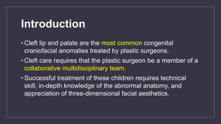
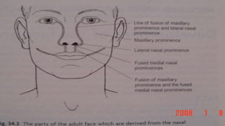
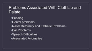
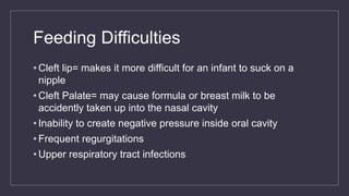
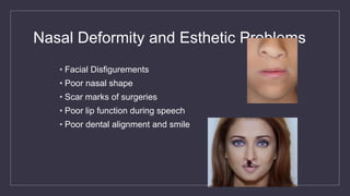
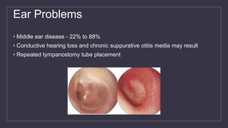
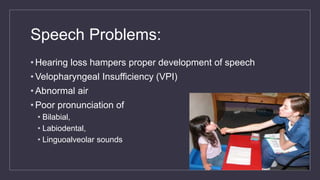
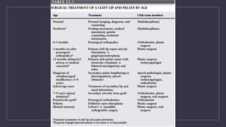
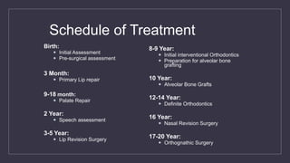
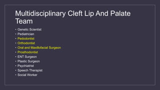
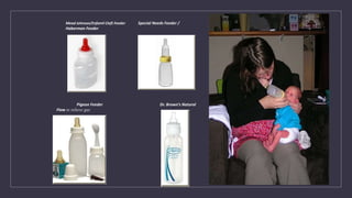
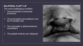
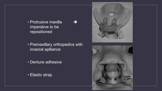
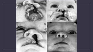
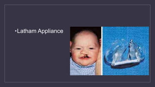
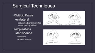
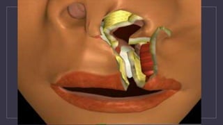
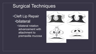
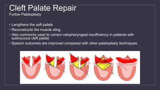
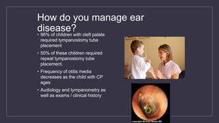
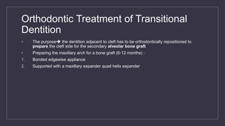
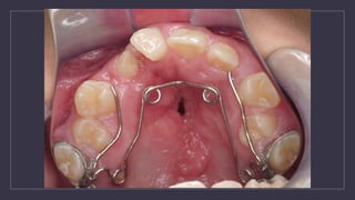
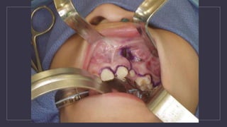
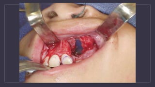
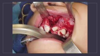
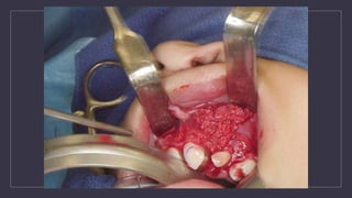
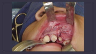
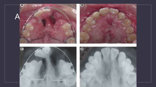
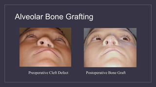
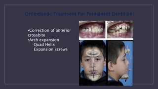
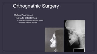
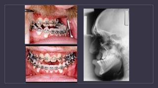
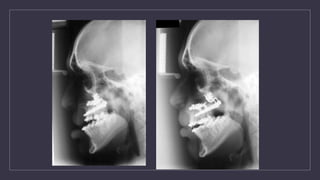
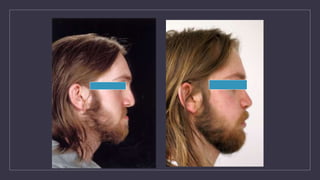
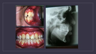
• Cleft lip and palate are the most common congenital
craniofacial anomalies treated by plastic surgeons.
• Cleft care requires that the plastic surgeon be a member of a
collaborative multidisciplinary team.
• Successful treatment of these children requires technical
skill, in-depth knowledge of the abnormal anatomy, and
appreciation of three-dimensional facial aesthetics.
EPIDEMIOLOGY
• the most common diagnosis is cleft lip and palate at 46%,
• Isolated cleft palate at 33%
• isolated cleft lip at 21%.
• Majority of bilateral cleft lips (86%) and unilateral cleft lips (68%) are associated with a cleft
palate.
• In the white population, cleft lip with or without cleft palate occurs in approximately 1 in 1,000
live births.
• twice as common in the Asian population
• half as common in African Americans
ETIOPATHOGENESIS
• Both environmental teratogens and genetic factors are implicated in the genesis of cleft lip and
palate
• Intrauterine exposure to the anticonvulsant phenytoin is associated with a 10-fold increase in
the incidence of cleft lip.
• Maternal smoking during pregnancy doubles the incidence of cleft lip.
• Genetic abnormalities can result in syndromes that include clefts of the primary or secondary
palates among the developmental fields affected.
• The most common syndrome associated with cleft lip and palate is van derWoude syndrome
• DiGeorge, or conotruncal anomaly syndromes are the most common diagnoses associated with
isolated cleft palate.
MULTIDISCIPLINARY CLEFT
CARE
• cleft lip and or palate require coordinated care from multiple specialties to
optimize treatment outcome.
• Typical members of a cleft team:
• audiologist, dentist, geneticist, nurse, nutritionist/dietitian, oral surgeon, orthodontist,
otolaryngologist, pediatrician, plastic surgeon, psychologist, social worker, and speech
pathologist.
• The goal of cleft care is to eliminate as many steps in the treatment plan as
possible by optimizing the outcome and benefit of each essential intervention.
• a child with a cleft requires a complex lengthy surgical treatment plan
Head and Neck of 4-Week Old Embryo
Trigeminal nerve
Facial nerve
Glossopharyngeal
nerve
Vagus nerve
Embryological Background
•Various theories have been given for its
development.
1. Alteration in intrinsic palatal shelf force
2. Failure of tongue to drop down
3. Non fusion of shelves
4. Rupture of cyst formed at the site of fusion
Classification
• We classify as the follows:
• its combined (cl+cp) or isolated cleft(cl or cp)?
• is it unilateral or bilateral?
• is it complete (if it cross the nasal philtrum) or incomplete ( if
it doesn’t cross the nasal philtrum.
Nagpur classification
• Group I - Cleft of lip
• Group I(A) - Cleft of lip & alveolus
• Group II - Cleft of palate
• Group II(S) - Submucous cleft of palate
• Group III - Cleft of lip & palate
Karnahan’s Classification
Millard’s Modification of
Karnahan’s Classifcation
Lip
Alveolus
Primary Palate
Hard Palate (Maxillary)
Hard Palate (Palatine)
Soft Palate
Kriens “LAHSHAL”
L = Lip (right)
A = Alveolus (right)
H = Hard Palate (right)
S = Soft Palate (median)
H = Hard Palate (left)
A = Alveolus (left)
L = Lip (left)
Capital letter = complete cleft
Lowercase letter = incomplete cleft
“.” or “-” = normal
Examples
LA….l = complete right cleft lip and alveolus,
incomplete left cleft lip
LAHS = complete right unilateral cleft lip, alveolus,
hard, and soft palate
Other Types of Clefts
• Microform Cleft:
• May look like
• a little dent in the red part of the lip
• a scar from the lip up to the nostril.
• Muscle tissue underneath the cleft can
be affected and may require surgery
• Submucous Cleft Palate:
• Midline deficiency or lack of muscular
tissue
• Often a submucous cleft palate is
associated with a bifid or cleft uvula
• Posterior nasal spine is almost always
missing
• Speech Problems are common
Prenatal Diagnosis
• Cleft lip can be easily diagnosed
by performing ultrasonography in
the second trimester
• Diagnosing a cleft palate with
ultrasonography is very difficult
• Three-dimensional imaging has
been introduced to prenatal
ultrasonography diagnostics of
cleft anomalies
Diagnosis
• Advantages of Prenatal
Diagnosis:
1. Time for parental education
2. Time for parental psychological
preparation
3. Opportunity to investigate other
associated anomalies
4. Gives parents the choice of
continuing the pregnancy
5. Opportunity for fetal surgery
Etiology
• “Actually no one knows exactly what causes clefts”
• Multiple factors may be involved, like:
• Genetics (inherited characteristic) from one or both parents
.
• Environmental factors
• Drugs: corticosteroids (anti-inflammatory), phenytoin
(anticonvulsant), retinoid.
• Infections: like rubella during pregnancy.
• Alcohol consumption, smoking, hypoxia during pregnancy,
some of dietary and vitamins deficiencies (like folic acid and
vitamin A deficiency)
• Maternal Age
GENETICS
CL/P
• Normal parents, one child with CL/P  4% risk CL/P in next child
• Normal parents, two kids with CL/P  9% risk CL/P in next child
• One parent CL/P, no affected kids  4% risk CL/P in next child
• One parent CL/P, one child CL/P  17% risk CL/P in next child
• Risk of CL/P in siblings increases with severity of deformity
• - child with unilateral CL  risk CL/P next child 2.5%
• - child with bilateral CL/P  risk CL/P next child 5.7%
CP
• Normal parents, one child with CP  2% risk of CP in next child
• Normal parents, 2 children with CP  7% risk of CP in next child
• Parent with CP, no affected children  6% risk for next child
• Parent with CP, one child with CP  15% risk for next child
• All infants with clefs must be evaluated for presence of other anomalies
• Associated nomalies are more common in infants with isolated CP
Nonsyndromic Cleft
Pierre Robin Sequence is the most
common associated nonsyndromic
anomaly
• is a relative term describing the small size
of the lower jaw ) and Glossoptosis (is a
medical condition and abnormality which
refers to the downward displacement or
retraction of the tongue)
Syndromic Cleft
Common Syndromes
Stickler (25% of syndromic CP) –
• autosomal dominant type 2 collagen gene
• Pierre Robin, ocular/hearing/joint malformations
Velocardiofacial (Shprintzen’s) – 15% of syndromic
CP
• Autosomal dominant, variable expressivity, CATCH
22 – Deletions at 22q11,
• Facial, cardiovascular, immunologic, developmental
anomalies
Van der Woude’s (19% of syndromic CL/P and CP)
• Autosomal dominant
• CL/P or CP with bilateral lower lip pits
Shprintzen’
Problems Associated With Cleft Lip and
Palate
•Feeding
•Dental problems
•Nasal Deformity and Esthetic Problems
•Ear Problems
•Speech Difficulties
•Associated Anomalies
Feeding Difficulties
• Cleft lip= makes it more difficult for an infant to suck on a
nipple
• Cleft Palate= may cause formula or breast milk to be
accidently taken up into the nasal cavity
• Inability to create negative pressure inside oral cavity
• Frequent regurgitations
• Upper respiratory tract infections
Dental Problems
• Local Dental Problems:
• Congenitally Missing teeth, Hypodontia, Hyperdontia, Oligodontia
• Presence of natal and neonatal teeth
• Anamalies of tooth morphology like microdontia, macrodontia etc
• Fused teeth
• Enamel Hypoplasia
• Poor periodontal support, early loss of teeth
• Gemination, Dilacerations
• Orthodontics Problems:
• Class III tendency
• Anterior and Posterior Cross bite
• Spacing and crowding
Nasal Deformity and Esthetic Problems
• Facial Disfigurements
• Poor nasal shape
• Scar marks of surgeries
• Poor lip function during speech
• Poor dental alignment and smile
Ear Problems
• Middle ear disease - 22% to 88%
• Conductive hearing loss and chronic suppurative otitis media may result
• Repeated tympanostomy tube placement
Why do cleft kids have eustachian
tube dysfunction?
•Abnormal curvature of the eustachian tube lumen
•Cephalometric data - width and angulation of the skull
base with respect to the eustachian tube are different
•Abnormal insertions of the tensor and levator veli
palatini muscles into the cartilages and skull base
•Palatal muscle dysfunction
Speech Problems:
• Hearing loss hampers proper development of speech
• Velopharyngeal Insufficiency (VPI)
• Abnormal air
• Poor pronunciation of
• Bilabial,
• Labiodental,
• Linguoalveolar sounds
C
MANAGEMENT OF
CLEFT LIP AND
PALATE
Schedule of Treatment
Birth:
 Initial Assessment
 Pre-surgical assessment
3 Month:
 Primary Lip repair
9-18 month:
 Palate Repair
2 Year:
 Speech assessment
3-5 Year:
 Lip Revision Surgery
8-9 Year:
 Initial interventional Orthodontics
 Preparation for alveolar bone
grafting
10 Year:
 Alveolar Bone Grafts
12-14 Year:
 Definite Orthodontics
16 Year:
 Nasal Revision Surgery
17-20 Year:
 Orthognathic Surgery
Multidisciplinary Cleft Lip And Palate
Team
• Genetic Scientist
• Pediatrician
• Pedodontist
• Orthodontist
• Oral and Maxillofacial Surgeon
• Prosthodontist
• ENT Surgeon
• Plastic Surgeon
• Psychiatrist
• Speech Therapist
• Social Worker
Feeding
• Cleft lip = makes it more difficult for an infant to suck on a
nipple
• use special nipples to allow the baby to latch properly
(either pump or use formula)
• Cleft Palate = may cause formula or breast milk to be
accidentally taken up into the nasal cavity
• don’t feed baby without palatal obturator (prosthetic palate)
• feed in an upright position to keep milk from coming out of
the nose
Mead Johnson/Enfamil Cleft Feeder Special Needs Feeder /
Haberman Feeder
Pigeon Feeder Dr. Brown’s Natural
Flow to relieve gas
Presurgical Orthopeadics:
1.Reduces the size of cleft; Aids in Surgery
2.Partial obturation aids in feeding
3.Parental Reassurance at a crucial time
• Maxillary Strapping
• Nasoalveolar Moulding Appliances (NAM)
 Require orthopedic repositioning
of the nasal cartilages, columella,
nasal tip, and lateral wall of the
vestibule
 Presurgical infant nasal
remodelling  nasal molding
 Nasal molding by post surgical
nasal stenting
 Facial taping  2 to 3 months
BILATERAL CLEFT LIP
The most challenging condition
1. The premaxilla is extremely
protrusive
2. The premaxilla and prolabium can
be of variable size
3. The columella is deficient/almost
nonexistent
4. The palatal shelves are collapsed
• Protrusive maxilla 
imperative to be
repositioned
• Premaxillary orthopedics with
inraoral aplliance
• Denture adhesive
• Elastic strap
•Latham Appliance
Rule of Ten
Primary repair- repaired at approximately 10 weeks
• The surgeon usually uses the “Rule of Ten”
• The child weighs 10 pounds
• The child has a hemoglobin of at least 10 grams
• The child has a white count of no higher than 10,000
• The child is at least 10 weeks of age
Surgical Techniques
• Cleft Lip Repair
•unilateral
• rotation-advancement flap
developed by Millard
• complications
•dehiscence
• infection
• excess tension
Surgical Techniques
•Cleft Lip Repair
•bilateral
• bilateral rotation
advancement with
attachment to
premaxilla mucosa
Cleft Palate Repair - Timing
• Dorf and Curtin
• 10% occurrence of articulation errors when palatoplasty was
completed by 1 year
• 86% incidence of articulation errors when repair was complete after 1
year
• Haapanen and Rantala –
• Significantly fewer children in the groups repaired before 18 months
had hypernasal speech, articulation errors, or required secondary
surgery to correct speech
Cleft Palate Repair
•Schweckendick’s Primary Veloplasty
•V-Y Pushback
•Von Langenbeck Palatal Repair
•Furlow Palatoplasty
Cleft Palate Repair
Schweckendick’s Primary Veloplasty
• Incisions made in soft palate
• Muscle bundles released from the posterior hard palate and rotated
• Reconstruction of levator sling
• Closure of mucosal layers separately
Cleft Palate Repair
V-Y Pushback (WARDILL OPERATION)
• Two uni-pedicled flaps (greater palatine artery) and one or two anteriorly based pedicled flaps
• Posterior flaps rotated in a V-Y advancement technique - increasing the length of the palate
• Nasal mucosa not closed
• Improved speech results compared with bipedicled techniques
• Indicated for incomplete clefts
Von Langenbeck Operation:
Cleft Palate Repair
Furlow Palatoplasty
• Lengthens the soft palate
• Reconstructs the muscle sling.
• Also commonly used to correct velopharyngeal insufficiency in patients with
submucous cleft palate
• Speech outcomes are improved compared with other palatoplasty techniques.
How do you manage ear
disease?• 96% of children with cleft palate
required tympanostomy tube
placement
• 50% of these children required
repeat tympanostomy tube
placement.
• Frequency of otitis media
decreases as the child with CP
ages
• Audiology and tympanometry as
well as exams / clinical history
Orthodontic Treatment of Transitional
Dentition
• The purpose the dentition adjacent to cleft has to be orthodontically repositioned to
prepare the cleft side for the secondary alveolar bone graft
• Preparing the maxillary arch for a bone graft (6-12 months) :
1. Bonded edgewise appliance
2. Supported with a maxillary expander quad helix expander
Alveolar Bone Grafting
• Primary Bone Grafting
• Bone graft done at the time of primary cheiloplasty
• Bone graft done during the first 2 years of life
• Bone graft done prior to the eruption of the primary canine
Secondary Bone Grafting
• Done before eruption of the permanent canine
• Usually when the root of the canine is 1/3 to 2/3 formed
• Usually between ages 8-10
• In CLP dental age is usually behind chronological age
Quadhelix to expand prior to ABG
angle brackets to keep roots
away from cleft
Alveolar Bone Grafting
1. Provide bone for the eruption and/or orthodontic repositioning of teeth
2. Closure of oro-nasal fistulas
3. Support and elevation of the alar base
4. Stabilization of the pre-maxilla in bilateral cases
5. Provide continuity of the alveolar ridge
Types of bone grafts
• Autogenous
• Cancellous- iliac crest
• Cortical- calvarium, mandible
• Cortico-cancellous- iliac, rib, tibia, mandible
• *Allogeneic
• Graft resorbs, remodels, may contribute to osteoinduction and osteoconduction
• **Alloplast
• Bone grows into, around alloplast
• No active osteoinduction but some osteoconduction
• Teeth do not erupt through alloplast
*Nique T, Fonseca RJ, et al: Particulate allogeneic bone grafts into maxillary alveolar clefts in humans- A preliminary report. J Oral
Maxillofac Surg 45: 386-392, 1987
**Horswell BB, El Deeb M: Nonporous HA in the repair of alveolar cleft defect in a primate model. J Oral Maxiilofac Surg 47:946-
952, 1989
Alveolar Bone Grafting
Preoperative Cleft Defect Postoperative Bone Graft
•Correction of anterior
crossbite
•Arch expansion
Quad Helix
Expansion screws
Orthodontic Treatment For Permanent Dentition
Orthognathic Surgery
• Midfacial Advancement
• LeForte osteotomies
• leave vascular pedicle attached in back
of maxilla - prevents necrosis
Rhinoplasty
• Rhinoplasty
• standard techniques
• tip projection
• alar rotation
• columellar length
• Age =17-20
Surgical Management of Velopharyngeal
Insufficiency
Major Goals of Surgery
• Close the gap or hole between the roof of the mouth and the nose.
• Reconnect the muscles that normally make the palate work.
• Make the repaired palate long enough so that when the muscles are working, the palate
can perform its function properly.
Velopharyngeal Insufficiency VPI
Wardill Operation: WY push back
• Dorrance and Brown’s – U shaped push back palatoplasty
Prosthetic Management of VPI
C
THANK YOU

Cleft lip and palate

  • 1.
    C CLEFT LIP AND PALATE DrSumer yadav MCh -Plastic and reconstructive surgery
  • 2.
    Introduction • Cleft lipand palate are the most common congenital craniofacial anomalies treated by plastic surgeons. • Cleft care requires that the plastic surgeon be a member of a collaborative multidisciplinary team. • Successful treatment of these children requires technical skill, in-depth knowledge of the abnormal anatomy, and appreciation of three-dimensional facial aesthetics.
  • 3.
    EPIDEMIOLOGY • the mostcommon diagnosis is cleft lip and palate at 46%, • Isolated cleft palate at 33% • isolated cleft lip at 21%. • Majority of bilateral cleft lips (86%) and unilateral cleft lips (68%) are associated with a cleft palate. • In the white population, cleft lip with or without cleft palate occurs in approximately 1 in 1,000 live births. • twice as common in the Asian population • half as common in African Americans
  • 4.
    ETIOPATHOGENESIS • Both environmentalteratogens and genetic factors are implicated in the genesis of cleft lip and palate • Intrauterine exposure to the anticonvulsant phenytoin is associated with a 10-fold increase in the incidence of cleft lip. • Maternal smoking during pregnancy doubles the incidence of cleft lip. • Genetic abnormalities can result in syndromes that include clefts of the primary or secondary palates among the developmental fields affected. • The most common syndrome associated with cleft lip and palate is van derWoude syndrome • DiGeorge, or conotruncal anomaly syndromes are the most common diagnoses associated with isolated cleft palate.
  • 5.
    MULTIDISCIPLINARY CLEFT CARE • cleftlip and or palate require coordinated care from multiple specialties to optimize treatment outcome. • Typical members of a cleft team: • audiologist, dentist, geneticist, nurse, nutritionist/dietitian, oral surgeon, orthodontist, otolaryngologist, pediatrician, plastic surgeon, psychologist, social worker, and speech pathologist. • The goal of cleft care is to eliminate as many steps in the treatment plan as possible by optimizing the outcome and benefit of each essential intervention. • a child with a cleft requires a complex lengthy surgical treatment plan
  • 6.
    Head and Neckof 4-Week Old Embryo Trigeminal nerve Facial nerve Glossopharyngeal nerve Vagus nerve Embryological Background
  • 11.
    Embryological Background •Various theorieshave been given for its development. 1. Alteration in intrinsic palatal shelf force 2. Failure of tongue to drop down 3. Non fusion of shelves 4. Rupture of cyst formed at the site of fusion
  • 12.
    Classification • We classifyas the follows: • its combined (cl+cp) or isolated cleft(cl or cp)? • is it unilateral or bilateral? • is it complete (if it cross the nasal philtrum) or incomplete ( if it doesn’t cross the nasal philtrum.
  • 13.
    Nagpur classification • GroupI - Cleft of lip • Group I(A) - Cleft of lip & alveolus • Group II - Cleft of palate • Group II(S) - Submucous cleft of palate • Group III - Cleft of lip & palate
  • 15.
    Karnahan’s Classification Millard’s Modificationof Karnahan’s Classifcation Lip Alveolus Primary Palate Hard Palate (Maxillary) Hard Palate (Palatine) Soft Palate
  • 16.
    Kriens “LAHSHAL” L =Lip (right) A = Alveolus (right) H = Hard Palate (right) S = Soft Palate (median) H = Hard Palate (left) A = Alveolus (left) L = Lip (left) Capital letter = complete cleft Lowercase letter = incomplete cleft “.” or “-” = normal Examples LA….l = complete right cleft lip and alveolus, incomplete left cleft lip LAHS = complete right unilateral cleft lip, alveolus, hard, and soft palate
  • 17.
    Other Types ofClefts • Microform Cleft: • May look like • a little dent in the red part of the lip • a scar from the lip up to the nostril. • Muscle tissue underneath the cleft can be affected and may require surgery • Submucous Cleft Palate: • Midline deficiency or lack of muscular tissue • Often a submucous cleft palate is associated with a bifid or cleft uvula • Posterior nasal spine is almost always missing • Speech Problems are common
  • 18.
    Prenatal Diagnosis • Cleftlip can be easily diagnosed by performing ultrasonography in the second trimester • Diagnosing a cleft palate with ultrasonography is very difficult • Three-dimensional imaging has been introduced to prenatal ultrasonography diagnostics of cleft anomalies
  • 19.
    Diagnosis • Advantages ofPrenatal Diagnosis: 1. Time for parental education 2. Time for parental psychological preparation 3. Opportunity to investigate other associated anomalies 4. Gives parents the choice of continuing the pregnancy 5. Opportunity for fetal surgery
  • 20.
    Etiology • “Actually noone knows exactly what causes clefts” • Multiple factors may be involved, like: • Genetics (inherited characteristic) from one or both parents . • Environmental factors • Drugs: corticosteroids (anti-inflammatory), phenytoin (anticonvulsant), retinoid. • Infections: like rubella during pregnancy. • Alcohol consumption, smoking, hypoxia during pregnancy, some of dietary and vitamins deficiencies (like folic acid and vitamin A deficiency) • Maternal Age
  • 21.
    GENETICS CL/P • Normal parents,one child with CL/P  4% risk CL/P in next child • Normal parents, two kids with CL/P  9% risk CL/P in next child • One parent CL/P, no affected kids  4% risk CL/P in next child • One parent CL/P, one child CL/P  17% risk CL/P in next child • Risk of CL/P in siblings increases with severity of deformity • - child with unilateral CL  risk CL/P next child 2.5% • - child with bilateral CL/P  risk CL/P next child 5.7% CP • Normal parents, one child with CP  2% risk of CP in next child • Normal parents, 2 children with CP  7% risk of CP in next child • Parent with CP, no affected children  6% risk for next child • Parent with CP, one child with CP  15% risk for next child • All infants with clefs must be evaluated for presence of other anomalies • Associated nomalies are more common in infants with isolated CP
  • 22.
    Nonsyndromic Cleft Pierre RobinSequence is the most common associated nonsyndromic anomaly • is a relative term describing the small size of the lower jaw ) and Glossoptosis (is a medical condition and abnormality which refers to the downward displacement or retraction of the tongue)
  • 23.
    Syndromic Cleft Common Syndromes Stickler(25% of syndromic CP) – • autosomal dominant type 2 collagen gene • Pierre Robin, ocular/hearing/joint malformations Velocardiofacial (Shprintzen’s) – 15% of syndromic CP • Autosomal dominant, variable expressivity, CATCH 22 – Deletions at 22q11, • Facial, cardiovascular, immunologic, developmental anomalies Van der Woude’s (19% of syndromic CL/P and CP) • Autosomal dominant • CL/P or CP with bilateral lower lip pits Shprintzen’
  • 24.
    Problems Associated WithCleft Lip and Palate •Feeding •Dental problems •Nasal Deformity and Esthetic Problems •Ear Problems •Speech Difficulties •Associated Anomalies
  • 25.
    Feeding Difficulties • Cleftlip= makes it more difficult for an infant to suck on a nipple • Cleft Palate= may cause formula or breast milk to be accidently taken up into the nasal cavity • Inability to create negative pressure inside oral cavity • Frequent regurgitations • Upper respiratory tract infections
  • 26.
    Dental Problems • LocalDental Problems: • Congenitally Missing teeth, Hypodontia, Hyperdontia, Oligodontia • Presence of natal and neonatal teeth • Anamalies of tooth morphology like microdontia, macrodontia etc • Fused teeth • Enamel Hypoplasia • Poor periodontal support, early loss of teeth • Gemination, Dilacerations • Orthodontics Problems: • Class III tendency • Anterior and Posterior Cross bite • Spacing and crowding
  • 27.
    Nasal Deformity andEsthetic Problems • Facial Disfigurements • Poor nasal shape • Scar marks of surgeries • Poor lip function during speech • Poor dental alignment and smile
  • 28.
    Ear Problems • Middleear disease - 22% to 88% • Conductive hearing loss and chronic suppurative otitis media may result • Repeated tympanostomy tube placement
  • 29.
    Why do cleftkids have eustachian tube dysfunction? •Abnormal curvature of the eustachian tube lumen •Cephalometric data - width and angulation of the skull base with respect to the eustachian tube are different •Abnormal insertions of the tensor and levator veli palatini muscles into the cartilages and skull base •Palatal muscle dysfunction
  • 30.
    Speech Problems: • Hearingloss hampers proper development of speech • Velopharyngeal Insufficiency (VPI) • Abnormal air • Poor pronunciation of • Bilabial, • Labiodental, • Linguoalveolar sounds
  • 31.
  • 33.
    Schedule of Treatment Birth: Initial Assessment  Pre-surgical assessment 3 Month:  Primary Lip repair 9-18 month:  Palate Repair 2 Year:  Speech assessment 3-5 Year:  Lip Revision Surgery 8-9 Year:  Initial interventional Orthodontics  Preparation for alveolar bone grafting 10 Year:  Alveolar Bone Grafts 12-14 Year:  Definite Orthodontics 16 Year:  Nasal Revision Surgery 17-20 Year:  Orthognathic Surgery
  • 34.
    Multidisciplinary Cleft LipAnd Palate Team • Genetic Scientist • Pediatrician • Pedodontist • Orthodontist • Oral and Maxillofacial Surgeon • Prosthodontist • ENT Surgeon • Plastic Surgeon • Psychiatrist • Speech Therapist • Social Worker
  • 35.
    Feeding • Cleft lip= makes it more difficult for an infant to suck on a nipple • use special nipples to allow the baby to latch properly (either pump or use formula) • Cleft Palate = may cause formula or breast milk to be accidentally taken up into the nasal cavity • don’t feed baby without palatal obturator (prosthetic palate) • feed in an upright position to keep milk from coming out of the nose
  • 36.
    Mead Johnson/Enfamil CleftFeeder Special Needs Feeder / Haberman Feeder Pigeon Feeder Dr. Brown’s Natural Flow to relieve gas
  • 37.
    Presurgical Orthopeadics: 1.Reduces thesize of cleft; Aids in Surgery 2.Partial obturation aids in feeding 3.Parental Reassurance at a crucial time • Maxillary Strapping • Nasoalveolar Moulding Appliances (NAM)
  • 38.
     Require orthopedicrepositioning of the nasal cartilages, columella, nasal tip, and lateral wall of the vestibule  Presurgical infant nasal remodelling  nasal molding  Nasal molding by post surgical nasal stenting  Facial taping  2 to 3 months
  • 39.
    BILATERAL CLEFT LIP Themost challenging condition 1. The premaxilla is extremely protrusive 2. The premaxilla and prolabium can be of variable size 3. The columella is deficient/almost nonexistent 4. The palatal shelves are collapsed
  • 40.
    • Protrusive maxilla imperative to be repositioned • Premaxillary orthopedics with inraoral aplliance • Denture adhesive • Elastic strap
  • 42.
  • 43.
    Rule of Ten Primaryrepair- repaired at approximately 10 weeks • The surgeon usually uses the “Rule of Ten” • The child weighs 10 pounds • The child has a hemoglobin of at least 10 grams • The child has a white count of no higher than 10,000 • The child is at least 10 weeks of age
  • 44.
    Surgical Techniques • CleftLip Repair •unilateral • rotation-advancement flap developed by Millard • complications •dehiscence • infection • excess tension
  • 46.
    Surgical Techniques •Cleft LipRepair •bilateral • bilateral rotation advancement with attachment to premaxilla mucosa
  • 47.
    Cleft Palate Repair- Timing • Dorf and Curtin • 10% occurrence of articulation errors when palatoplasty was completed by 1 year • 86% incidence of articulation errors when repair was complete after 1 year • Haapanen and Rantala – • Significantly fewer children in the groups repaired before 18 months had hypernasal speech, articulation errors, or required secondary surgery to correct speech
  • 48.
    Cleft Palate Repair •Schweckendick’sPrimary Veloplasty •V-Y Pushback •Von Langenbeck Palatal Repair •Furlow Palatoplasty
  • 49.
    Cleft Palate Repair Schweckendick’sPrimary Veloplasty • Incisions made in soft palate • Muscle bundles released from the posterior hard palate and rotated • Reconstruction of levator sling • Closure of mucosal layers separately
  • 50.
    Cleft Palate Repair V-YPushback (WARDILL OPERATION) • Two uni-pedicled flaps (greater palatine artery) and one or two anteriorly based pedicled flaps • Posterior flaps rotated in a V-Y advancement technique - increasing the length of the palate • Nasal mucosa not closed • Improved speech results compared with bipedicled techniques • Indicated for incomplete clefts
  • 51.
  • 52.
    Cleft Palate Repair FurlowPalatoplasty • Lengthens the soft palate • Reconstructs the muscle sling. • Also commonly used to correct velopharyngeal insufficiency in patients with submucous cleft palate • Speech outcomes are improved compared with other palatoplasty techniques.
  • 53.
    How do youmanage ear disease?• 96% of children with cleft palate required tympanostomy tube placement • 50% of these children required repeat tympanostomy tube placement. • Frequency of otitis media decreases as the child with CP ages • Audiology and tympanometry as well as exams / clinical history
  • 54.
    Orthodontic Treatment ofTransitional Dentition • The purpose the dentition adjacent to cleft has to be orthodontically repositioned to prepare the cleft side for the secondary alveolar bone graft • Preparing the maxillary arch for a bone graft (6-12 months) : 1. Bonded edgewise appliance 2. Supported with a maxillary expander quad helix expander
  • 55.
    Alveolar Bone Grafting •Primary Bone Grafting • Bone graft done at the time of primary cheiloplasty • Bone graft done during the first 2 years of life • Bone graft done prior to the eruption of the primary canine
  • 56.
    Secondary Bone Grafting •Done before eruption of the permanent canine • Usually when the root of the canine is 1/3 to 2/3 formed • Usually between ages 8-10 • In CLP dental age is usually behind chronological age
  • 57.
    Quadhelix to expandprior to ABG
  • 58.
    angle brackets tokeep roots away from cleft
  • 59.
    Alveolar Bone Grafting 1.Provide bone for the eruption and/or orthodontic repositioning of teeth 2. Closure of oro-nasal fistulas 3. Support and elevation of the alar base 4. Stabilization of the pre-maxilla in bilateral cases 5. Provide continuity of the alveolar ridge
  • 60.
    Types of bonegrafts • Autogenous • Cancellous- iliac crest • Cortical- calvarium, mandible • Cortico-cancellous- iliac, rib, tibia, mandible • *Allogeneic • Graft resorbs, remodels, may contribute to osteoinduction and osteoconduction • **Alloplast • Bone grows into, around alloplast • No active osteoinduction but some osteoconduction • Teeth do not erupt through alloplast *Nique T, Fonseca RJ, et al: Particulate allogeneic bone grafts into maxillary alveolar clefts in humans- A preliminary report. J Oral Maxillofac Surg 45: 386-392, 1987 **Horswell BB, El Deeb M: Nonporous HA in the repair of alveolar cleft defect in a primate model. J Oral Maxiilofac Surg 47:946- 952, 1989
  • 69.
  • 70.
    Alveolar Bone Grafting PreoperativeCleft Defect Postoperative Bone Graft
  • 71.
    •Correction of anterior crossbite •Archexpansion Quad Helix Expansion screws Orthodontic Treatment For Permanent Dentition
  • 72.
    Orthognathic Surgery • MidfacialAdvancement • LeForte osteotomies • leave vascular pedicle attached in back of maxilla - prevents necrosis
  • 77.
    Rhinoplasty • Rhinoplasty • standardtechniques • tip projection • alar rotation • columellar length • Age =17-20
  • 78.
    Surgical Management ofVelopharyngeal Insufficiency Major Goals of Surgery • Close the gap or hole between the roof of the mouth and the nose. • Reconnect the muscles that normally make the palate work. • Make the repaired palate long enough so that when the muscles are working, the palate can perform its function properly.
  • 79.
  • 80.
  • 81.
    • Dorrance andBrown’s – U shaped push back palatoplasty
  • 82.
  • 83.