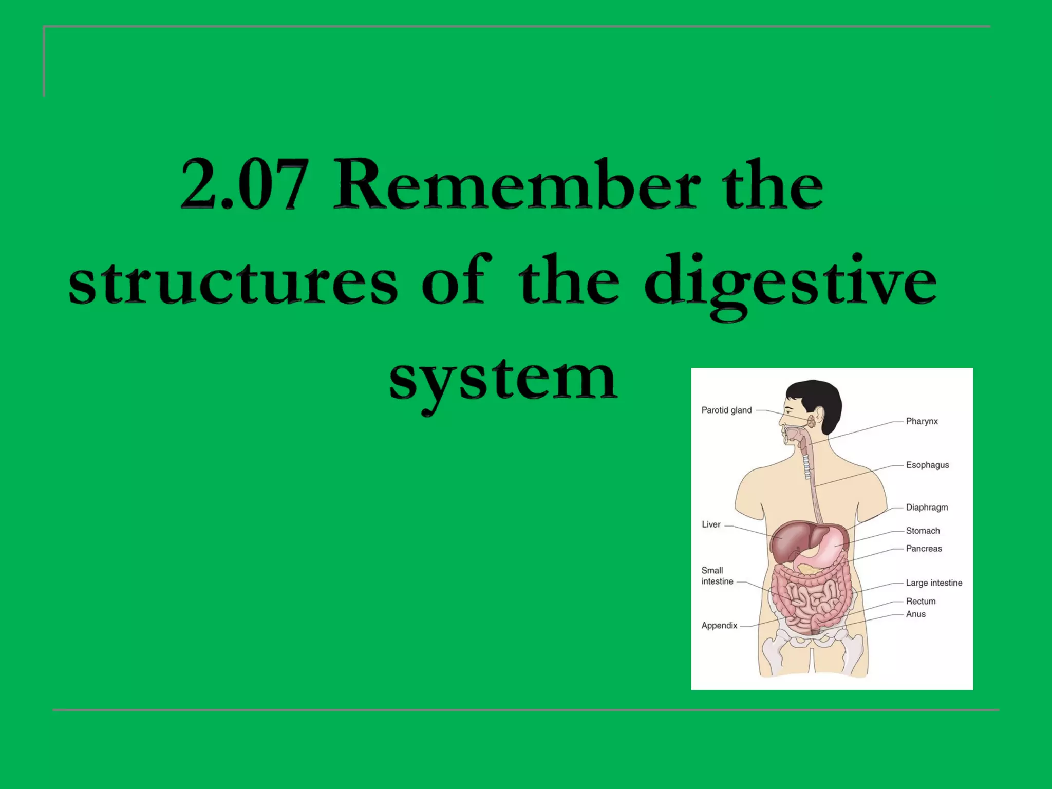
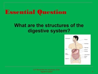
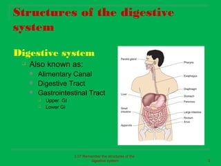
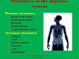
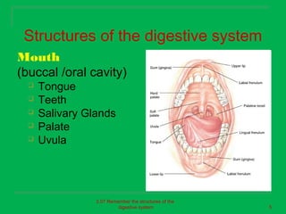
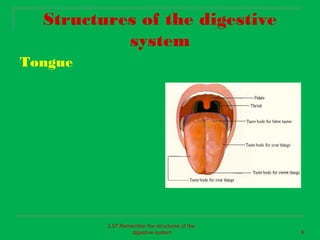
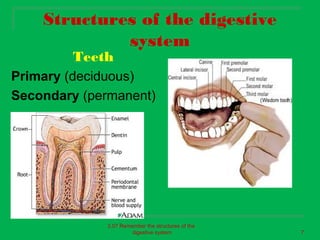
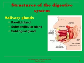
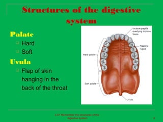
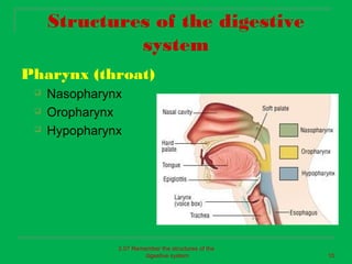
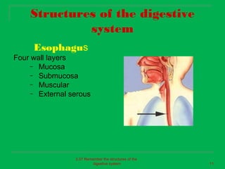
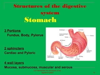
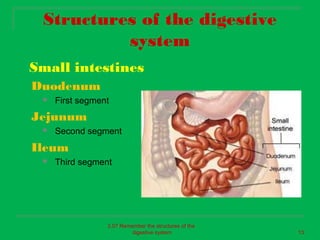
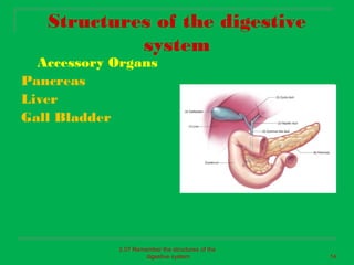
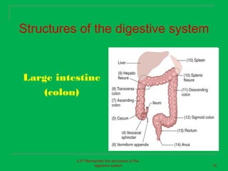
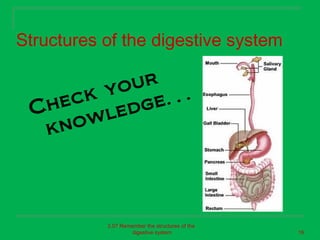
2. Essential Question
What are the structures of the
digestive system?
2.07 Remember the structures of the
digestive system 2
3. Structures of the digestive
system
Digestive system
Also known as:
Alimentary Canal
Digestive Tract
Gastrointestinal Tract
Upper GI
Lower GI
2.07 Remember the structures of the
digestive system 3
4. Structures of the digestive
system
Primary structures
Mouth (oral cavity)
Esophagus (gullet)
Stomach
Small intestine
Large intestine (colon)
Accessory structures
Tongue
Teeth
Salivary glands
Pancreas
Liver
Gall bladder
2.07 Remember the structures of the
digestive system 4
5. Structures of the digestive system
Mouth
(buccal /oral cavity)
Tongue
Teeth
Salivary Glands
Palate
Uvula
2.07 Remember the structures of the
digestive system 5
6. Structures of the digestive
system
Tongue
2.07 Remember the structures of the
digestive system 6
7. Structures of the digestive
system
Teeth
Primary (deciduous)
Secondary (permanent)
2.07 Remember the structures of the
digestive system 7
8. Structures of the digestive
system
Salivary glands
Parotid gland
Submandibular gland
Sublingual gland
2.07 Remember the structures of the
digestive system 8
9. Structures of the digestive
system
Palate
Hard
Soft
Uvula
Flap of skin
hanging in the
back of the throat
2.07 Remember the structures of the
digestive system 9
10. Structures of the digestive
system
Pharynx (throat)
Nasopharynx
Oropharynx
Hypopharynx
2.07 Remember the structures of the
digestive system 10
11. Structures of the digestive
system
Esophagus
Four wall layers
– Mucosa
– Submucosa
– Muscular
– External serous
2.07 Remember the structures of the
digestive system 11
12. Structures of the digestive
system
Stomach
3 Portions
Fundus, Body, Pylorus
2 sphincters
Cardiac and Pyloric
4 wall layers
Mucosa, submucosa, muscular and serous
2.07 Remember the structures of the
digestive system 12
13. Structures of the digestive
system
Small intestines
Duodenum
First segment
Jejunum
Second segment
Ileum
Third segment
2.07 Remember the structures of the
digestive system 13
14. Structures of the digestive
system
Accessory Organs
Pancreas
Liver
Gall Bladder
2.07 Remember the structures of the
digestive system 14
15. Structures of the digestive system
Large intestine
(colon)
2.07 Remember the structures of the
digestive system 15
16. Structures of the digestive system
yo ur
h eck g e…
C
ow l ed
kn
2.07 Remember the structures of the
digestive system 16
17. Essential Question
What are the structures of the
digestive system?
2.07 Remember the structures of the
digestive system 17
Editor's Notes
Approximately 30’ in length from mouth to anus
Lips protect the opening of your mouth. The inside of your mouth is lined with a mucous membrane
Attached to floor of mouth to help in both chewing and swallowing. The tongue is made from skeletal muscles. The tongue is attached to four bones- hyoid, mandible, and the left and right temporal bones. On the surface of the tongue are projections called papillae. There are nerve endings in the papillae which form the sense organs of taste- TASTE BUDS. Taste buds are able to respond to Sweet Sour Bitter and Salty. Taste buds are also sensitive to cold, heat, and pressure.
The gums (gingivae) support and protect the teeth. The gums are made up of fleshy tissue and is covered with mucous membranes. This membrane surrounds the narrow portions of the teeth and covers structures in the upper and lower jaw. 20 Deciduous (baby) teeth start to erupt at about 6 months of age and continue until around 2 years of age. There are 4 incisors (sharp for biting) 2 canines (pointed for tearing), and 4 molars (ridges for crushing and grinding). These teeth will last until about age 12. 32 Permanent teeth start developing around age 6. They push out the deciduous (baby) teeth. The last of the teeth to erupt are the third molars (wisdom teeth) which come in around 17-25 years of age. Each tooth is divided into three parts-the crown, the neck, and the root. The crown is the part of the tooth that is visible. The neck is where the tooth enters the gumline. The root is embedded into the alveolar process of the jaw. The periodontal membrane helps anchor the tooth into place. Inside the tooth is the pulp cavity which contains nerves and blood supply to the tooth. This cavity is surrounded by calcified tissue called dentin. In the crown portion, the dentin is covered by enamel. Enamel is the hardest substance in the body.
Saliva is secreted into the oral cavity by 3 pairs of salivary glands- parotid, submandibular, and sublingual. The parotid glands are located on both sides of the face (in front and below the ears). They are the largest of the salivary glands. Submandibular salivary gland is located below the parotids and near the angle of the lower jaw. It is about the size of a walnut. Sublingual salivary gland is the smallest of the three salivary glands. They are located under the sides of the tongue.
The hard palate is formed by the maxillary and the palatine bones and is covered with a mucous membrane. The soft palate is an arch shaped structure which separates the mouth from the nasopharynx. The soft palate is made from a movable mucous membrane fold.
The esophagus is a muscular tube which is 10 inches long. It begins at the lower end of the pharynx behind the trachea. It continues downward towards the mediastinum, in front of the spinal column and passes through the diaphragm. From there, the esophagus enters the upper part of the stomach. The muscles in the upper third of the esophagus are skeletal muscle (voluntary) and the lower portion is smooth muscle (involuntary).
Located in the upper part of the abdominal cavity just to the left of and below the diaphragm. The shape and position are determined by several factors- the amount of food contained within the stomach, the stage of digestion, the position of the person’s body, and the pressure exerted upon the stomach from the intestines below. Divided into three parts- upper (fundus) middle (body or greater curvature) lower (pylorus) The opening from the esophagus into the stomach is through a circle of muscle called cardiac sphincter which controls the passage of food into the stomach. The pyloric sphincter control the passage of food from the stomach into the first part of the small intestine. Wall Layers Mucous - the innermost layer. It is a thick layer made up of gastric glands embedded in connective tissue. When the stomach is not distended with food, the gastric mucosa is thrown into folds called rugae. Submucosa - made of loose areolar connective tissue Muscular - three layers of smooth muscle. Helps the stomach to perform peristalsis which pushes food into the small intestine. Serosa - thick outer layer covering the stomach.
Has the same four wall layers as the stomach- mucosa, submucosa, muscle layer, and the serosa. It is the coiled portion of the digective tract. 1 st section- duodenum is about 12 inches long and curves around the head of the pancreas. 2 nd section-jejunum is about 8 feet long 3 rd section- ileum is about 10-12 feet long.
Pancreas- feather shaped organ and is located behind the stomach Liver- largest organ in the body. Located below the diaphragm in the upper right quadrant of the abdominal cavity Gall bladder- small green organ in the inferior surface of the liver
About 5 feet long and 2 inches in diameter. Cecum- first portion of the colon Ascending colon- upward section on the right side of the abdomen Transverse colon- runs horizontally below the diaphragm Descending colon- travels downward on the left side of the abdomen Sigmoid colon- the s shaped portion of the colon Rectum- the muscular storage area for undigested wastes, and makes up the final portion of the digestive tract. The opening to the outside of the body is called the anus. Two sphincters- internal is smooth muscle involuntary and external is skeletal muscle voluntary.