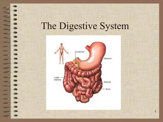
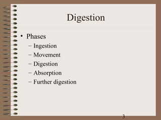
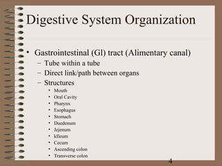
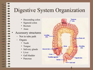
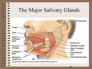
The Digestive System


                       1
Digestion
• Processing of food
• Types
   – Mechanical (physical)
      •   Chew
      •   Tear
      •   Grind
      •   Mash
      •   Mix
   – Chemical
      • Catabolic reactions
      • Enzymatic hydrolysis
           – Carbohydrate
           – Protein
           – Lipid
                                        2
Digestion
• Phases
  –   Ingestion
  –   Movement
  –   Digestion
  –   Absorption
  –   Further digestion


                               3
Digestive System Organization

• Gastrointestinal (Gl) tract (Alimentary canal)
   – Tube within a tube
   – Direct link/path between organs
   – Structures
      •   Mouth
      •   Oral Cavity
      •   Pharynx
      •   Esophagus
      •   Stomach
      •   Duedenum
      •   Jejenum
      •   kIleum
      •   Cecum
      •   Ascending colon
      •   Transverse colon
                                         4
Digestive System Organization
       •   Descending colon
       •   Sigmoid colon
       •   Rectum
       •   Anus
• Accessory structures
   – Not in tube path
   – Organs
       •   Teeth
       •   Tongue
       •   Salivary glands
       •   Liver
       •   Gall bladder
       •   Pancreas


                              5
Anatomy of the Mouth and
        Throat


                    6
Human Deciduous and
  Permanent Teeth


                 7
Dorsal Surface of the Tongue


                      8
The Major Salivary Glands


                     9
Deglutition (swallowing)
• Sequence
   – Voluntary stage
       • Push food to back of
         mouth
   – Pharyngeal stage
       • Raise
           – Soft palate
           – Larynx + hyoid
           – Tongue to soft palate
   – Esophageal stage
       • Contract pharyngeal
         muscles
       • Open esophagus
       • Start peristalsis


                                     10
Deglutition (swallowing)
• Control
   – Nerves
      • Glossopharyngeal
      • Vagus
      • Accessory
   – Brain stem
      • Deglutition center
            – Medulla oblongata
            – Pons
   – Disorders
      • Dysphagia
      • Aphagia


                                  11
Esophagus
• Usually collapsed (closed)
• 3 constrictions
   – Aortic arch
   – Left primary bronchus
   – Diaphragm
• Surrounded by
   – SNS plexus
   – Blood vessels
• Functions
   – Secrete mucous
   – Transport food
                                 12
Peristalsis and Segmentation


                       13
Esophagus
• Sphincters
  – Upper
  – Lower
• Abnormalities
  –   Achalasia
  –   Atresia
  –   Hernia
  –   Barret’s esophagus
  –   Esophageal varices


                               14
Stomach
• Usually “J” shaped
• Left side, anterior to the spleen
• Mucous membrane
   – G cells – make gastrin
   – Goblet cells – make mucous
   – Gastric pit – Oxyntic gland – Parietal cells – Make
     HCl
   – Chief cells – Zymogenic cells
      • Pepsin
      • Gastric lipase


                                              15
Anatomy of the Stomach


                   16
Stomach
• 3 muscle layers
     – Oblique
     – Circular
     – Longitudinal
• Regions
     –   Cardiac sphincter
     –   Fundus
     –   Antrum (pylorus)
     –   Pyloric sphincter
• Vascular
• Inner surface thrown into
  folds – Rugae
•   Contains enzymes that work
    best at pH 1-2
                                       17
Stomach
• Functions                       – Absorbs
   – Mix food                        •   Alcohol
   – Reservoir                       •   Water
   – Start digestion of
                                     •   Lipophilic acid
       • Protein
       • Nucleic acids
                                     •   B 12
       • Fats
   – Activates some enzymes
   – Destroy some bacteria
   – Makes intrinsic factor – B
     12 absorption
   – Destroys some bacteria


                                                18
Small Intestine
• Extends from pyloric
  sphincter  ileocecal
  valve
• Regions
   – Duodenum
   – Jejenum
   – Ileum
• Movements
   – Segmentation
   – Peristalsis


                                  19
Small Intestine
• Histology
  –   Intestinal glands – Intestinal enzymes
  –   Duodenal glands – Alkaline mucous
  –   Paneth cells – Lysozyme
  –   Microvilli
  –   Lacteals
  –   Plica circularis
  –   Smooth muscle
  –   Lymphatic tissue – GALT
  –   Vascular


                                               20
Small Intestine
• Absorbs                      – Lipids
   –   80% ingested water         •   Monoglycerides
   –   Electrolytes               •   Fatty acids
   –   Vitamins                   •   Micelles
   –   Minerals
                                  •   Chylomicrons
   –   Carbonates
        • Active/facilitated
          transport
        • Monosaccharides
   – Proteins
        • Di-/tripeptides
        • Amino acids


                                            21
Structure of the Villi in the
      Small Intestine


                        22
Small Intestine
• Secretes digestive
  enzymes
   – Peptidases
        • Amino-
        • Di-
        • Tri-
   –   Sucrases
   –   Maltase
   –   Lactase
   –   Saccharidases
        • Di-
        • Tri-
   – Lipase
   – Nucleases
                                     23
Small Intestine
• Control
• Requires pancreatic
  enzymes & bile to
  complete digestion


                               24
Large Intestine
• Extends from ileocecal valve to anus
• Regions
   – Cecum – Appendix
   – Colon
      • Ascending
      • Transverse
      • Descending
   – Rectum
   – Anal canal


                                         25
Anatomy of the Large Intestine


                       26
Large Intestine
• Histology
  – No villi
  – No permanent circular folds
  – Smooth muscle
     • Taeniae coli
     • Haustra
  – Epiploic appendages
  – Otherwise like rest of Gl tract


                                      27
Large Intestine
• Functions
  – Mechanical digestion       – Absorbs
     • Haustral churning          •More water
     • Peristalsis                •Vitamins
                                      –B
     • Reflexes
                                      –K
         – Gastroileal
         – Gastrocolic         – Concentrate/eliminate
                               wastes
  – Chemical digestion –
    Bacterial digestion
     • Ferment carbohydrates
     • Protein/amino acid
       breakdown

                                           28
Feces Formation and Defecation
• Chyme dehydrated to            • Control
  form feces
                                   – Parasympathetic
• Feces composition
   –   Water                       – Voluntary
   –   Inorganic salts
   –   Epithelial cells
   –   Bacteria
   –   Byproducts of digestion
• Defecation
   – Peristalsis pushes feces
     into rectum
   – Rectal walls stretch


                                             29
Liver
• Location
   – R. Hypochondrium
   – Epigastric region
• 4 Lobes
   –   Left
   –   Quadrate
   –   Caudate
   –   Right
• Each lobe has lobules – Contains hepatocytes –
  Surround sinusoids – Feed into central vein

                                       30
Liver
• Functions
   – Makes bile
      • Detergent – emulsifies
        fats
      • Release promoted by:
          – Vagus n.
          – CCK
          – Secretin
      • Contains
          –   Water
          –   Bile salts
          –   Bile pigments
          –   Electrolytes
          –   Cholesterol
          –   Lecithin


                                      31
Liver
– Detoxifies/removes
     • Drugs
     • Alcohol
– Stores
     •   Gycolgen
     •   Vitamins (A, D, E, K)
     •   Fe and other minerals
     •   Cholesterol
–   Activates vitamin D
–   Fetal RBC production
–   Phagocytosis
–   Metabolizes absorbed food
    molecules
     • Carbohydrates
     • Proteins
     • Lipids
                                         32
Liver
• Dual blood supply
  – Hepatic portal vein
     • Direct input from small
       intestine
  – Hepatic artery/vein
     • Direct links to heart


                                   33
The Duodenum and Related
        Organs


                   34
The Organs and Positions in the
      Abdominal Cavity


                        35
Structures of the Alimentary
           Canal


                       36

Digestive system

  • 1.
  • 2.
    Digestion • Processing offood • Types – Mechanical (physical) • Chew • Tear • Grind • Mash • Mix – Chemical • Catabolic reactions • Enzymatic hydrolysis – Carbohydrate – Protein – Lipid 2
  • 3.
    Digestion • Phases – Ingestion – Movement – Digestion – Absorption – Further digestion 3
  • 4.
    Digestive System Organization •Gastrointestinal (Gl) tract (Alimentary canal) – Tube within a tube – Direct link/path between organs – Structures • Mouth • Oral Cavity • Pharynx • Esophagus • Stomach • Duedenum • Jejenum • kIleum • Cecum • Ascending colon • Transverse colon 4
  • 5.
    Digestive System Organization • Descending colon • Sigmoid colon • Rectum • Anus • Accessory structures – Not in tube path – Organs • Teeth • Tongue • Salivary glands • Liver • Gall bladder • Pancreas 5
  • 6.
    Anatomy of theMouth and Throat 6
  • 7.
    Human Deciduous and Permanent Teeth 7
  • 8.
    Dorsal Surface ofthe Tongue 8
  • 9.
  • 10.
    Deglutition (swallowing) • Sequence – Voluntary stage • Push food to back of mouth – Pharyngeal stage • Raise – Soft palate – Larynx + hyoid – Tongue to soft palate – Esophageal stage • Contract pharyngeal muscles • Open esophagus • Start peristalsis 10
  • 11.
    Deglutition (swallowing) • Control – Nerves • Glossopharyngeal • Vagus • Accessory – Brain stem • Deglutition center – Medulla oblongata – Pons – Disorders • Dysphagia • Aphagia 11
  • 12.
    Esophagus • Usually collapsed(closed) • 3 constrictions – Aortic arch – Left primary bronchus – Diaphragm • Surrounded by – SNS plexus – Blood vessels • Functions – Secrete mucous – Transport food 12
  • 13.
  • 14.
    Esophagus • Sphincters – Upper – Lower • Abnormalities – Achalasia – Atresia – Hernia – Barret’s esophagus – Esophageal varices 14
  • 15.
    Stomach • Usually “J”shaped • Left side, anterior to the spleen • Mucous membrane – G cells – make gastrin – Goblet cells – make mucous – Gastric pit – Oxyntic gland – Parietal cells – Make HCl – Chief cells – Zymogenic cells • Pepsin • Gastric lipase 15
  • 16.
    Anatomy of theStomach 16
  • 17.
    Stomach • 3 musclelayers – Oblique – Circular – Longitudinal • Regions – Cardiac sphincter – Fundus – Antrum (pylorus) – Pyloric sphincter • Vascular • Inner surface thrown into folds – Rugae • Contains enzymes that work best at pH 1-2 17
  • 18.
    Stomach • Functions – Absorbs – Mix food • Alcohol – Reservoir • Water – Start digestion of • Lipophilic acid • Protein • Nucleic acids • B 12 • Fats – Activates some enzymes – Destroy some bacteria – Makes intrinsic factor – B 12 absorption – Destroys some bacteria 18
  • 19.
    Small Intestine • Extendsfrom pyloric sphincter  ileocecal valve • Regions – Duodenum – Jejenum – Ileum • Movements – Segmentation – Peristalsis 19
  • 20.
    Small Intestine • Histology – Intestinal glands – Intestinal enzymes – Duodenal glands – Alkaline mucous – Paneth cells – Lysozyme – Microvilli – Lacteals – Plica circularis – Smooth muscle – Lymphatic tissue – GALT – Vascular 20
  • 21.
    Small Intestine • Absorbs – Lipids – 80% ingested water • Monoglycerides – Electrolytes • Fatty acids – Vitamins • Micelles – Minerals • Chylomicrons – Carbonates • Active/facilitated transport • Monosaccharides – Proteins • Di-/tripeptides • Amino acids 21
  • 22.
    Structure of theVilli in the Small Intestine 22
  • 23.
    Small Intestine • Secretesdigestive enzymes – Peptidases • Amino- • Di- • Tri- – Sucrases – Maltase – Lactase – Saccharidases • Di- • Tri- – Lipase – Nucleases 23
  • 24.
    Small Intestine • Control •Requires pancreatic enzymes & bile to complete digestion 24
  • 25.
    Large Intestine • Extendsfrom ileocecal valve to anus • Regions – Cecum – Appendix – Colon • Ascending • Transverse • Descending – Rectum – Anal canal 25
  • 26.
    Anatomy of theLarge Intestine 26
  • 27.
    Large Intestine • Histology – No villi – No permanent circular folds – Smooth muscle • Taeniae coli • Haustra – Epiploic appendages – Otherwise like rest of Gl tract 27
  • 28.
    Large Intestine • Functions – Mechanical digestion – Absorbs • Haustral churning •More water • Peristalsis •Vitamins –B • Reflexes –K – Gastroileal – Gastrocolic – Concentrate/eliminate wastes – Chemical digestion – Bacterial digestion • Ferment carbohydrates • Protein/amino acid breakdown 28
  • 29.
    Feces Formation andDefecation • Chyme dehydrated to • Control form feces – Parasympathetic • Feces composition – Water – Voluntary – Inorganic salts – Epithelial cells – Bacteria – Byproducts of digestion • Defecation – Peristalsis pushes feces into rectum – Rectal walls stretch 29
  • 30.
    Liver • Location – R. Hypochondrium – Epigastric region • 4 Lobes – Left – Quadrate – Caudate – Right • Each lobe has lobules – Contains hepatocytes – Surround sinusoids – Feed into central vein 30
  • 31.
    Liver • Functions – Makes bile • Detergent – emulsifies fats • Release promoted by: – Vagus n. – CCK – Secretin • Contains – Water – Bile salts – Bile pigments – Electrolytes – Cholesterol – Lecithin 31
  • 32.
    Liver – Detoxifies/removes • Drugs • Alcohol – Stores • Gycolgen • Vitamins (A, D, E, K) • Fe and other minerals • Cholesterol – Activates vitamin D – Fetal RBC production – Phagocytosis – Metabolizes absorbed food molecules • Carbohydrates • Proteins • Lipids 32
  • 33.
    Liver • Dual bloodsupply – Hepatic portal vein • Direct input from small intestine – Hepatic artery/vein • Direct links to heart 33
  • 34.
    The Duodenum andRelated Organs 34
  • 35.
    The Organs andPositions in the Abdominal Cavity 35
  • 36.
    Structures of theAlimentary Canal 36