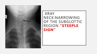
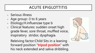
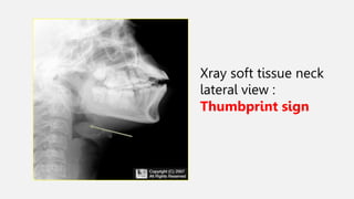
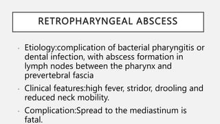
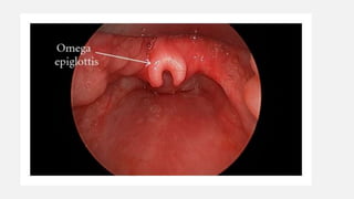
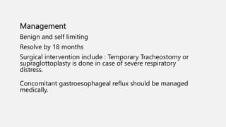
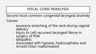
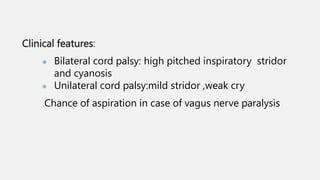
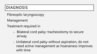
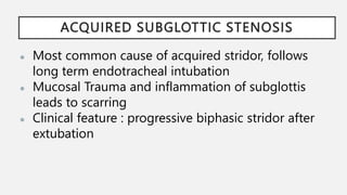
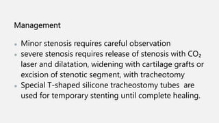
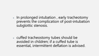
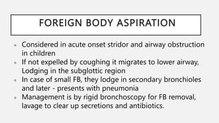
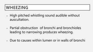
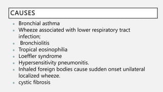
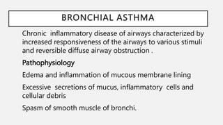
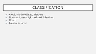
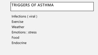
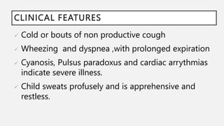
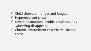
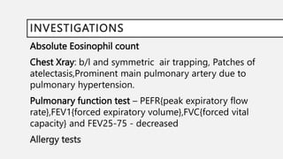
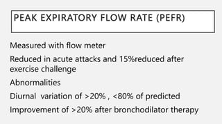
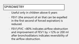
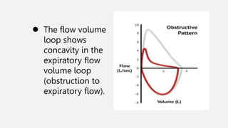
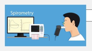
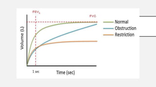
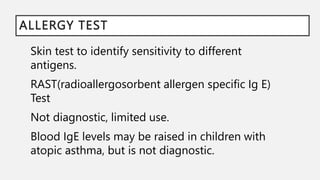
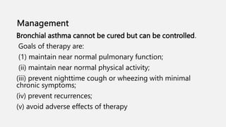
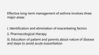
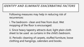
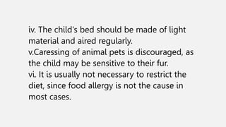
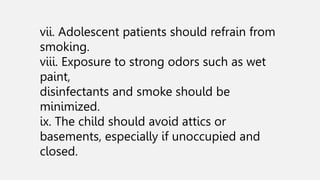
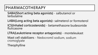
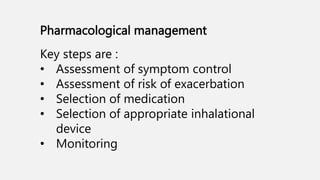
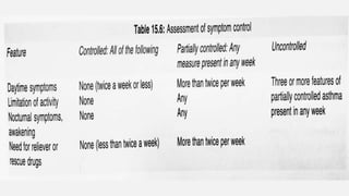
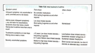
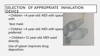
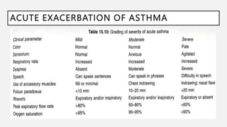
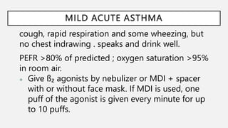
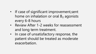
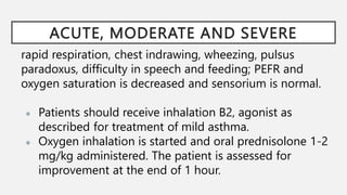
WHEEZING AND
STRIDOR IN CHILDREN
SHIFANA MD
MUHAMMED SUHAIL
STRIDOR
Audible respiratory noises produced by turbulent
airflow through an obstructed upper airway.
● Etiologies:
1. Infections
● Acute laryngotracheobronchitis(croup)
● Acute epiglottitis
● Bacterial tracheitis
● Retropharyngeal abscess
2.Congenital causes
● Laryngomalacia
● Vocal cord paralysis
3.Iatrogenic causes
● Acquired sub glottic stenosis
4.FB aspiration
ACUTE LARYNGOTRACHEOBRONCHITIS
(CROUP)
● Viral URTI:
○ parainfluenzae type 1,2,3 RSV( respiratory
syncytial virus ), adenovirus,
influenza,rhinovirus
● 6months to 3years
● Clinical features
○ Low grade fever, common cold
○ Biphasic stridor
○ Barking cough
ASSESSMENT OF SEVERITY OF CROUP
XRAY
NECK:NARROWING
OF THE SUBGLOTTIC
REGION “STEEPLE
SIGN”
MANAGEMENT
● Conservative : reassurance,cool mist and oral
hydration
● Nebulized epinephrine ( 1:1000 dilution 0.1- 0.5
ml/kg, to a maximum of 5 mL) provide
symptomatic relief
● single dose of dexamethasone (0.3-0.6 mg/kg,
intramuscular) reduces severity, if given within the
first 24 hours.
● Inhaled budesonide (1 mg twice a day for two
days)
antibiotics against Staphylococcus and H. influenzae
are indicated, if the child fails to improve .
ACUTE EPIGLOTTITIS
● Serious illness
● Age group:-3 to 6 years
● Etiology:H.influenzae type b
● Clinical features: sudden onset high
grade fever, sore throat, muffled voice,
inspiratory stridor, dysphagia.
Releiving factor:Child Sits in a leaning
forward position “tripod position” with
his neck extended and saliva dribbling.
DIAGNOSIS:
Airway management - Intubation or Rigid
broncoscopy , followed by tracheostomy
Humidified oxygen by hood, fluids to maintain
hydration, Ceftriaxone or cefotaxime 100mg/kg/day
for 7 days
Atypical presentation:
Flexible endoscopy – edema,erythema of supraglottic
structures
Xray soft tissue neck
lateral view :
Thumbprint sign
RETROPHARYNGEAL ABSCESS
• Etiology:complication of bacterial pharyngitis or
dental infection, with abscess formation in
lymph nodes between the pharynx and
prevertebral fascia
• Clinical features:high fever, stridor, drooling and
reduced neck mobility.
• Complication:Spread to the mediastinum is
fatal.
Management:
● parenteral antibiotics
● surgical drainage of abscess by transoral or
transcervical approach.
● Tracheostomy may be necessary to secure the
airway.
Bacterial tracheitis
Etiology: S. aureus, affects younger children and
follows a viral upper respiratory tract infection.
The child appears toxic ,brassy cough with biphasic
or expiratory stridor.
X-ray neck shows an irregular tracheal wall.
Diagnosis:Bronchoscopy ; culture and removal of
purulent tracheal secretions.
LARYNGOMALACIA
Most common congenital anomaly
Chief cause of chronic stridor in infancy
Clinical features: inspiratory stridor; increase in supine or crying
position
Flexible endoscopy:
● Omega shaped epiglottis
● Short aryepiglottic folds
● Partial collapse of a flaccid supraglottic airway with
inspiration
Management
Benign and self limiting
Resolve by 18 months
Surgical intervention include : Temporary Tracheostomy or
supraglottoplasty is done in case of severe respiratory
distress.
Concomitant gastroesophageal reflux should be managed
medically.
VOCAL CORD PARALYSIS
Second most common congenital laryngeal anomaly
Causes:
● excessive stretching of the neck during vaginal
delivery
● Injury to Left recurrent laryngeal Nerve in
surgery of PDA
● Idiopathic
● Associated with hypoxia ,hydrocephalus and
Arnold Chiari malformation
Clinical features:
● Bilateral cord palsy: high pitched inspiratory stridor
and cyanosis
● Unilateral cord palsy:mild stridor ,weak cry
Chance of aspiration in case of vagus nerve paralysis
DIAGNOSIS
Fibreoptic laryngoscopy
Management
Treatment required in
● Bilateral cord palsy: tracheostomy to secure
airway
● Unilateral cord palsy without aspiration, do not
need active management as hoarseness improves
with time
ACQUIRED SUBGLOTTIC STENOSIS
● Most common cause of acquired stridor, follows
long term endotracheal intubation
● Mucosal Trauma and inflammation of subglottis
leads to scarring
● Clinical feature : progressive biphasic stridor after
extubation
Management
● Minor stenosis requires careful observation
● severe stenosis requires release of stenosis with CO₂
laser and dilatation, widening with cartilage grafts or
excision of stenotic segment, with tracheotomy
● Special T-shaped silicone tracheostomy tubes are
used for temporary stenting until complete healing.
● In prolonged intubation , early tracheotomy
prevents the complication of post-intubation
subglottic stenosis.
● cuffed tracheostomy tubes should be
avoided in children; if a cuffed tube is
essential, intermittent deflation is advised.
FOREIGN BODY ASPIRATION
● Considered in acute onset stridor and airway obstruction
in children
● If not expelled by coughing it migrates to lower airway,
Lodging in the subglottic region
● In case of small FB, they lodge in secondary bronchioles
and later - presents with pneumonia
● Management is by rigid bronchoscopy for FB removal,
lavage to clear up secretions and antibiotics.
WHEEZING
● High pitched whistling sound audible without
auscultation.
● Partial obstruction of bronchi and bronchioles
leading to narrowing produces wheezing.
● Due to causes within lumen or in walls of bronchi
CAUSES
● Bronchial asthma
● Wheeze associated with lower respiratory tract
infection;
● Bronchiolitis
● Tropical eosinophilia
● Loeffler syndrome
● Hypersensitivity pneumonitis.
● Inhaled foreign bodies cause sudden onset unilateral
localized wheeze.
● cystic fibrosis
BRONCHIAL ASTHMA
Chronic inflammatory disease of airways characterized by
increased responsiveness of the airways to various stimuli
and reversible diffuse airway obstruction .
Pathophysiology
Edema and inflammation of mucous membrane lining
Excessive secretions of mucus, inflammatory cells and
cellular debris
Spasm of smooth muscle of bronchi.
CLASSIFICATION
● Atopic – IgE mediated, allergens
● Non atopic – non IgE mediated, infections
● Mixed
● Exercise induced
TRIGGERS OF ASTHMA
Infections ( viral )
Exercise
Weather
Emotions : stress
Food
Endocrine
CLINICAL FEATURES
 Cold or bouts of non productive cough
 Wheezing and dyspnea ,with prolonged expiration
 Cyanosis, Pulsus paradoxus and cardiac arrythmias
indicate severe illness.
 Child sweats profusely and is apprehensive and
restless.
 Child shows air hunger and fatigue.
 Hyperresonant chest
 severe obstruction : feeble breath sounds
,wheezing disappears.
 Chronic intermittent cases:barrel shaped
chest
DIAGNOSIS
Clinical
 Family h/o asthma or atopic dermatitis or allergic
rhinitis
 Recurrent episodes that resolved
 Relatively afebrile episode
 Presence of trigger factors :nocturnal or seasonal
exacerbations,exercise induced
 Clear and mucoid sputum sometimes yellow.
INVESTIGATIONS
Absolute Eosinophil count
Chest Xray: b/l and symmetric air trapping, Patches of
atelectasis,Prominent main pulmonary artery due to
pulmonary hypertension.
Pulmonary function test – PEFR{peak expiratory flow
rate},FEV1{forced expiratory volume},FVC{forced vital
capacity} and FEV25-75 - decreased
Allergy tests
PEAK EXPIRATORY FLOW RATE (PEFR)
Measured with flow meter
Reduced in acute attacks and 15%reduced after
exercise challenge
Abnormalities
Diurnal variation of >20% , <80% of predicted
Improvement of >20% after bronchodilator therapy
SPIROMETRY
• Useful only in children above 6 years
• FEV1 (the amount of air that can be expelled
in the first second of forced expiration) is
reduced.
• FEV1/FVC <80% indicates airflow obstruction
and improvement of FEV1 by >12% or 200 ml
after bronchodilators indicates reversibility of
the airflow obstruction.
 The flow volume
loop shows
concavity in the
expiratory flow
volume loop
(obstruction to
expiratory flow).
ALLERGY TEST
Skin test to identify sensitivity to different
antigens.
RAST(radioallergosorbent allergen specific Ig E)
Test
Not diagnostic, limited use.
Blood IgE levels may be raised in children with
atopic asthma, but is not diagnostic.
Bronchial asthma cannot be cured but can be controlled.
Goals of therapy are:
(1) maintain near normal pulmonary function;
(ii) maintain near normal physical activity;
(iii) prevent nighttime cough or wheezing with minimal
chronic symptoms;
(iv) prevent recurrences;
(v) avoid adverse effects of therapy
Effective long-term management of asthma involves three
major areas:
i. Identification and elimination of exacerbating factors
ii. Pharmacological therapy
iii. Education of patient and parents about nature of disease
and steps to avoid acute exacerbation
IDENTIFY AND ELIMINATE EXACERBATING FACTORS
Following measures may help in reducing risk of
recurrences:
i. The bedroom- clean and free from dust. Wet
mopping of the floor is encouraged.
ii. Since heavy tapestry attracts dust, light plain cloth
sheet to be used as curtains in the child's bedroom.
iii. Periodic cleaning of carpets, stuffed furniture, loose
clothing and hangings, calendars and books.
iv. The child's bed should be made of light
material and aired regularly.
v.Caressing of animal pets is discouraged, as
the child may be sensitive to their fur.
vi. It is usually not necessary to restrict the
diet, since food allergy is not the cause in
most cases.
vii. Adolescent patients should refrain from
smoking.
viii. Exposure to strong odors such as wet
paint,
disinfectants and smoke should be
minimized.
ix. The child should avoid attics or
basements, especially if unoccupied and
closed.
PHARMACOTHERAPY
Pharmacological management
Key steps are :
• Assessment of symptom control
• Assessment of risk of exacerbation
• Selection of medication
• Selection of appropriate inhalational
device
• Monitoring
SELECTION OF APPROPRIATE INHALATION
DEVICE
• Children <4-year-old: MDI with spacer
with
face mask
• Children >4-year-old: MDI with spacer
preferred
• Children>12-year-old: MDI used
directly.
Use of spacer improves drug
deposition.
Monitoring and Modification of Treatment
• After initiating treatment, patients should be
seen every 4 -12 weeks.
• history regarding frequency of symptoms,
sleep disturbance, physical activity, school
absenteeism, visit to a doctor and need for
bronchodilators, and PEFR is recorded.
• The inhalation technique and compliance is
checked.
• Patient is assessed as controlled, partially
controlled or uncontrolled.
• If partially controlled or uncontrolled, cause
mostly would be poor compliance, wrong
technique, Associated infections,
inappropriate doses etc,
• If no cause detected then step up
• Step down - control is Sustained for 3-6
Months.
• Description of asthma in plain language should
be made.
• The spectrum of severity of the illness, likely
course and satisfactory outcome is explained.
• Avoiding environmental triggers
• Avoidance of all kinds of smoke at home,
including tobacco smoke, wood burning and
kerosene stove is emphasized.
Education of parents
ACUTE EXACERBATION OF ASTHMA
MILD ACUTE ASTHMA
cough, rapid respiration and some wheezing, but
no chest indrawing . speaks and drink well.
PEFR >80% of predicted ; oxygen saturation >95%
in room air.
● Give ß₂ agonists by nebulizer or MDI + spacer
with or without face mask. If MDI is used, one
puff of the agonist is given every minute for up
to 10 puffs.
• If case of significant improvement,sent
home on inhalation or oral B₂ agonists
every 6-8 hours
• Review After 1-2 weeks for reassessment
and long term treatment.
• In case of unsatisfactory response, the
patient should be treated as moderate
exacerbation.
ACUTE, MODERATE AND SEVERE
rapid respiration, chest indrawing, wheezing, pulsus
paradoxus, difficulty in speech and feeding; PEFR and
oxygen saturation is decreased and sensorium is normal.
● Patients should receive inhalation B2, agonist as
described for treatment of mild asthma.
● Oxygen inhalation is started and oral prednisolone 1-2
mg/kg administered. The patient is assessed for
improvement at the end of 1 hour.
● Improvement, continued on inhaled B₂
agonists every 30 minutes, and the interval
gradually increased to 4-6 hourly.
● Oxygen inhalation is stopped, if patient is
able to maintain oxygen saturation >95%.
● Prednisolone is continued once daily for 5-
7 days, and then stopped without tapering.
● The patient is discharged from hospital when the
need for bronchodilators is every 4-6 hours, able
to feed and speak well, maintains oxygen
saturation >95% in room air and PEFR >75% of
predicted.
● These patients should be educated about the
disease, need for regular follow-up and avoidance
of triggers.
● They should be assessed for long-term treatment.
● No improvement at the end of 1 hour,
inhalation of salbutamol is continued and
inhaled ipratropium 250 mg given every 20
minutes.
● An injection of hydrocortisone 10 mg/kg is
given and reassessed at end of 2 hours. If
satisfactory response is obtained, the patient is
treated like early responders.
● If case of no response, IV theophylline bolus is
followed by continuous infusion.
• Such patients respond well to magnesium
infusion at a dose of 50 mg/kg (with dextrose
over 30 minutes). If no improvement :possible
mechanical ventilation.
• transfer to an intensive care unit include
worsening hypoxia or hypercapnia,
exhaustion, feeble respiration, confusion,
drowsiness, coma or respiratory arrest.
BRONCHIOLITIS
• Occurs with in first 2 years, highest incidence in first 6
months.
• Usually in winter Or spring
• Repeated attack indicate viral infection (RSV)
associated with wheezing, or asthma.
• Chest xray shows hyperinflation with scattered areas
of infiltration.
• Infants with bronchiolitis and atopic dermatitis, high
IgE levels Or family history of allergy need follow up
for later development of asthma
CONGENITAL MALFORMATIONS:
Cause obstruction by;
 vascular rings due to aberrant right subclavian
artery or double aortic arch
 bronchogenic cysts
 tracheomalacia
ASPIRATION OF FOREIGN BODY
• Cause localized area of wheezing, hyperresonance
and reduced air entry.
• History of aspiration may be forgotten.
• Frequent infection in lung may occur.
CYSTIC FIBROSIS
• Recurrent wheezing, clubbing and malabsorption.
• X ray chest shows hyperinflation, peribronchial
cuffing and pneumonia.
THANK YOU

stridor and wheeze-1-1.pptx

  • 1.
    WHEEZING AND STRIDOR INCHILDREN SHIFANA MD MUHAMMED SUHAIL
  • 2.
    STRIDOR Audible respiratory noisesproduced by turbulent airflow through an obstructed upper airway. ● Etiologies: 1. Infections ● Acute laryngotracheobronchitis(croup) ● Acute epiglottitis ● Bacterial tracheitis ● Retropharyngeal abscess
  • 3.
    2.Congenital causes ● Laryngomalacia ●Vocal cord paralysis 3.Iatrogenic causes ● Acquired sub glottic stenosis 4.FB aspiration
  • 4.
    ACUTE LARYNGOTRACHEOBRONCHITIS (CROUP) ● ViralURTI: ○ parainfluenzae type 1,2,3 RSV( respiratory syncytial virus ), adenovirus, influenza,rhinovirus ● 6months to 3years ● Clinical features ○ Low grade fever, common cold ○ Biphasic stridor ○ Barking cough
  • 5.
  • 6.
  • 7.
    MANAGEMENT ● Conservative :reassurance,cool mist and oral hydration ● Nebulized epinephrine ( 1:1000 dilution 0.1- 0.5 ml/kg, to a maximum of 5 mL) provide symptomatic relief ● single dose of dexamethasone (0.3-0.6 mg/kg, intramuscular) reduces severity, if given within the first 24 hours. ● Inhaled budesonide (1 mg twice a day for two days) antibiotics against Staphylococcus and H. influenzae are indicated, if the child fails to improve .
  • 8.
    ACUTE EPIGLOTTITIS ● Seriousillness ● Age group:-3 to 6 years ● Etiology:H.influenzae type b ● Clinical features: sudden onset high grade fever, sore throat, muffled voice, inspiratory stridor, dysphagia. Releiving factor:Child Sits in a leaning forward position “tripod position” with his neck extended and saliva dribbling.
  • 9.
    DIAGNOSIS: Airway management -Intubation or Rigid broncoscopy , followed by tracheostomy Humidified oxygen by hood, fluids to maintain hydration, Ceftriaxone or cefotaxime 100mg/kg/day for 7 days Atypical presentation: Flexible endoscopy – edema,erythema of supraglottic structures
  • 10.
    Xray soft tissueneck lateral view : Thumbprint sign
  • 11.
    RETROPHARYNGEAL ABSCESS • Etiology:complicationof bacterial pharyngitis or dental infection, with abscess formation in lymph nodes between the pharynx and prevertebral fascia • Clinical features:high fever, stridor, drooling and reduced neck mobility. • Complication:Spread to the mediastinum is fatal.
  • 13.
    Management: ● parenteral antibiotics ●surgical drainage of abscess by transoral or transcervical approach. ● Tracheostomy may be necessary to secure the airway.
  • 14.
    Bacterial tracheitis Etiology: S.aureus, affects younger children and follows a viral upper respiratory tract infection. The child appears toxic ,brassy cough with biphasic or expiratory stridor. X-ray neck shows an irregular tracheal wall. Diagnosis:Bronchoscopy ; culture and removal of purulent tracheal secretions.
  • 15.
    LARYNGOMALACIA Most common congenitalanomaly Chief cause of chronic stridor in infancy Clinical features: inspiratory stridor; increase in supine or crying position Flexible endoscopy: ● Omega shaped epiglottis ● Short aryepiglottic folds ● Partial collapse of a flaccid supraglottic airway with inspiration
  • 17.
    Management Benign and selflimiting Resolve by 18 months Surgical intervention include : Temporary Tracheostomy or supraglottoplasty is done in case of severe respiratory distress. Concomitant gastroesophageal reflux should be managed medically.
  • 18.
    VOCAL CORD PARALYSIS Secondmost common congenital laryngeal anomaly Causes: ● excessive stretching of the neck during vaginal delivery ● Injury to Left recurrent laryngeal Nerve in surgery of PDA ● Idiopathic ● Associated with hypoxia ,hydrocephalus and Arnold Chiari malformation
  • 19.
    Clinical features: ● Bilateralcord palsy: high pitched inspiratory stridor and cyanosis ● Unilateral cord palsy:mild stridor ,weak cry Chance of aspiration in case of vagus nerve paralysis
  • 20.
    DIAGNOSIS Fibreoptic laryngoscopy Management Treatment requiredin ● Bilateral cord palsy: tracheostomy to secure airway ● Unilateral cord palsy without aspiration, do not need active management as hoarseness improves with time
  • 21.
    ACQUIRED SUBGLOTTIC STENOSIS ●Most common cause of acquired stridor, follows long term endotracheal intubation ● Mucosal Trauma and inflammation of subglottis leads to scarring ● Clinical feature : progressive biphasic stridor after extubation
  • 22.
    Management ● Minor stenosisrequires careful observation ● severe stenosis requires release of stenosis with CO₂ laser and dilatation, widening with cartilage grafts or excision of stenotic segment, with tracheotomy ● Special T-shaped silicone tracheostomy tubes are used for temporary stenting until complete healing.
  • 23.
    ● In prolongedintubation , early tracheotomy prevents the complication of post-intubation subglottic stenosis. ● cuffed tracheostomy tubes should be avoided in children; if a cuffed tube is essential, intermittent deflation is advised.
  • 24.
    FOREIGN BODY ASPIRATION ●Considered in acute onset stridor and airway obstruction in children ● If not expelled by coughing it migrates to lower airway, Lodging in the subglottic region ● In case of small FB, they lodge in secondary bronchioles and later - presents with pneumonia ● Management is by rigid bronchoscopy for FB removal, lavage to clear up secretions and antibiotics.
  • 25.
    WHEEZING ● High pitchedwhistling sound audible without auscultation. ● Partial obstruction of bronchi and bronchioles leading to narrowing produces wheezing. ● Due to causes within lumen or in walls of bronchi
  • 26.
    CAUSES ● Bronchial asthma ●Wheeze associated with lower respiratory tract infection; ● Bronchiolitis ● Tropical eosinophilia ● Loeffler syndrome ● Hypersensitivity pneumonitis. ● Inhaled foreign bodies cause sudden onset unilateral localized wheeze. ● cystic fibrosis
  • 27.
    BRONCHIAL ASTHMA Chronic inflammatorydisease of airways characterized by increased responsiveness of the airways to various stimuli and reversible diffuse airway obstruction . Pathophysiology Edema and inflammation of mucous membrane lining Excessive secretions of mucus, inflammatory cells and cellular debris Spasm of smooth muscle of bronchi.
  • 28.
    CLASSIFICATION ● Atopic –IgE mediated, allergens ● Non atopic – non IgE mediated, infections ● Mixed ● Exercise induced
  • 29.
    TRIGGERS OF ASTHMA Infections( viral ) Exercise Weather Emotions : stress Food Endocrine
  • 30.
    CLINICAL FEATURES  Coldor bouts of non productive cough  Wheezing and dyspnea ,with prolonged expiration  Cyanosis, Pulsus paradoxus and cardiac arrythmias indicate severe illness.  Child sweats profusely and is apprehensive and restless.
  • 31.
     Child showsair hunger and fatigue.  Hyperresonant chest  severe obstruction : feeble breath sounds ,wheezing disappears.  Chronic intermittent cases:barrel shaped chest
  • 32.
    DIAGNOSIS Clinical  Family h/oasthma or atopic dermatitis or allergic rhinitis  Recurrent episodes that resolved  Relatively afebrile episode  Presence of trigger factors :nocturnal or seasonal exacerbations,exercise induced  Clear and mucoid sputum sometimes yellow.
  • 33.
    INVESTIGATIONS Absolute Eosinophil count ChestXray: b/l and symmetric air trapping, Patches of atelectasis,Prominent main pulmonary artery due to pulmonary hypertension. Pulmonary function test – PEFR{peak expiratory flow rate},FEV1{forced expiratory volume},FVC{forced vital capacity} and FEV25-75 - decreased Allergy tests
  • 34.
    PEAK EXPIRATORY FLOWRATE (PEFR) Measured with flow meter Reduced in acute attacks and 15%reduced after exercise challenge Abnormalities Diurnal variation of >20% , <80% of predicted Improvement of >20% after bronchodilator therapy
  • 35.
    SPIROMETRY • Useful onlyin children above 6 years • FEV1 (the amount of air that can be expelled in the first second of forced expiration) is reduced. • FEV1/FVC <80% indicates airflow obstruction and improvement of FEV1 by >12% or 200 ml after bronchodilators indicates reversibility of the airflow obstruction.
  • 36.
     The flowvolume loop shows concavity in the expiratory flow volume loop (obstruction to expiratory flow).
  • 39.
    ALLERGY TEST Skin testto identify sensitivity to different antigens. RAST(radioallergosorbent allergen specific Ig E) Test Not diagnostic, limited use. Blood IgE levels may be raised in children with atopic asthma, but is not diagnostic.
  • 40.
    Bronchial asthma cannotbe cured but can be controlled. Goals of therapy are: (1) maintain near normal pulmonary function; (ii) maintain near normal physical activity; (iii) prevent nighttime cough or wheezing with minimal chronic symptoms; (iv) prevent recurrences; (v) avoid adverse effects of therapy
  • 41.
    Effective long-term managementof asthma involves three major areas: i. Identification and elimination of exacerbating factors ii. Pharmacological therapy iii. Education of patient and parents about nature of disease and steps to avoid acute exacerbation
  • 42.
    IDENTIFY AND ELIMINATEEXACERBATING FACTORS Following measures may help in reducing risk of recurrences: i. The bedroom- clean and free from dust. Wet mopping of the floor is encouraged. ii. Since heavy tapestry attracts dust, light plain cloth sheet to be used as curtains in the child's bedroom. iii. Periodic cleaning of carpets, stuffed furniture, loose clothing and hangings, calendars and books.
  • 43.
    iv. The child'sbed should be made of light material and aired regularly. v.Caressing of animal pets is discouraged, as the child may be sensitive to their fur. vi. It is usually not necessary to restrict the diet, since food allergy is not the cause in most cases.
  • 44.
    vii. Adolescent patientsshould refrain from smoking. viii. Exposure to strong odors such as wet paint, disinfectants and smoke should be minimized. ix. The child should avoid attics or basements, especially if unoccupied and closed.
  • 45.
  • 46.
    Pharmacological management Key stepsare : • Assessment of symptom control • Assessment of risk of exacerbation • Selection of medication • Selection of appropriate inhalational device • Monitoring
  • 49.
    SELECTION OF APPROPRIATEINHALATION DEVICE • Children <4-year-old: MDI with spacer with face mask • Children >4-year-old: MDI with spacer preferred • Children>12-year-old: MDI used directly. Use of spacer improves drug deposition.
  • 50.
    Monitoring and Modificationof Treatment • After initiating treatment, patients should be seen every 4 -12 weeks. • history regarding frequency of symptoms, sleep disturbance, physical activity, school absenteeism, visit to a doctor and need for bronchodilators, and PEFR is recorded. • The inhalation technique and compliance is checked.
  • 51.
    • Patient isassessed as controlled, partially controlled or uncontrolled. • If partially controlled or uncontrolled, cause mostly would be poor compliance, wrong technique, Associated infections, inappropriate doses etc, • If no cause detected then step up • Step down - control is Sustained for 3-6 Months.
  • 52.
    • Description ofasthma in plain language should be made. • The spectrum of severity of the illness, likely course and satisfactory outcome is explained. • Avoiding environmental triggers • Avoidance of all kinds of smoke at home, including tobacco smoke, wood burning and kerosene stove is emphasized. Education of parents
  • 54.
  • 55.
    MILD ACUTE ASTHMA cough,rapid respiration and some wheezing, but no chest indrawing . speaks and drink well. PEFR >80% of predicted ; oxygen saturation >95% in room air. ● Give ß₂ agonists by nebulizer or MDI + spacer with or without face mask. If MDI is used, one puff of the agonist is given every minute for up to 10 puffs.
  • 56.
    • If caseof significant improvement,sent home on inhalation or oral B₂ agonists every 6-8 hours • Review After 1-2 weeks for reassessment and long term treatment. • In case of unsatisfactory response, the patient should be treated as moderate exacerbation.
  • 57.
    ACUTE, MODERATE ANDSEVERE rapid respiration, chest indrawing, wheezing, pulsus paradoxus, difficulty in speech and feeding; PEFR and oxygen saturation is decreased and sensorium is normal. ● Patients should receive inhalation B2, agonist as described for treatment of mild asthma. ● Oxygen inhalation is started and oral prednisolone 1-2 mg/kg administered. The patient is assessed for improvement at the end of 1 hour.
  • 58.
    ● Improvement, continuedon inhaled B₂ agonists every 30 minutes, and the interval gradually increased to 4-6 hourly. ● Oxygen inhalation is stopped, if patient is able to maintain oxygen saturation >95%. ● Prednisolone is continued once daily for 5- 7 days, and then stopped without tapering.
  • 59.
    ● The patientis discharged from hospital when the need for bronchodilators is every 4-6 hours, able to feed and speak well, maintains oxygen saturation >95% in room air and PEFR >75% of predicted. ● These patients should be educated about the disease, need for regular follow-up and avoidance of triggers. ● They should be assessed for long-term treatment.
  • 60.
    ● No improvementat the end of 1 hour, inhalation of salbutamol is continued and inhaled ipratropium 250 mg given every 20 minutes. ● An injection of hydrocortisone 10 mg/kg is given and reassessed at end of 2 hours. If satisfactory response is obtained, the patient is treated like early responders. ● If case of no response, IV theophylline bolus is followed by continuous infusion.
  • 61.
    • Such patientsrespond well to magnesium infusion at a dose of 50 mg/kg (with dextrose over 30 minutes). If no improvement :possible mechanical ventilation. • transfer to an intensive care unit include worsening hypoxia or hypercapnia, exhaustion, feeble respiration, confusion, drowsiness, coma or respiratory arrest.
  • 62.
    BRONCHIOLITIS • Occurs within first 2 years, highest incidence in first 6 months. • Usually in winter Or spring • Repeated attack indicate viral infection (RSV) associated with wheezing, or asthma. • Chest xray shows hyperinflation with scattered areas of infiltration. • Infants with bronchiolitis and atopic dermatitis, high IgE levels Or family history of allergy need follow up for later development of asthma
  • 63.
    CONGENITAL MALFORMATIONS: Cause obstructionby;  vascular rings due to aberrant right subclavian artery or double aortic arch  bronchogenic cysts  tracheomalacia
  • 64.
    ASPIRATION OF FOREIGNBODY • Cause localized area of wheezing, hyperresonance and reduced air entry. • History of aspiration may be forgotten. • Frequent infection in lung may occur. CYSTIC FIBROSIS • Recurrent wheezing, clubbing and malabsorption. • X ray chest shows hyperinflation, peribronchial cuffing and pneumonia.
  • 65.