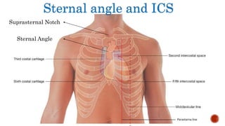
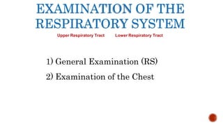
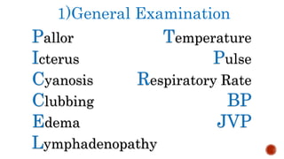
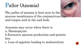
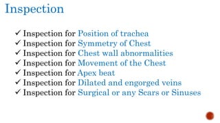
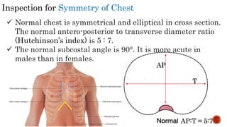
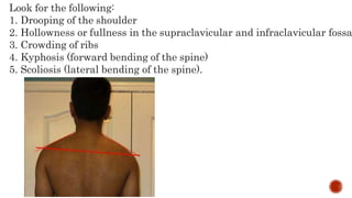
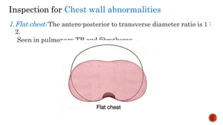
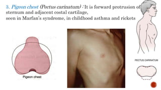
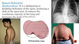
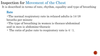
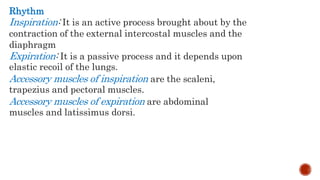
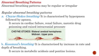
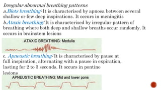
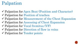
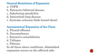
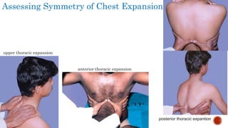
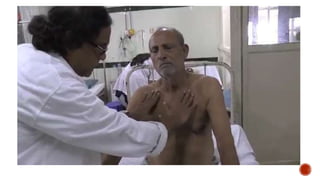
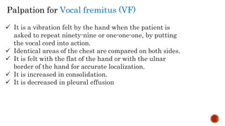
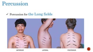
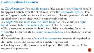
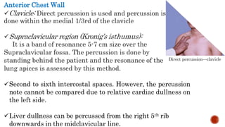
Clinical Examination of the
Respiratory System
Nalukwago Dorcus
&
Asiimwe Anne
The history
Most patients with respiratory disease will presentwith breathlessness, cough,
excess sputum, haemoptysis, wheeze or chest pain
Breathlessness
Everyone becomes breathless on strenuous exertion. Breathlessness
inappropriate to the level of physical exertion, or even occurring at rest, is
called dyspnoea.
People with cardiac disease and even non-cardiorespiratory conditions
such as anaemia, thyrotoxicosis or metabolic acidosis may become dyspnoeic
as well as those with primarily respiratory problems.
Cont…….
An important assessment is exercise tolerance, ask wether there are any times
of day or night that are usually worse than others. Variable airways
obstruction due to asthma is very often worse at night and in the early
morning.
By contrast, people with predominantly irreversible airways obstruction due
to chronic obstructive pulmonary disease (COPD) will often say that as long
as they are sitting in bed, they feel quite normal; it is exercise that troubles
them.
Cough
A cough may be dry or productive of sputum.
• How long has the cough been present?
A cough lasting a few days following a cold has less significance than one lasting several
weeks in a middle-aged smoker, which may be the first sign of a malignancy.
• Is the cough worse at any time of day or night?
A dry cough at night may be an early symptom of asthma, as may a cough that comes in
spasms lasting several minutes.
Cont…….
• Is the cough aggravated by anything? , eg. allergic triggers such as dust,
animals or pollen, or non-specific triggers like exercise or cold air? The
increased reactivity of the airways seen in asthma, and in some normal
people for several weeks after viral respiratory infections, may present in this
way.
Severe coughing, whatever its cause, may be followed by vomiting.
SPUTUM
Is sputum produced?
What does it look like?
ask for a description of its colour and consistency. Yellow or green sputum is
usually purulent. People with asthma may produce small amounts of very thick or
jelly-like sputum, sometimes in the shape of a cast of the airways. Eosinophils may
accumulate in the sputum in asthma, causing a purulent appearance even when no
infection is present.
How much is produced?
sputum produced daily in bronchiectasis often exceeded a cupful. chronic bronchitis
causes the production of smaller amounts of sputum.
HAEMOPTYSIS
Haemoptysis means the coughing up of blood in the Sputum.
• Ask if it is fresh or altered blood, how much is produced, when it started and
how often it happens.
• They should always be asked about associated conditions such as epistaxis
(nose bleeds), or the subsequent development of melaena (altered blood in
the stool), which occurs in the case of upper gastrointestinal bleeding.
WHEEZING
• Always ask whether the patient hears any noises coming from the chest. Even if a
wheeze is not present when you examine the patient, it is useful to know that he has
noticed it on occasions.
• Sometimes, wheezing will have been noticed by others (especially by a partner at
night, when asthma is worse) but not by the patient. Sometimes stridor may be
mistaken for wheezing by both patient and doctor. This serious finding usually
indicates narrowing of the larynx, trachea or main bronchi.
PAIN IN THE CHEST
• Consequent upon prolonged bouts of coughing, chest pain caused by lung disease usually
arises from the pleura. Pleuritic pain is sharp and stabbing, and is made worse by deep
breathing or coughing. It occurs when the pleura is inflamed, most commonly by infection in
the underlying lung.
• More constant pain, unrelated to breathing, may be caused by local invasion of the chest
wall by a lung or pleural tumour.
• A spontaneous pneumothorax causes pain which is worse on breathing but which may have
more of an aching character than the stabbing pain of pleurisy.
• If a pulmonary embolus causes infarction of the lung, pleurisy and hence pleuritic pain may
occur, but an acute pulmonary embolus can also cause pain which is not stabbing in nature.
A large pulmonary embolus causing haemodynamic disturbance may cause cardiac-type
Other aspects of the history that are particularly relevant to the respiratory
system
 Ask Questions related to the ear, nose and throat are relevant
 A change in the voice may indicate involvement of the left recurrent laryngeal nerve by a
carcinoma of the lung
 Sometimes patients using inhaled corticosteroids for asthma develop oropharyngeal
candidiasis or even hoarseness or weakness of the voice
 The smoking history
 The family history
 The occupational history
Sternal angle and ICS
Suprasternal Notch
Sternal Angle
EXAMINATION OF THE
RESPIRATORY SYSTEM
Upper Respiratory Tract Lower Respiratory Tract
1) General Examination (RS)
2) Examination of the Chest
1)General Examination
Pallor
Icterus
Cyanosis
Clubbing
Edema
Temperature
Pulse
Respiratory Rate
BP
JVP
Lymphadenopathy
Pallor (Anemia)
The pallor of anemia is best seen in the
mucous membranes of the conjunctivae, lips
and tongue and in the nail beds
Anaemia may occur when there is
a. Haemoptysis
b.Excessive sputum production and protein
loss
c. Loss of appetite leading to malnutrition
Cyanosis
This is a blue discoloration of the skin and mucous membranes
caused by increased concentration of reduced hemoglobin (5g/dl)
Central cyanosis may result from the reduced arterial oxygen saturation
caused by cardiac or pulmonary disease. Intracardiac or extracardiac
shunting.
Impaired pulmonary function
a. Alveolar hypoventilation
b. Ventilation—Perfusion mismatch
c. Impaired oxygen diffusion.
Bulbous enlargement of the distal portion
of the digit due to increased subungual soft
tissue.
Clubbing
Grading of Clubbing
Grade I
Grade II
Positive nail bed fluctuation
Obliteration of the Lovibond angle
Grade III Parrot beak / Drumstick appearance
Grade IV Hypertrophic osteoarthropathy.
Pulmonary and Thoracic Causes
a. Bronchogenic carcinoma (rare in adenocarcinoma)
b. Metastatic lung cancer
c. Suppurative lung disease
1. Bronchiectasis
2. Cystic fibrosis
3. Lung abscess
4. Empyema
d. Interstitial lung disease
e. Longstanding pulmonary tuberculosis
f. Chronic bronchitis
g. Mesothelioma
h. Neurogenic diaphragmatic tumour
i. Pulmonary AV malformation
j. Sarcoidosis.
Hypertrophic Osteoarthropathy
It is a painful swelling of the wrist, elbow, knee, ankle,
with radiographic evidence of sub-periosteal new bone
formation. It can be familial or idiopathic.
common disorders that can produce it are:
a. Bronchogenic carcinoma
b. Cystic fibrosis
c. Neurofibroma
d. A-V malformation.
Lymphadenopathy
Scalene lymph node enlargement
1.Large and fixed in secondary involvement from a
primary lung malignancy
2.Hard and craggy, matted, with or without sinus
formation in healed and calcified tuberculous
lymphadenopathy.
Blood Pressure
Pulsus Paradoxus
 Systolic blood pressure normally falls during quiet inspiration in
normal individuals.
 Pulsus paradoxus is defined as a fall of systolic blood pressure of
>10 mmHg during the inspiratory phase.
 severe acute asthma or exacerbations of chronic obstructive
pulmonary disease.
Examination of the Neck Veins
Jugular Venous Pulse
COPD/cor pulmonale
 Bilateral non-pulsatile
SVC obstruction
Massive right sided pleural effusion
2) Examination of the Chest
 Inspection
 Palpation
 Percussion
 Auscultation
The subject should be examined in the Standing or Sitting position in an
erect, and in good light.
All the findings in the clinical examination should
be compared on both sides in the following areas:
1. Supraclavicular area
2. Infraclavicular area
3. Mammary region
4. Inframammary region
5. Axillary region
6. Infra-axillary region
7. Suprascapular region
8. Interscapular region
9. Infrascapular region.
Inspection
 Inspection for Position of trachea
 Inspection for Symmetry of Chest
 Inspection for Chest wall abnormalities
 Inspection for Movement of the Chest
 Inspection for Apex beat
 Inspection for Dilated and engorged veins
 Inspection for Surgical or any Scars or Sinuses
Inspection for Position of trachea
Trail’s sign: It is the undue prominence of the clavicular head of
sternomastoid on the side to which the trachea is deviated.
Position of Apex Beat
The apex beat is shifted to the side of mediastinal shift.
Inspection for Symmetry of Chest
 Normal chest is symmetrical and elliptical in cross section.
The normal antero-posterior to transverse diameter ratio
(Hutchinson’s index) is 5 : 7.
 The normal subcostal angle is 90°. It is more acute in
males than in females.
AP
T
AP:T = 5:7
Look for the following:
1. Drooping of the shoulder
2. Hollowness or fullness in the supraclavicular and infraclavicular fossa
3. Crowding of ribs
4. Kyphosis (forward bending of the spine)
5. Scoliosis (lateral bending of the spine).
Inspection for Chest wall abnormalities
1.Flat chest: The antero-posterior to transverse diameter ratio is 1 :
2.
Seen in pulmonary TB and fibrothorax
2.Barrel chest: The anteroposterior to transverse diameter
ratio is 1 : 1.
Seen in physiological states like infancy and old age and in
pathological states like COPD (emphysema)
3. Pigeon chest (Pectus carinatum) : It is forward protrusion of
sternum and adjacent costal cartilage,
seen in Marfan’s syndrome, in childhood asthma and rickets
4.Pectus excavatum (funnel chest, cobbler’s chest)
It is the exaggeration of the normal hollowness over the
lower end of the sternum. It is a developmental defect.
The apex beat shifted further to the left and the ventilatory
capacity of the lung is restricted.
It is seen in Marfan’s syndrome
5. Harrison’s sulcus: It is due to the indrawing of ribs to form
symmetrical horizontal grooves above the costal margin, along the line of
attachment of diaphragm
occurs in chronic respiratory
disease in childhood,
childhood asthma, rickets and
blocked nasopharynx due to
adenoid enlargement
6. Scorbutic rosary: It is the sharp
angulation, with or without beading or
rosary formation, of the ribs, arising as
a result of backward displacement or
pushing in of the sternum,
e.g. Vitamin C deficiency.
7. Rickety rosary: It is a bead like
enlargement of costochondral junction,
e.g. rickets
Spinal Deformity
Kyphoscoliosis : It is a disfiguring or
disabling deformity of the spine, producing a
shift of the apex beat. It reduces the
ventilatory capacity of the lung and
increases the work of breathing.
Inspection for Movement of the Chest
It is described in terms of rate, rhythm, equality and type of breathing
Rate
•The normal respiratory rate in relaxed adults is 14-18
breaths per minute
•The type of breathing in women is thoraco-abdominal
and in men is abdomino-thoracic
• The ratio of pulse rate to respiratory rate is 4 : 1.
Tachypnoea: It is an increase in respiratory rate more
than 20 per minute(Adult). Conditions causing tachypnoea
are:
a. Nervousness
b. Exertion
c. Fever
d. Hypoxia
e. Respiratory conditions
i. Acute pulmonary oedema
ii. Pneumonia
iii. Pulmonary embolism
iv.ARDS
v. Metabolic acidosis
Bradypnoea: It is a decrease in the rate of respiration.
Conditions causing bradypnoea are:
a. Alkalosis
b. Hypothyroidism (myxoedema)
c. Narcotic drug poisoning
d. Raised intracranial tension.
Hyperpnoea: It is an increase in depth of respiration.
Conditions causing hyperpnoea are:
a. Acidosis
b. Brainstem lesion
c. Hysteria.
Rhythm
Inspiration: It is an active process brought about by the
contraction of the external intercostal muscles and the
diaphragm
Expiration: It is a passive process and it depends upon
elastic recoil of the lungs.
Accessory muscles of inspiration are the scaleni,
trapezius and pectoral muscles.
Accessory muscles of expiration are abdominal
muscles and latissimus dorsi.
Abnormal Breathing Patterns
Abnormal breathing patterns may be regular or irregular
Regular abnormal breathing patterns
a. Cheyne-Stokes breathing: It is characterised by hyperpnoea
followed by apnoea.
It occurs in cardiac failure, renal failure, narcotic drug
poisoning and raised intracranial pressure
b. Kussmaul’s breathing: It is characterised by increase in rate and
depth of breathing.
It occurs in metabolic acidosis and pontine lesions.
Irregular abnormal breathing patterns
a.Biots breathing: It is characterised by apnoea between several
shallow or few deep inspirations. It occurs in meningitis
b.Ataxic breathing: It is characterised by irregular pattern of
breathing where both deep and shallow breaths occur randomly. It
occurs in brainstem lesions
c. Apneustic breathing: It is characterised by pause at
full inspiration, alternating with a pause in expiration,
lasting for 2 to 3 seconds. It occurs in pontine
lesions
Palpation
 Palpation for Apex Beat (Position and Character)
 Palpation for Position of trachea
 Palpation for Measurement of the Chest Expansion
 Palpation for Assessing of Chest Expansion
 Palpation for Vocal fremitus (VF)
 Palpation for Direction of flow in veins
 Palpation for Tender points
The position of the trachea is confirmed by slightly flexing the neck
so that the chin remains in the midline.
The index finger is then inserted in the suprasternal notch and the
tracheal ring is felt.
Slight shift of trachea to the right is normal
Palpation for Position of trachea
Measurement of the Chest Expansion
The expansion of the chest should be measured with a tape
measure placed around the chest just below the level of the
nipples/inferior angle of scapula.
 Chest circumference in full expiration
 Chest circumference at full inspiration
 Chest expansion
 Right/Left Hemithorax
Normal expansion of the chest is 5-8 cm
In severe emphysema, it is less than 1 cm
General Restriction of Expansion
a. COPD
b. Extensive bilateral disease
c. Ankylosing spondylitis
d. Interstitial lung disease
e. Systemic sclerosis (hide bound chest).
Asymmetrical Expansion of the Chest
a. Pleural effusion
b. Pneumothorax
c. Extensive consolidation
d. Collapse
e. Fibrosis.
In all these above conditions, diminished
expansion occurs on the affected side.
Assessing Symmetry of Chest Expansion
upper thoracic expansion
anterior thoracic expansion
posterior thoracic expantion
 It is a vibration felt by the hand when the patient is
asked to repeat ninety-nine or one-one-one, by putting
the vocal cord into action.
 Identical areas of the chest are compared on both sides.
 It is felt with the flat of the hand or with the ulnar
border of the hand for accurate localization.
 It is increased in consolidation.
 It is decreased in pleural effusion
Palpation for Vocal fremitus (VF)
Tenderness over the Chest Wall
It may be due to:
1. Empyema
2.Local inflammation of parietal pleura, soft tissue and
osteomyelitis
3. Infiltration with tumor
4. Non-respiratory cause (amoebic liver abscess).
Percussion
 Percussion for the Lung fields
Cardinal Rules of Percussion
a. The pleximeter: The middle finger of the examiner’s left hand should
be opposed tightly over the chest wall, over the intercostal spaces. The
other fingers should not touch the chest wall. Greater pressure should be
applied over a thick chest wall to remove air pockets
b.The plexor: The middle or the index finger of the examiner’s right
hand is used to hit the middle phalanx of the pleximeter
c.The percussion movement should be sudden, originating from the
wrist. The finger should be removed immediately after striking to avoid
damping
d.Proceed from the area of normal resonance to the area of impaired or
dull note, as the difference is then easily appreciated
e.The long axis of the pleximeter is kept parallel to the border of the
organ to be percussed.
Direct percussion—clavicle
Anterior Chest Wall
Clavicle: Direct percussion is used and percussion is
done within the medial 1/3rd of the clavicle
Supraclavicular region (Kronig’s isthumus):
It is a band of resonance 5-7 cm size over the
Supraclavicular fossa. The percussion is done by
standing behind the patient and the resonance of the
lung apices is assessed by this method.
Second to sixth intercostal spaces. However, the percussion
note cannot be compared due to relative cardiac dullness on
the left side.
Liver dullness can be percussed from the right 5th rib
downwards in the midclavicular line.
Lateral Chest Wall
Fourth to seventh intercostal spaces.
Liver dullness can be percussed from the right 8th rib
downwards in the midaxillary line.
Posterior Chest Wall
a. Suprascapular (above the spine of the scapula)
b. Interscapular region
c. Infrascapular region up to the eleventh rib.
Liver dullness can be percussed from the right 10th rib
downwards in the midscapular line.
Tidal Percussion
 This is done to differentiate upward enlargement of liver or
subdiaphragmatic abscess from right sided parenchymal or pleural
disorder.
 If on deep inspiration, the previous dull note in the fifth right
intercostal space on the mid clavicular line becomes resonant, it
indicates that the dullness was due to the liver, which had been
pushed down by the right hemidiaphragm with deep inspiration.
 If the dullness persists on the other hand, it indicates underlying
right sided parenchymal or pleural pathology, in the absence of
diaphragmatic paralysis.
Shifting Dullness
This is done to demonstrate the shift of fluid in hydropneumothorax.
The immediate shift of fluid can be demonstrated by the dull area
percussed in the axilla in the sitting posture, becoming resonant on
lying down on the healthy side.
Auscultation
 Auscultation for Breath Sounds
 Auscultation for Vocal Resonance
Listen with the patient relaxed and breathing deeply
through his open mouth.
Auscultate each side alternately, comparing findings over a
large number of equivalent positions to ensure that you do
not miss localised abnormalities.
Listen:
■ anteriorly from above the clavicle down to the sixth rib
■ laterally from the axilla to the eighth rib
■ posteriorly down to the level of the 11th rib.
■Assess the quality and amplitude of the breath sounds.
Identifyany gap between inspiration and expiration, and
listen for added sounds.
Avoid auscultation within 3 cm of the midline anteriorly or
posteriorly, as these areas may transmit soundsdirectly from
the trachea or main bronchi.
Vesicular breath sounds
 low pitched, rustling in
nature
 produced by attenuating and
filtering effect of the lung
parenchyma.
 Duration of the inspiratory
phase is longer than the
expiratory phase in a ratio of
3 : 1.
 There is no pause between
the end of inspiration and the
beginning of expiration.
Bronchial breath sounds
 It is loud and high pitched,
with an aspirate or guttural
quality.
 It is produced by passage of
air through the trachea and
large bronchi
 The duration of inspiration is
shortened whereas that of
expiration is prolonged or
equal
 There is a pause between
inspiration and expiration.
INVESTIGATIONS
1. Sputum examination
Mucoid sputum is characteristic in patients with chronic bronchitis when there is no active
infection. It is clear and sticky and not necessarily produced in a large volume. Sputum may
become mucopurulent or purulent when bacterial infection is present in patients with bronchitis,
pneumonia, bronchiectasis or a lung abscess. In these last two conditions, the quantities
may be large and the sputum is often foul
smelling.
INVESTIGATIONS
 Lung function tests
 Arterial blood sampling
 The chest X-ray
 The computed tomography scan
 Magnetic resonance imaging
 Ultrasound
 Radioisotope imaging
 Positron emission tomography (PET)
scanning
 Flexible bronchoscopy
 Immunological tests
References
Breath Sounds Made Incredibly Easy (2005)– Lippincott
Williams and Wilkins
Gleadle J(2012)History and Clinical Examination at a
Glance Wiley Blackwell
Hogan-Quigley, Palm, Bickley (2012) Bates Nursing Guide
to Physical Examination and History Taking Lippincott
Williams and Wilkins

Examinating the Resipiratory System.pptx

  • 1.
    Clinical Examination ofthe Respiratory System Nalukwago Dorcus & Asiimwe Anne
  • 2.
    The history Most patientswith respiratory disease will presentwith breathlessness, cough, excess sputum, haemoptysis, wheeze or chest pain Breathlessness Everyone becomes breathless on strenuous exertion. Breathlessness inappropriate to the level of physical exertion, or even occurring at rest, is called dyspnoea. People with cardiac disease and even non-cardiorespiratory conditions such as anaemia, thyrotoxicosis or metabolic acidosis may become dyspnoeic as well as those with primarily respiratory problems.
  • 3.
    Cont……. An important assessmentis exercise tolerance, ask wether there are any times of day or night that are usually worse than others. Variable airways obstruction due to asthma is very often worse at night and in the early morning. By contrast, people with predominantly irreversible airways obstruction due to chronic obstructive pulmonary disease (COPD) will often say that as long as they are sitting in bed, they feel quite normal; it is exercise that troubles them.
  • 4.
    Cough A cough maybe dry or productive of sputum. • How long has the cough been present? A cough lasting a few days following a cold has less significance than one lasting several weeks in a middle-aged smoker, which may be the first sign of a malignancy. • Is the cough worse at any time of day or night? A dry cough at night may be an early symptom of asthma, as may a cough that comes in spasms lasting several minutes.
  • 5.
    Cont……. • Is thecough aggravated by anything? , eg. allergic triggers such as dust, animals or pollen, or non-specific triggers like exercise or cold air? The increased reactivity of the airways seen in asthma, and in some normal people for several weeks after viral respiratory infections, may present in this way. Severe coughing, whatever its cause, may be followed by vomiting.
  • 6.
    SPUTUM Is sputum produced? Whatdoes it look like? ask for a description of its colour and consistency. Yellow or green sputum is usually purulent. People with asthma may produce small amounts of very thick or jelly-like sputum, sometimes in the shape of a cast of the airways. Eosinophils may accumulate in the sputum in asthma, causing a purulent appearance even when no infection is present. How much is produced? sputum produced daily in bronchiectasis often exceeded a cupful. chronic bronchitis causes the production of smaller amounts of sputum.
  • 7.
    HAEMOPTYSIS Haemoptysis means thecoughing up of blood in the Sputum. • Ask if it is fresh or altered blood, how much is produced, when it started and how often it happens. • They should always be asked about associated conditions such as epistaxis (nose bleeds), or the subsequent development of melaena (altered blood in the stool), which occurs in the case of upper gastrointestinal bleeding.
  • 8.
    WHEEZING • Always askwhether the patient hears any noises coming from the chest. Even if a wheeze is not present when you examine the patient, it is useful to know that he has noticed it on occasions. • Sometimes, wheezing will have been noticed by others (especially by a partner at night, when asthma is worse) but not by the patient. Sometimes stridor may be mistaken for wheezing by both patient and doctor. This serious finding usually indicates narrowing of the larynx, trachea or main bronchi.
  • 9.
    PAIN IN THECHEST • Consequent upon prolonged bouts of coughing, chest pain caused by lung disease usually arises from the pleura. Pleuritic pain is sharp and stabbing, and is made worse by deep breathing or coughing. It occurs when the pleura is inflamed, most commonly by infection in the underlying lung. • More constant pain, unrelated to breathing, may be caused by local invasion of the chest wall by a lung or pleural tumour. • A spontaneous pneumothorax causes pain which is worse on breathing but which may have more of an aching character than the stabbing pain of pleurisy. • If a pulmonary embolus causes infarction of the lung, pleurisy and hence pleuritic pain may occur, but an acute pulmonary embolus can also cause pain which is not stabbing in nature. A large pulmonary embolus causing haemodynamic disturbance may cause cardiac-type
  • 10.
    Other aspects ofthe history that are particularly relevant to the respiratory system  Ask Questions related to the ear, nose and throat are relevant  A change in the voice may indicate involvement of the left recurrent laryngeal nerve by a carcinoma of the lung  Sometimes patients using inhaled corticosteroids for asthma develop oropharyngeal candidiasis or even hoarseness or weakness of the voice  The smoking history  The family history  The occupational history
  • 12.
    Sternal angle andICS Suprasternal Notch Sternal Angle
  • 13.
    EXAMINATION OF THE RESPIRATORYSYSTEM Upper Respiratory Tract Lower Respiratory Tract 1) General Examination (RS) 2) Examination of the Chest
  • 14.
  • 15.
    Pallor (Anemia) The pallorof anemia is best seen in the mucous membranes of the conjunctivae, lips and tongue and in the nail beds Anaemia may occur when there is a. Haemoptysis b.Excessive sputum production and protein loss c. Loss of appetite leading to malnutrition
  • 16.
    Cyanosis This is ablue discoloration of the skin and mucous membranes caused by increased concentration of reduced hemoglobin (5g/dl) Central cyanosis may result from the reduced arterial oxygen saturation caused by cardiac or pulmonary disease. Intracardiac or extracardiac shunting. Impaired pulmonary function a. Alveolar hypoventilation b. Ventilation—Perfusion mismatch c. Impaired oxygen diffusion.
  • 17.
    Bulbous enlargement ofthe distal portion of the digit due to increased subungual soft tissue. Clubbing
  • 18.
    Grading of Clubbing GradeI Grade II Positive nail bed fluctuation Obliteration of the Lovibond angle Grade III Parrot beak / Drumstick appearance Grade IV Hypertrophic osteoarthropathy.
  • 19.
    Pulmonary and ThoracicCauses a. Bronchogenic carcinoma (rare in adenocarcinoma) b. Metastatic lung cancer c. Suppurative lung disease 1. Bronchiectasis 2. Cystic fibrosis 3. Lung abscess 4. Empyema d. Interstitial lung disease e. Longstanding pulmonary tuberculosis f. Chronic bronchitis g. Mesothelioma h. Neurogenic diaphragmatic tumour i. Pulmonary AV malformation j. Sarcoidosis.
  • 20.
    Hypertrophic Osteoarthropathy It isa painful swelling of the wrist, elbow, knee, ankle, with radiographic evidence of sub-periosteal new bone formation. It can be familial or idiopathic. common disorders that can produce it are: a. Bronchogenic carcinoma b. Cystic fibrosis c. Neurofibroma d. A-V malformation.
  • 21.
    Lymphadenopathy Scalene lymph nodeenlargement 1.Large and fixed in secondary involvement from a primary lung malignancy 2.Hard and craggy, matted, with or without sinus formation in healed and calcified tuberculous lymphadenopathy.
  • 22.
    Blood Pressure Pulsus Paradoxus Systolic blood pressure normally falls during quiet inspiration in normal individuals.  Pulsus paradoxus is defined as a fall of systolic blood pressure of >10 mmHg during the inspiratory phase.  severe acute asthma or exacerbations of chronic obstructive pulmonary disease.
  • 23.
    Examination of theNeck Veins Jugular Venous Pulse COPD/cor pulmonale  Bilateral non-pulsatile SVC obstruction Massive right sided pleural effusion
  • 24.
    2) Examination ofthe Chest  Inspection  Palpation  Percussion  Auscultation The subject should be examined in the Standing or Sitting position in an erect, and in good light.
  • 25.
    All the findingsin the clinical examination should be compared on both sides in the following areas: 1. Supraclavicular area 2. Infraclavicular area 3. Mammary region 4. Inframammary region 5. Axillary region 6. Infra-axillary region 7. Suprascapular region 8. Interscapular region 9. Infrascapular region.
  • 26.
    Inspection  Inspection forPosition of trachea  Inspection for Symmetry of Chest  Inspection for Chest wall abnormalities  Inspection for Movement of the Chest  Inspection for Apex beat  Inspection for Dilated and engorged veins  Inspection for Surgical or any Scars or Sinuses
  • 27.
    Inspection for Positionof trachea Trail’s sign: It is the undue prominence of the clavicular head of sternomastoid on the side to which the trachea is deviated. Position of Apex Beat The apex beat is shifted to the side of mediastinal shift.
  • 28.
    Inspection for Symmetryof Chest  Normal chest is symmetrical and elliptical in cross section. The normal antero-posterior to transverse diameter ratio (Hutchinson’s index) is 5 : 7.  The normal subcostal angle is 90°. It is more acute in males than in females. AP T AP:T = 5:7
  • 29.
    Look for thefollowing: 1. Drooping of the shoulder 2. Hollowness or fullness in the supraclavicular and infraclavicular fossa 3. Crowding of ribs 4. Kyphosis (forward bending of the spine) 5. Scoliosis (lateral bending of the spine).
  • 30.
    Inspection for Chestwall abnormalities 1.Flat chest: The antero-posterior to transverse diameter ratio is 1 : 2. Seen in pulmonary TB and fibrothorax
  • 31.
    2.Barrel chest: Theanteroposterior to transverse diameter ratio is 1 : 1. Seen in physiological states like infancy and old age and in pathological states like COPD (emphysema)
  • 32.
    3. Pigeon chest(Pectus carinatum) : It is forward protrusion of sternum and adjacent costal cartilage, seen in Marfan’s syndrome, in childhood asthma and rickets
  • 33.
    4.Pectus excavatum (funnelchest, cobbler’s chest) It is the exaggeration of the normal hollowness over the lower end of the sternum. It is a developmental defect. The apex beat shifted further to the left and the ventilatory capacity of the lung is restricted. It is seen in Marfan’s syndrome
  • 34.
    5. Harrison’s sulcus:It is due to the indrawing of ribs to form symmetrical horizontal grooves above the costal margin, along the line of attachment of diaphragm occurs in chronic respiratory disease in childhood, childhood asthma, rickets and blocked nasopharynx due to adenoid enlargement
  • 35.
    6. Scorbutic rosary:It is the sharp angulation, with or without beading or rosary formation, of the ribs, arising as a result of backward displacement or pushing in of the sternum, e.g. Vitamin C deficiency. 7. Rickety rosary: It is a bead like enlargement of costochondral junction, e.g. rickets
  • 37.
    Spinal Deformity Kyphoscoliosis :It is a disfiguring or disabling deformity of the spine, producing a shift of the apex beat. It reduces the ventilatory capacity of the lung and increases the work of breathing.
  • 38.
    Inspection for Movementof the Chest It is described in terms of rate, rhythm, equality and type of breathing Rate •The normal respiratory rate in relaxed adults is 14-18 breaths per minute •The type of breathing in women is thoraco-abdominal and in men is abdomino-thoracic • The ratio of pulse rate to respiratory rate is 4 : 1.
  • 39.
    Tachypnoea: It isan increase in respiratory rate more than 20 per minute(Adult). Conditions causing tachypnoea are: a. Nervousness b. Exertion c. Fever d. Hypoxia e. Respiratory conditions i. Acute pulmonary oedema ii. Pneumonia iii. Pulmonary embolism iv.ARDS v. Metabolic acidosis
  • 40.
    Bradypnoea: It isa decrease in the rate of respiration. Conditions causing bradypnoea are: a. Alkalosis b. Hypothyroidism (myxoedema) c. Narcotic drug poisoning d. Raised intracranial tension. Hyperpnoea: It is an increase in depth of respiration. Conditions causing hyperpnoea are: a. Acidosis b. Brainstem lesion c. Hysteria.
  • 41.
    Rhythm Inspiration: It isan active process brought about by the contraction of the external intercostal muscles and the diaphragm Expiration: It is a passive process and it depends upon elastic recoil of the lungs. Accessory muscles of inspiration are the scaleni, trapezius and pectoral muscles. Accessory muscles of expiration are abdominal muscles and latissimus dorsi.
  • 42.
    Abnormal Breathing Patterns Abnormalbreathing patterns may be regular or irregular Regular abnormal breathing patterns a. Cheyne-Stokes breathing: It is characterised by hyperpnoea followed by apnoea. It occurs in cardiac failure, renal failure, narcotic drug poisoning and raised intracranial pressure b. Kussmaul’s breathing: It is characterised by increase in rate and depth of breathing. It occurs in metabolic acidosis and pontine lesions.
  • 43.
    Irregular abnormal breathingpatterns a.Biots breathing: It is characterised by apnoea between several shallow or few deep inspirations. It occurs in meningitis b.Ataxic breathing: It is characterised by irregular pattern of breathing where both deep and shallow breaths occur randomly. It occurs in brainstem lesions c. Apneustic breathing: It is characterised by pause at full inspiration, alternating with a pause in expiration, lasting for 2 to 3 seconds. It occurs in pontine lesions
  • 44.
    Palpation  Palpation forApex Beat (Position and Character)  Palpation for Position of trachea  Palpation for Measurement of the Chest Expansion  Palpation for Assessing of Chest Expansion  Palpation for Vocal fremitus (VF)  Palpation for Direction of flow in veins  Palpation for Tender points
  • 45.
    The position ofthe trachea is confirmed by slightly flexing the neck so that the chin remains in the midline. The index finger is then inserted in the suprasternal notch and the tracheal ring is felt. Slight shift of trachea to the right is normal Palpation for Position of trachea
  • 48.
    Measurement of theChest Expansion The expansion of the chest should be measured with a tape measure placed around the chest just below the level of the nipples/inferior angle of scapula.  Chest circumference in full expiration  Chest circumference at full inspiration  Chest expansion  Right/Left Hemithorax Normal expansion of the chest is 5-8 cm In severe emphysema, it is less than 1 cm
  • 49.
    General Restriction ofExpansion a. COPD b. Extensive bilateral disease c. Ankylosing spondylitis d. Interstitial lung disease e. Systemic sclerosis (hide bound chest). Asymmetrical Expansion of the Chest a. Pleural effusion b. Pneumothorax c. Extensive consolidation d. Collapse e. Fibrosis. In all these above conditions, diminished expansion occurs on the affected side.
  • 50.
    Assessing Symmetry ofChest Expansion upper thoracic expansion anterior thoracic expansion posterior thoracic expantion
  • 52.
     It isa vibration felt by the hand when the patient is asked to repeat ninety-nine or one-one-one, by putting the vocal cord into action.  Identical areas of the chest are compared on both sides.  It is felt with the flat of the hand or with the ulnar border of the hand for accurate localization.  It is increased in consolidation.  It is decreased in pleural effusion Palpation for Vocal fremitus (VF)
  • 53.
    Tenderness over theChest Wall It may be due to: 1. Empyema 2.Local inflammation of parietal pleura, soft tissue and osteomyelitis 3. Infiltration with tumor 4. Non-respiratory cause (amoebic liver abscess).
  • 54.
  • 55.
    Cardinal Rules ofPercussion a. The pleximeter: The middle finger of the examiner’s left hand should be opposed tightly over the chest wall, over the intercostal spaces. The other fingers should not touch the chest wall. Greater pressure should be applied over a thick chest wall to remove air pockets b.The plexor: The middle or the index finger of the examiner’s right hand is used to hit the middle phalanx of the pleximeter c.The percussion movement should be sudden, originating from the wrist. The finger should be removed immediately after striking to avoid damping d.Proceed from the area of normal resonance to the area of impaired or dull note, as the difference is then easily appreciated e.The long axis of the pleximeter is kept parallel to the border of the organ to be percussed.
  • 56.
    Direct percussion—clavicle Anterior ChestWall Clavicle: Direct percussion is used and percussion is done within the medial 1/3rd of the clavicle Supraclavicular region (Kronig’s isthumus): It is a band of resonance 5-7 cm size over the Supraclavicular fossa. The percussion is done by standing behind the patient and the resonance of the lung apices is assessed by this method. Second to sixth intercostal spaces. However, the percussion note cannot be compared due to relative cardiac dullness on the left side. Liver dullness can be percussed from the right 5th rib downwards in the midclavicular line.
  • 57.
    Lateral Chest Wall Fourthto seventh intercostal spaces. Liver dullness can be percussed from the right 8th rib downwards in the midaxillary line. Posterior Chest Wall a. Suprascapular (above the spine of the scapula) b. Interscapular region c. Infrascapular region up to the eleventh rib. Liver dullness can be percussed from the right 10th rib downwards in the midscapular line.
  • 59.
    Tidal Percussion  Thisis done to differentiate upward enlargement of liver or subdiaphragmatic abscess from right sided parenchymal or pleural disorder.  If on deep inspiration, the previous dull note in the fifth right intercostal space on the mid clavicular line becomes resonant, it indicates that the dullness was due to the liver, which had been pushed down by the right hemidiaphragm with deep inspiration.  If the dullness persists on the other hand, it indicates underlying right sided parenchymal or pleural pathology, in the absence of diaphragmatic paralysis. Shifting Dullness This is done to demonstrate the shift of fluid in hydropneumothorax. The immediate shift of fluid can be demonstrated by the dull area percussed in the axilla in the sitting posture, becoming resonant on lying down on the healthy side.
  • 60.
    Auscultation  Auscultation forBreath Sounds  Auscultation for Vocal Resonance
  • 61.
    Listen with thepatient relaxed and breathing deeply through his open mouth. Auscultate each side alternately, comparing findings over a large number of equivalent positions to ensure that you do not miss localised abnormalities. Listen: ■ anteriorly from above the clavicle down to the sixth rib ■ laterally from the axilla to the eighth rib ■ posteriorly down to the level of the 11th rib. ■Assess the quality and amplitude of the breath sounds. Identifyany gap between inspiration and expiration, and listen for added sounds. Avoid auscultation within 3 cm of the midline anteriorly or posteriorly, as these areas may transmit soundsdirectly from the trachea or main bronchi.
  • 62.
    Vesicular breath sounds low pitched, rustling in nature  produced by attenuating and filtering effect of the lung parenchyma.  Duration of the inspiratory phase is longer than the expiratory phase in a ratio of 3 : 1.  There is no pause between the end of inspiration and the beginning of expiration. Bronchial breath sounds  It is loud and high pitched, with an aspirate or guttural quality.  It is produced by passage of air through the trachea and large bronchi  The duration of inspiration is shortened whereas that of expiration is prolonged or equal  There is a pause between inspiration and expiration.
  • 65.
    INVESTIGATIONS 1. Sputum examination Mucoidsputum is characteristic in patients with chronic bronchitis when there is no active infection. It is clear and sticky and not necessarily produced in a large volume. Sputum may become mucopurulent or purulent when bacterial infection is present in patients with bronchitis, pneumonia, bronchiectasis or a lung abscess. In these last two conditions, the quantities may be large and the sputum is often foul smelling.
  • 66.
    INVESTIGATIONS  Lung functiontests  Arterial blood sampling  The chest X-ray  The computed tomography scan  Magnetic resonance imaging  Ultrasound  Radioisotope imaging  Positron emission tomography (PET) scanning  Flexible bronchoscopy  Immunological tests
  • 67.
    References Breath Sounds MadeIncredibly Easy (2005)– Lippincott Williams and Wilkins Gleadle J(2012)History and Clinical Examination at a Glance Wiley Blackwell Hogan-Quigley, Palm, Bickley (2012) Bates Nursing Guide to Physical Examination and History Taking Lippincott Williams and Wilkins