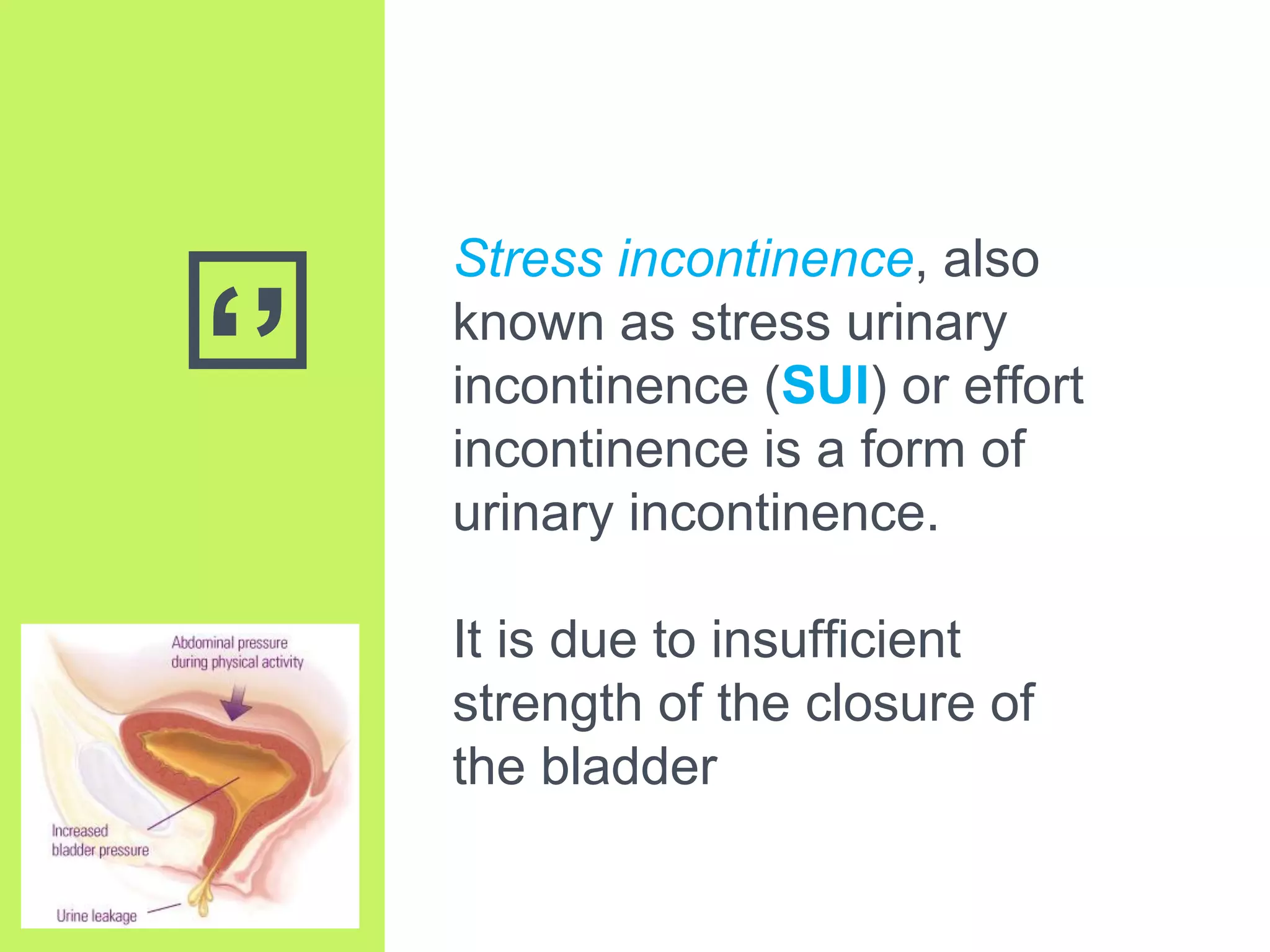
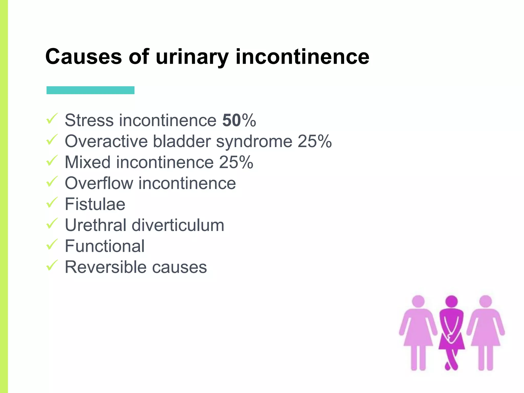
2. Contents
Defining SUI and types
Symptoms & etiology
Pathophysiology
Prevention
Diagnostic approach
Non-surgical and surgical management
Conclusion
3. ‘’
Stress incontinence, also
known as stress urinary
incontinence (SUI) or effort
incontinence is a form of
urinary incontinence.
It is due to insufficient
strength of the closure of
the bladder
4. Urinary incontinence
Urinary incontinence is a condition
in which involuntary loss of urine is
a social or hygienic problem and is
objectively demonstrable
International Continence Society
(ICS), 2002
“The complaint of any involuntary
leakage of urine.”
6. Prevalence
After detailed assessment of data from 48
epidemiological studies, conducted over 40 years,
hampel at al found that over all prevalence of UI ranged
from 4.5 – 53%.
SUI is most common type of urinary incontinence in
women, with 78% of incontinent women presenting with
the symptoms of SUI.
8. Stress urinary incontinence
Sir. Eardly Holland in 1922
introduced the term SUI
Stress urinary incontinence is defined
by the international continence
society (ICS) as:
“the complaint of involuntary leakage
of urine on effort or exertion, or on
sneezing or coughing”
9. Genuine stress urinary incontinence
Urinary loss which occurs with
sudden elevation of the intra
abdominal pressure without
detrusor contraction is called
stress urinary incontinence
10. SUI – symptoms
o Involuntary leakage of urine on effort or exertion, or
on sneezing or coughing
o Usually small amounts
o Pressure in the bladder exceeds the urethral
pressure
o No bladder contraction
12. SUI – grading
Grade 0
Incontinence without leakage
Grade 1
Incontinence with only severe stress, such as coughing,
sneezing, and jogging
Grade 2
Incontinence with moderate stress, such as fast walk,
going up and down the stairs
Grade 3
Incontinence with mild stress such as standing
13. SUI – types
Type 1
Incontinence due to loss of posterior urethrovesical
angle alone
Type 2
Incontinence due to loss of posterior urethrovesical
angle as well as urethral hypermobility
Type 3
Incontinence due to ISD (intrinsic sphincteric deficiency)
15. SUI – Types
Types During Rest During Stress
Bladder base
position (IMPS)
Bladder neck &
Proximal urethra
Bladder base
position
Bladder neck &
Proximal urethra
Type 0 Normal position closed
Rotational
descent No leakage
Type 1 Normal position closed
Descent
2cm
Open
leakage ve
Type 2A Normal position closed Rotational descent
Open
Leakage
Type 2B At or below closed Further descent
Open
Leakage
Type 3 or
ISD
Normal position Open No descent Open and leakage
17. The Pathophysiology of Stress Urinary
Incontinence: A Historical Perspective
▣ Urethrocele is dislocation of urethra and it is the
cause of incontinence (Mann’s American System of Gynecology 19 cent.)
▣ Kelly invented cystoscope in 1914
“The cystoscopic picture presents a gaping internal
sphincter orifice which closes sluggishly”
▣ Bonney attributed SUI to vesical neck funneling and
he hypothesized cause of this is:
loss of elasticity of urethral and vesical sphincter.
▣ Incontinence caused by sagging of pubocervial
muscle sheet which interfere with the sphincter
mechanism (bonney 1923)
18. The Pathophysiology of Stress Urinary
Incontinence: A Historical Perspective
▣Kennedy, 1923 suggested injury to the urethral
sphincter as the principal etiology of SUI
▣Funneling of the bladder floor towards the urethra and
flattening of urethro vesical angle of the bladder showed
in sagittal image in cystogram (1937)
▣SUI is now thought to be due to abnormality in urethra.
On MRI of the pelvic floor SUI was associated with
unequal movement of anterior and posterior wall of
bladder neck and urethra in the presence of increased
intra abdominal pressure
19. Sphincteric Dysfunction Theory
Agency for Health Care Policy and Research, 1992
SUI: the condition of ISD “intrinsic sphincteric
deficiencyʼʼ
In this condition, the
urethral sphincter is
unable to generate
enough resistance to
retain urine in the
bladder especially during
stress maneuver
20. Hammock hypothesis
In 1996, De Lancey proposed a consolidated theory
He hypothesized that the pubocervical fascia
provides a hammock like support for the vesical neck
and there by creates a backboard for the
compression of proximal urethra during increased
intra abdominal pressure.
Loss of this support would compromise equal
transmission of intra abdominal pressure.
campbell walsh urology: expert consult
23. Prevention of SUI
▣3 months postpartum the women in the planned
cesarean delivery group reported less urinary
incontinence than those in the planned vaginal birth
group (4.5% versus 7.3%; relative risk 0.62, 95% CI
0.41– 0.93)
▣In a prospective randomized trial, 17 episiotomy did
not protect women from developing stress urinary
incontinence after delivery.
Outcomes at 3 months after planned cesarean vs planned
vaginal delivery for breech presentation at term: the International
Randomized Term Breech Trial. JAMA 2002;287:1822–31.
Sleep J, Grant A, Garcia J, Elbourne D, Spencer J, Chalmers
I. West Berkshire perineal management trial. Br J Med
1984;289:587–90.
24. Prevention of SUI
▣Follow-up study 5 years after a randomized trial
comparing forceps and vacuum-assisted deliveries,
symptoms of urinary incontinence were common and
did not differ between the groups.
▣At this time, there is insufficient evidence to determine
whether pelvic floor muscle training can prevent stress
incontinence.
Maternal and child health after assisted vaginal delivery: five-
year follow up of a randomized controlled study comparing
forceps and ventouse. Br J Obstet Gynecol 1999;106:544 –9.
Hay-Smith J, Herbison P, Morkved S. Physical therapies
for prevention of urinary and faecal incontinence in adults
(Cochrane Review). In: The Cochrane Library, Issue 2, 2002.
26. The primary goals of evaluation of
women presenting with symptoms of SUI
1. Provide a clinical diagnosis of stress incontinence
versus overactive bladder symptoms
2. Determine factors that may contribute to symptoms
or that may require further evaluation
3. Assess whether coexisting pelvic floor disorders,
such as pelvic organ prolapse or anal incontinence,
are present
4. Establish baseline stress urinary incontinence
severity to aid in assessing treatment effects,
5. Determine the impact of the patient’s symptoms on
her quality of life.
27. The primary goals of evaluation of
women presenting with symptoms of SUI
It is also important to determine:
which treatment options are acceptable to each
patient
what her own therapeutic goals are
and to provide her with appropriate education
regarding these goals.
29. Basic evaluation: Careful history
1. Frequency and amount of leakage
2. Precipitating factors
3. Impact of the leakage on daily life and pad use
4. Pelvic floor symptoms, such as a sensation of bulge
or pressure in the vagina, urinary urgency or
frequency, nocturia, hematuria, recurrent urinary
tract infections, voiding problems, anal incontinence,
and defecating dysfunction
30. Basic evaluation: physical examination
1. Pelvic examination to rule out pelvic or abdominal
masses, pelvic organ prolapse, and vaginal atrophy.
2. A positive cough stress test, in which leakage is
visualized at the moment of the cough, is helpful to
confirm the diagnosis
3. On vaginal or rectal examination, check the pelvic
floor muscles quality (symmetry and bulk) and
whether or not, and to what degree, a woman can
volitionally contract her muscles
31. Basic evaluation: physical examination
4. Cotton swab test
(test of urethral mobility)
5. To rule out urinary retention and overflow
incontinence, we assess the postvoid
residual volume by either direct
catheterization or by ultrasonography
(Most experts agree that a postvoid residual volume less
than 50 mL is normal and more than 200 mL is abnormal)
32. Basic evaluation: physical examination
6. Finally, a urinalysis is done to rule out urinary tract
infection as a transient cause of stress urinary
incontinence.
33. Advanced evaluation
Further evaluation is needed after basic testing if we are
still left with an uncertain diagnosis:
Major discrepancies between the history, the voiding diary
and symptom scales
When Pt is considered for surgery
If the patient has hematuria in the absence of an infection
An elevated postvoid residual volume, a neurologic
condition that may complicate treatment (such as multiple
sclerosis),
marked pelvic organ prolapse, or numerous prior surgical
attempts at correction
34. Advanced evaluation for SUI
Urethroscopy
Provides Information about:
Openning pressure
Urethritis
Diverticula
Rigid urethra
Urethrovesical junction
35. Advanced evaluation for SUI
Uroflowmetry
Volume of urine passed is plotted over time.
Flow time, peak flow rate, and time to peak flow
increase with the volume voided.
36. Advanced evaluation for SUI
Cystometrogram / cystometry
done to assess bladder and urethral function during
bladder filling.
Cystometry is termed simple when only bladder
pressure is recorded and complex (or multichannel)
when both bladder and abdominal pressure
(measured via a rectal or vaginal catheter) are
recorded.
37. Advanced evaluation for SUI
Cystometrogram / cystometry
Multichannel
cystometrogram showing
detrusor overactivity. Note
that at the peak of the
detrusor contractions, the
patient leaked urine, and
the detrusor pressure then
gradually decreased. No
increase in abdominal
pressure over baseline is
seen.
38. Advanced evaluation for SUI
Cystometrogram / cystometry
Multichannel cystometrogram showing
urodynamic stress incontinence. Each
spike represents a Valsalva effort.
The abdominal pressure generated by
the Valsalva is transmitted to the
bladder as seen by the increased
vesical pressure.
However, the detrusor pressure
remains low, and leakage (black
arrows) occurs in the absence of a
detrusor contraction.
39. Advanced evaluation for SUI
Urodynamic studies
o Cystometry: measurement of pressure within the
bladder and urethra during artificial filling
o Uroflowmetry: urine flow rate and volume
o Micturition cystourethrography: for posterior urethro
vesical angle
40. Advanced evaluation for SUI
Other studies
o Ultrasound: for bladder volume and residual urine
o Video-cystourethrography: it combines the pressure
studies with video position of bladder neck and
urethrovesical angle
o MRI: to detect the defect in pelvic floor muscle and
supporting fasciae
42. Management of SUI
A. Non-surgical treatment of SUI
i. Lifestyle interventions
ii. Pelvic floor muscle training
iii. Medications
iv. Devices
B. Surgical treatment of SUI
i. Use of injectable bulking agents,
ii. Laparoscopic suspensions
iii. Midurethral slings
iv. Pubovaginal slings
v. Open retropubic suspensions
43. Non-surgical treatment of SUI:
Life style interventions
Weight reduction
Postural changes (such as crossing legs) often
prevent stress urinary incontinence.
Fluid intake and voiding habits (decreasing the fluid intake
is helpful in for patient with high fluid consumption & voiding
prior to strenous activity beneficial in mild SUI)
44. Non-surgical treatment of SUI:
Pelvic floor muscle training
Medical evidence from well-designed randomized
clinical trials show that supervised pelvic floor muscle
training (Kegel exercises) is an effective treatment for
stress urinary incontinence.
Should be offered as first-line conservative
management to women.
45. Non-surgical treatment of SUI:
Pelvic floor muscle training
Several factors are important in maximizing the chance
that pelvic muscle training will alleviate SUI:
The woman must do the exercises correctly,
regularly, and for an adequate duration.
Many physical therapists recommend training
sessions 3–4 times per week, with 3 repetitions of 8–
10 sustained contractions each time.
46. Non-surgical treatment of SUI:
Medications
Estrogen
Estrogen has tropical effects on urethral epithelium
subepithelial vascular plexus and connective tissues
Fentl at al reviewed 23 articles and found patients
had subjective improvement but not there is no
improvement in objective parameters
47. Non-surgical treatment of SUI:
Medications
Alpha-adrenoreceptor agonist
Ephedrine, Norephedrine and Midorine
Have shown only modest effect in small trials
Nygaard IE, Kreder KJ. Pharmacologic therapy of lower
urinary tract dysfunction. Clin Obstet Gynecol 2004;47:
83–92.
48. Non-surgical treatment of SUI:
Medications
Serotonin and norepinephrine reuptake inhibitors
Norton et al: multicenter, double-blind, placebo-
controlled randomized trial to study the safety and
effectiveness of duloxetine chloride for the treatment
of SUI
There was an 18–23% net difference in the
percentage reduction in incontinence episode
frequency comparing duloxetine chloride 40 mg/d
(59% reduction) and 80 mg/d (64% reduction) with
placebo (40% reduction). Norton P, Zinner N, Ilker Y, Bump R. Duloxetine versus
placebo in the treatment of stress urinary incontinence.
Am J Obstet Gynecol 2002;187:40–8.
49. Non-surgical treatment of SUI:
Devices
▣Ring pessary
The pessary compresses the
urethra against the symphysis
pubis and elevates the bladder
neck.
For some women this may reduce
stress leakage
50. Minimally invasive and surgical options
For women in whom Kegel exercises are ineffective and
who desire defenitive surgery, 5 procedures are
endorsed by the American Urological Association:
1. Use of injectable bulking agents,
2. Laparoscopic suspensions (laparoscopic “Burch”
colposuspension)
3. Midurethral slings
4. Pubovaginal slings
5. Open retropubic suspensions
51. Minimally invasive and surgical options:
Injectable bulking agents
▣ Bulking agents are injectable materials placed at the
bladder neck to improve continence.
▣ Several different bulking agents are available
including silicone particles, carbon beads, calcium
hydroxyapatite, ethylene vinyl alcohol copolymer,
porcine dermal implants, etc
▣ Although less effective than surgery, these agents
are a reasonable option for women with multiple
comorbidities who are poor surgical candidates and
desire short term symptomatic relief.
53. Minimally invasive and surgical options:
Burch colposuspension and fascial slings
▣ The Burch procedure involves suspending the
anterior vaginal wall to the ileopectineal (Cooper’s)
ligament
▣ In women with stress urinary incontinence, success
rates were higher for those in the pubovaginal sling
group (66% vs 49%; P<0.001)
55. Minimally invasive and surgical options:
Midurethral synthetic slings
▣ In recent years, midurethral synthetic slings have
replaced pubovaginal slings as the gold standard for
surgical correction of stress urinary incontinence
▣ The placement of a sling is minimally invasive and is
usually performed in an outpatient setting.
▣ They can be placed either retropubically, as in the
classic tension-free vaginal tape procedure, or
through the transobturator tape approach
57. Choice of sling
A systematic review and meta-analysis of sling surgery for SUI
recommends the use of either tension-free vaginal tape or
transobturator tape slings for objective and subjective cure
Pubovaginal slings are recommended over Burch procedures to
maximize cure
Midurethral slings are recommended over pubovaginal slings for
better subjective cure
Single incision mini-slings have gained popularity as an option
with potentially fewer complications. However, mini-slings have
lower subjective and objective cure rates and higher reoperation
rates when compared with traditional midurethral slings
58. To conclude,
Stress urinary incontinence is common in women and may
impact their activity levels and quality of life.
Conservative management should precede surgery.
Minimally invasive treatment measures can readily be initiated
by primary care providers, with referral to a specialist when
conservative management is not effective
Many gaps remain in our knowledge of this disorder. Further
research is also needed to study prevention of leakage, factors
that impact treatment success, and longevity of various
therapies.
New therapies should be studied in randomized clinical trials
before general clinical use.