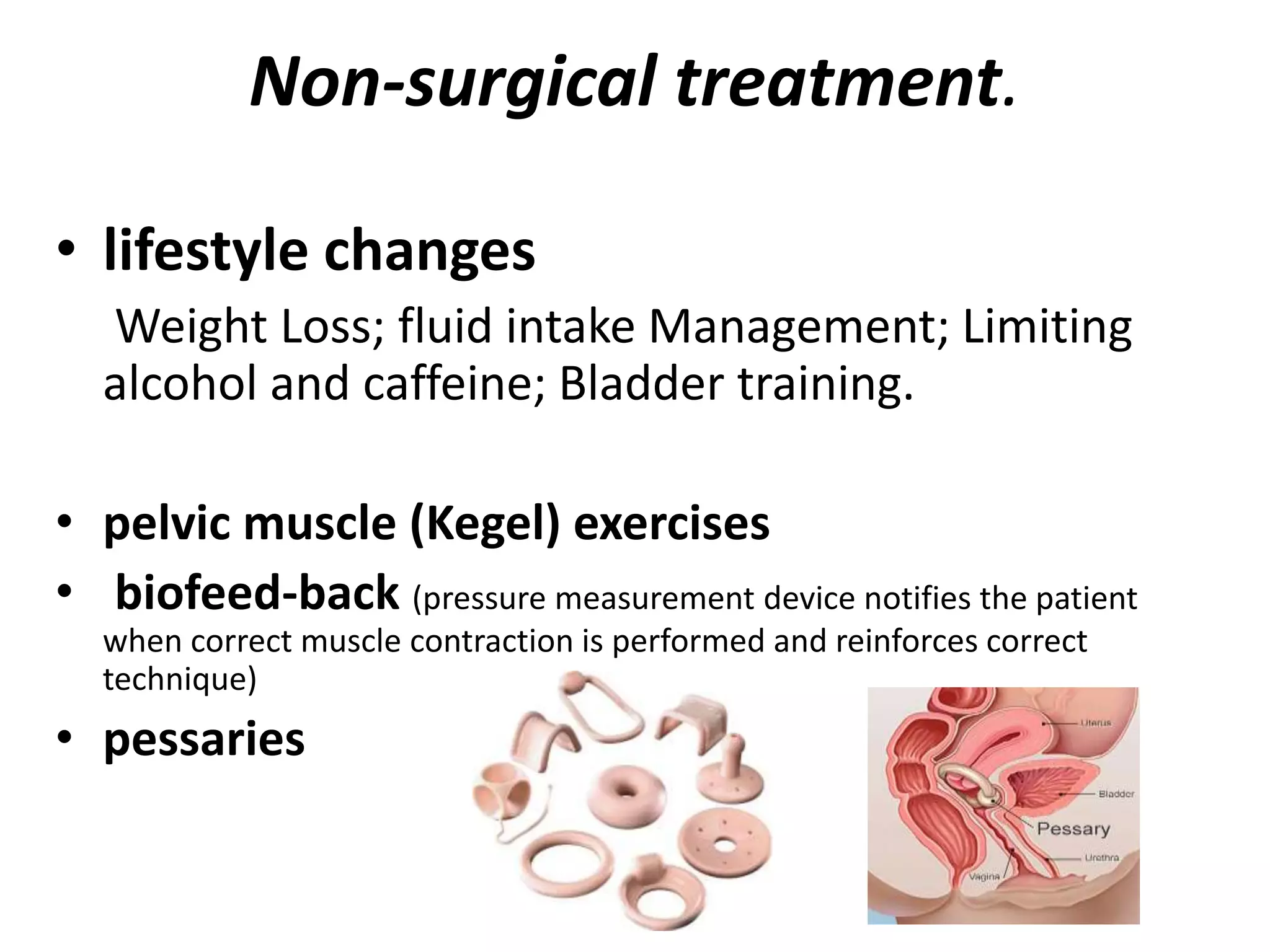
Urinary ncontinence
Definition
• Urinary incontinence simply means
involuntary leaking of urine.
• Incontinence can range from leaking
just a few drops of urine to
complete emptying of the bladder.
• Social and hygienic problem.
Prevᵃlence
• Incidence: increases with age: 4-8 %
ultimately seek medical attention.
• One of three women over 60y has bladder
control problems.
Mechanism
• Continence and urination involve balance
between urethral closure and detrusor (bladder
smooth muscle) activity.
• Urethral pressure normally exceed bladder
pressure, resulting in urine remaining in the
bladder. Intra-abdominal pressure increases
(coughing, sneezing) normally are transmitted in
both urethra and bladder equally, maintaining
continence. Disruption of this balance leads to
various types of incontinence.
causes
• Urinary tract infection (UTI)—UTIs sometimes cause leakage and are
treated with antibiotics.
• Diuretic medications, caffeine, or alcohol—Incontinence may be a side
effect of substances that cause your body to make more urine.
• Pelvic floor disorders—These disorders are caused by
weakening of the muscles and tissues of the pelvic
floor and include urinary incontinence, accidental
bowel leakage, and pelvic organ prolapse.
• Constipation—Long-term constipation often is present in women with
urinary incontinence, especially in older women.
• Neuromuscular problems—When nerve (neurologic) signals from the
brain to the bladder and urethra are disrupted, the muscles that control
those organs can malfunction, allowing urine to leak.
• Anatomical problems—The outlet of the bladder into the urethra can
become blocked by bladder stones or other growths.
What other symptoms occur with urinary
incontinence?
It is common for other symptoms to occur along with
urinary incontinence:
• Urgency !! —Having a strong
urge to urinate
• Frequency—Urinating (also
called voiding) more often than
what is usual for the patient
• Dysuria—Painful urination
• Nocturia—Waking from sleep
to urinate
• Nocturnal enuresis—Leaking
urine while sleeping
Diagnosis (6 basis components)
▲ History - severity of symptoms, rule out medication cause, stress
correlates with amount of urine loss.
▲ Physical examination;
• General examination
• Neurologic screening examination –reflexes
• Urogynecologic examination: may reveal severe vulvar excoriation from
continual dampness. Vaginal tissue atrophy, stenosis.. The patient is asked
to cough or Valsalva repeatedly with a full bladder in the lithotomy or
standing position to induce urine leakage.
▲ Urinalysis and urine culture. To rule out inf before further
evaluation.
▲ Residual urine volume after voiding. Catheterization
▲ Frecuency – volume bladder chart.,,,
▲ Urodynamics: Cystometry, Uroflowmetry, Complex urodynamic tests.
Urodynamics
– Simple cystometry involves placing a catheter and gradually
filling the bladder with sterile water. Involuntary “detrusor”
contractions are demonstrated by a rise in water level during
filling due to back-pressure. Normally, the first sensation to
void occurs at 150 mL and bladder capacity is typically 400–
600 mL.
– Uroflowmetry is used to determine the urinary flow rate and
flow time to screen for the presence of outflow obstruction
and abnormal detrusor contractility. Normally, women
achieve a peak flow rate of 15–20 mL/s with a voided volume
of 150–200 mL.
– Complex urodynamic testing requires placement of an
intravesical catheter to measure detrusor pressures and a
vaginal or rectal catheter to indirectly measure
intraabdominal pressures.
types of urinary incontinence
• Stress urinary incontinence (SUI)
• Urge incontinence
• Overflow incontinence
Stress urinary incontinence
(SUI)
• Patients have loss of small amounts of urine with
coughing, laughing, sneezing, exercising, or other
movements that increase intra-abdominal pressure and
thus increase pressure on the bladder.
Etiology
• Physical changes resulting from pregnancy, childbirth,
and menopause often result in weaknesses in the
pelvic floor, and urethral support structures and nerve
damage
Stress urinary incontinence
(SUI)
• Mechanism. If the fascial support is weakened, the
urethra can move downward at times of increased
abdominal pressure, causing bladder pressure to
exceed urethral sphincter closure pressure (hyper-
mobile urethra). Incomplete urethral closure may be
due to scarring or neuromuscular damage, and cause a
more severe form of stress urinary incontinence –
intrinsic sphincter deficiency.
• Diagnosis. SUI is suggested by history, physical
examination, and a positive stress test (demonstrable
loss of urine while the patient is being examined).
Non-surgical treatment.
• lifestyle changes
Weight Loss; fluid intake Management; Limiting
alcohol and caffeine; Bladder training.
• pelvic muscle (Kegel) exercises
• biofeed-back (pressure measurement device notifies the patient
when correct muscle contraction is performed and reinforces correct
technique)
• pessaries
pelvic muscle (Kegel) exercises
• Find the right muscles. To identify your pelvic floor muscles, stop
urination in midstream. If you succeed, you've got the right
muscles. Once you've identified your pelvic floor muscles you can
do the exercises in any position, although you might find it easiest
to do them lying down at first.
• Perfect your technique. Tighten your pelvic floor muscles, hold the
contraction for five seconds, and then relax for five seconds. Try it
four or five times in a row. Work up to keeping the muscles
contracted for 10 seconds at a time, relaxing for 10 seconds
between contractions.
• Maintain your focus. For best results, focus on tightening only your
pelvic floor muscles. Be careful not to flex the muscles in your
abdomen, thighs or buttocks. Avoid holding your breath. Instead,
breathe freely during the exercises.
• Repeat three times a day. Aim for at least three sets of 10
repetitions a day.
Surgical treatment
• Tension-free transvaginal tape (TVT) or
transobturator tape (TOT) are minimally invasive
suburethral sling procedures that are rapidly
becoming the “gold standard”
• Burch colposuspension
• Anterior colporrhaphy
• Collagen periurethral
Urge incontinence
• Patients experience involuntary leakage for no apparent
reason while suddenly feeling an urgent need to urinate.
This may be accompanied by urinary frequency and
nocturia, and patients often describe their bladder as
“spastic” or “overactive”.
• Etiology. Involuntary detrusor muscle contractions.
Detrusor hyperactivity can be due to loss of central nervous
system (CNS) inhibitory pathways, local irritants, or bladder
outlet obstruction.
• Mechanism. Frequently idiopathic, but results from
damage to the nerves of the bladder, the nervous system
(spinal cord and brain), or the muscles themselves.
Urge incontinence
• Treatment. Behavior modification (bladder
drills, biofeedback) and/or pharmacologic
therapy (oxybutynin chloride, imipramine,
mirabegron), injection of the detrusor muscle
with botulinum toxin A, neuromodulation.
Overflow incontinence
• Patients experience continuous, unstoppable
dribbling of urine, or continuing to dribble for
some time after they have passed urine.
• Etiology. The bladder is always full and
overflows, resulting in frequent or continuous
urine leakage.
Overflow incontinence
• Mechanism. Weak bladder detrusor muscles,
resulting in incomplete emptying, or a blocked
urethra (outflow obstruction) due to pelvic
organ prolapse, or after an anti-incontinence
procedure that has overcorrected the problem.
• Treatment. Catheter drainage, followed by
treatment of the underlying condition.
pelvic organ prolapse
• Descent of one or more pelvic organs (uterine
cervix, vaginal apex, anterior vagina, posterior
vagina, or cul de sac peritoneum) through the
pelvic flor into the vaginal canal.
• ½ of parous women have prolapse on
examination
• 10% will undergo surgery for prolapse or urinary
incontinence in their lifetime
• Prolapse is the most common indication for
hysterectomy in women after age 55.
Cystocele
• Surgical options
• Anterior colporrhaphy involves vaginally plicating
the endopelvic fascia in the midline to provide
support and raise the bladder to correct its
anatomic position.
• Paravaginal repair replaces the anterolateral
vaginal wall to its anatomic position.
• The McCall culdoplasty shortens the uterosacral
ligaments and reattaches them to the vaginal
apex.
Rectocele
• Surgical options
• Posterior colporrhaphy mimics the anterior procedure with a
midline plication of endopelvic fascia. Perineorrhaphy is commonly
required due to an attenuated perineal body or widened genital
hiatus.
• Enterocele
• Surgical options
• As an enterocele is a true herniation of the peritoneal cavity at the
pouch of Douglas which bulges into the rectovaginal septum, repair
is usually performed at the same time as posterior colporrhaphy.
The hernia sac is visualized as the vagina is separated from the
rectum and it must be dissected free of underlying tissue. The neck
of the hernia is then isolated and sutured. Fixing the uterosacral
ligaments to the sac will help prevent recurrence.
Uterine procidentia
(Prolapse)
Surgical options
• TVH is common, but anterior and posterior colporrhaphy
generally do not provide sufficient long-term apical support
• Sacrospinous ligament suspension (SSLS) may be
concomitantly performed vaginally by suspending the fascia
of the apex to one or both ligaments.
• Abdominal sacrocolpopexy with total abdominal
hysterectomy (TAH) is another reasonable option that has less
apical failure, post- operative dyspareunia, and stress
incontinence than SSLS, but is associated with longer surgical
time, longer patient recovery, and more short- and long-term
complications. Laparoscopic and robotic-assisted techniques
are the preferred option due to reduced recovery times.
• Colpocleisis (Lefort procedure
Thank You

Urine incompet

  • 1.
  • 2.
    Definition • Urinary incontinencesimply means involuntary leaking of urine. • Incontinence can range from leaking just a few drops of urine to complete emptying of the bladder. • Social and hygienic problem.
  • 3.
    Prevᵃlence • Incidence: increaseswith age: 4-8 % ultimately seek medical attention. • One of three women over 60y has bladder control problems.
  • 4.
    Mechanism • Continence andurination involve balance between urethral closure and detrusor (bladder smooth muscle) activity. • Urethral pressure normally exceed bladder pressure, resulting in urine remaining in the bladder. Intra-abdominal pressure increases (coughing, sneezing) normally are transmitted in both urethra and bladder equally, maintaining continence. Disruption of this balance leads to various types of incontinence.
  • 5.
    causes • Urinary tractinfection (UTI)—UTIs sometimes cause leakage and are treated with antibiotics. • Diuretic medications, caffeine, or alcohol—Incontinence may be a side effect of substances that cause your body to make more urine. • Pelvic floor disorders—These disorders are caused by weakening of the muscles and tissues of the pelvic floor and include urinary incontinence, accidental bowel leakage, and pelvic organ prolapse. • Constipation—Long-term constipation often is present in women with urinary incontinence, especially in older women. • Neuromuscular problems—When nerve (neurologic) signals from the brain to the bladder and urethra are disrupted, the muscles that control those organs can malfunction, allowing urine to leak. • Anatomical problems—The outlet of the bladder into the urethra can become blocked by bladder stones or other growths.
  • 7.
    What other symptomsoccur with urinary incontinence? It is common for other symptoms to occur along with urinary incontinence: • Urgency !! —Having a strong urge to urinate • Frequency—Urinating (also called voiding) more often than what is usual for the patient • Dysuria—Painful urination • Nocturia—Waking from sleep to urinate • Nocturnal enuresis—Leaking urine while sleeping
  • 8.
    Diagnosis (6 basiscomponents) ▲ History - severity of symptoms, rule out medication cause, stress correlates with amount of urine loss. ▲ Physical examination; • General examination • Neurologic screening examination –reflexes • Urogynecologic examination: may reveal severe vulvar excoriation from continual dampness. Vaginal tissue atrophy, stenosis.. The patient is asked to cough or Valsalva repeatedly with a full bladder in the lithotomy or standing position to induce urine leakage. ▲ Urinalysis and urine culture. To rule out inf before further evaluation. ▲ Residual urine volume after voiding. Catheterization ▲ Frecuency – volume bladder chart.,,, ▲ Urodynamics: Cystometry, Uroflowmetry, Complex urodynamic tests.
  • 9.
    Urodynamics – Simple cystometryinvolves placing a catheter and gradually filling the bladder with sterile water. Involuntary “detrusor” contractions are demonstrated by a rise in water level during filling due to back-pressure. Normally, the first sensation to void occurs at 150 mL and bladder capacity is typically 400– 600 mL. – Uroflowmetry is used to determine the urinary flow rate and flow time to screen for the presence of outflow obstruction and abnormal detrusor contractility. Normally, women achieve a peak flow rate of 15–20 mL/s with a voided volume of 150–200 mL. – Complex urodynamic testing requires placement of an intravesical catheter to measure detrusor pressures and a vaginal or rectal catheter to indirectly measure intraabdominal pressures.
  • 10.
    types of urinaryincontinence • Stress urinary incontinence (SUI) • Urge incontinence • Overflow incontinence
  • 11.
    Stress urinary incontinence (SUI) •Patients have loss of small amounts of urine with coughing, laughing, sneezing, exercising, or other movements that increase intra-abdominal pressure and thus increase pressure on the bladder. Etiology • Physical changes resulting from pregnancy, childbirth, and menopause often result in weaknesses in the pelvic floor, and urethral support structures and nerve damage
  • 12.
    Stress urinary incontinence (SUI) •Mechanism. If the fascial support is weakened, the urethra can move downward at times of increased abdominal pressure, causing bladder pressure to exceed urethral sphincter closure pressure (hyper- mobile urethra). Incomplete urethral closure may be due to scarring or neuromuscular damage, and cause a more severe form of stress urinary incontinence – intrinsic sphincter deficiency. • Diagnosis. SUI is suggested by history, physical examination, and a positive stress test (demonstrable loss of urine while the patient is being examined).
  • 13.
    Non-surgical treatment. • lifestylechanges Weight Loss; fluid intake Management; Limiting alcohol and caffeine; Bladder training. • pelvic muscle (Kegel) exercises • biofeed-back (pressure measurement device notifies the patient when correct muscle contraction is performed and reinforces correct technique) • pessaries
  • 14.
    pelvic muscle (Kegel)exercises • Find the right muscles. To identify your pelvic floor muscles, stop urination in midstream. If you succeed, you've got the right muscles. Once you've identified your pelvic floor muscles you can do the exercises in any position, although you might find it easiest to do them lying down at first. • Perfect your technique. Tighten your pelvic floor muscles, hold the contraction for five seconds, and then relax for five seconds. Try it four or five times in a row. Work up to keeping the muscles contracted for 10 seconds at a time, relaxing for 10 seconds between contractions. • Maintain your focus. For best results, focus on tightening only your pelvic floor muscles. Be careful not to flex the muscles in your abdomen, thighs or buttocks. Avoid holding your breath. Instead, breathe freely during the exercises. • Repeat three times a day. Aim for at least three sets of 10 repetitions a day.
  • 15.
    Surgical treatment • Tension-freetransvaginal tape (TVT) or transobturator tape (TOT) are minimally invasive suburethral sling procedures that are rapidly becoming the “gold standard” • Burch colposuspension • Anterior colporrhaphy • Collagen periurethral
  • 18.
    Urge incontinence • Patientsexperience involuntary leakage for no apparent reason while suddenly feeling an urgent need to urinate. This may be accompanied by urinary frequency and nocturia, and patients often describe their bladder as “spastic” or “overactive”. • Etiology. Involuntary detrusor muscle contractions. Detrusor hyperactivity can be due to loss of central nervous system (CNS) inhibitory pathways, local irritants, or bladder outlet obstruction. • Mechanism. Frequently idiopathic, but results from damage to the nerves of the bladder, the nervous system (spinal cord and brain), or the muscles themselves.
  • 19.
    Urge incontinence • Treatment.Behavior modification (bladder drills, biofeedback) and/or pharmacologic therapy (oxybutynin chloride, imipramine, mirabegron), injection of the detrusor muscle with botulinum toxin A, neuromodulation.
  • 20.
    Overflow incontinence • Patientsexperience continuous, unstoppable dribbling of urine, or continuing to dribble for some time after they have passed urine. • Etiology. The bladder is always full and overflows, resulting in frequent or continuous urine leakage.
  • 21.
    Overflow incontinence • Mechanism.Weak bladder detrusor muscles, resulting in incomplete emptying, or a blocked urethra (outflow obstruction) due to pelvic organ prolapse, or after an anti-incontinence procedure that has overcorrected the problem. • Treatment. Catheter drainage, followed by treatment of the underlying condition.
  • 22.
    pelvic organ prolapse •Descent of one or more pelvic organs (uterine cervix, vaginal apex, anterior vagina, posterior vagina, or cul de sac peritoneum) through the pelvic flor into the vaginal canal. • ½ of parous women have prolapse on examination • 10% will undergo surgery for prolapse or urinary incontinence in their lifetime • Prolapse is the most common indication for hysterectomy in women after age 55.
  • 24.
    Cystocele • Surgical options •Anterior colporrhaphy involves vaginally plicating the endopelvic fascia in the midline to provide support and raise the bladder to correct its anatomic position. • Paravaginal repair replaces the anterolateral vaginal wall to its anatomic position. • The McCall culdoplasty shortens the uterosacral ligaments and reattaches them to the vaginal apex.
  • 25.
    Rectocele • Surgical options •Posterior colporrhaphy mimics the anterior procedure with a midline plication of endopelvic fascia. Perineorrhaphy is commonly required due to an attenuated perineal body or widened genital hiatus. • Enterocele • Surgical options • As an enterocele is a true herniation of the peritoneal cavity at the pouch of Douglas which bulges into the rectovaginal septum, repair is usually performed at the same time as posterior colporrhaphy. The hernia sac is visualized as the vagina is separated from the rectum and it must be dissected free of underlying tissue. The neck of the hernia is then isolated and sutured. Fixing the uterosacral ligaments to the sac will help prevent recurrence.
  • 26.
    Uterine procidentia (Prolapse) Surgical options •TVH is common, but anterior and posterior colporrhaphy generally do not provide sufficient long-term apical support • Sacrospinous ligament suspension (SSLS) may be concomitantly performed vaginally by suspending the fascia of the apex to one or both ligaments. • Abdominal sacrocolpopexy with total abdominal hysterectomy (TAH) is another reasonable option that has less apical failure, post- operative dyspareunia, and stress incontinence than SSLS, but is associated with longer surgical time, longer patient recovery, and more short- and long-term complications. Laparoscopic and robotic-assisted techniques are the preferred option due to reduced recovery times. • Colpocleisis (Lefort procedure
  • 28.

Editor's Notes

  • #14 In overweight women, losing even a small amount of weight (less than 10% of total body weight) may decrease urine leakage. If the patient has leakage in the early morning or at night, sche may want to limit her intake of fluids several hours before bedtime. Limiting the amount of fluids she drinks also may be useful (no more than 2 liters total a day). The goal of bladder training is to learn how to control the urge to empty the bladder and increase the time span between urinating to normal intervals (every 3–4 hours during the day and every 4–8 hours at night). Biofeedback is a training technique that may help the patient locate the correct muscles. In one type of biofeedback, sensors are placed inside or outside the vagina that measure the force of pelvic muscle contraction. When she contracts the right muscles, the measurement will appear on a monitor. A pessary is a device that is inserted into the vagina to treat pelvic support problems and SUI. Pessaries support the walls of the vagina to lift the bladder and urethra. They come in many shapes and sizes. Usually the patient can insert and remove a support pessary herself. Pessaries may provide relief of symptoms without surgery. An over-the-counter tampon-like device also is available that is designed specifically to help prevent bladder leaks.
  • #16 Burch colposuspension involves suture placement at the Cooper ligament. The Marshall–Marchetti–Krantz (MMK) variation has sutures going through the periosteum of the pubic symphysis. Anterior colporrhaphy has a poor long-term success rate. Collagen periurethral injections (Coaptite, Macroplastique) are designed as a treatment for SUI resulting from intrinsic sphincter deficiency.
  • #20 Drugs that control muscle spasms or unwanted bladder contractions can help prevent leakage from urgency urinary incontinence and relieve the symptoms of urgency and frequency. Mirabegron is a drug that relaxes the bladder muscle and allows the bladder to store more urine. This drug is used to treat urgency urinary incontinence and relieve the symptoms of urgency and frequency. Injection of a drug called onabotulinumtoxinA into the muscle of the bladder helps stop unwanted bladder muscle contractions. The effects last for about 3–9 months. Sacral neuromodulation—This is a technique in which a thin wire is placed under the skin of the low back and close to the nerve that controls the bladder. The wire is attached to a battery device placed under the skin nearby. The device sends a mild electrical signal along the wire to improve bladder function. Percutaneous tibial nerve stimulation (PTNS)—PTNS is a procedure that is similar to acupuncture. In PTNS, a slender needle is inserted near a nerve in the ankle and connected to a special machine. A signal is sent through the needle to the nerve, which sends the signal to the pelvic floor. PTNS usually involves weekly 30-minute office sessions for a few months.